A 37 years old woman presented upper gastrointestinal bleeding. Upper endoscopy and biopsy confirmed a gastric MALT lymphoma (Figure 1). She also presented headache without localization signs at neurological examination. Cerebral MRI showed an expanding tissue tumor process of the left choroid plexus associated with peri-lesional edema (Figure 2). The biopsy could not be realized because high risk of bleeding due to the localization of the tumor.
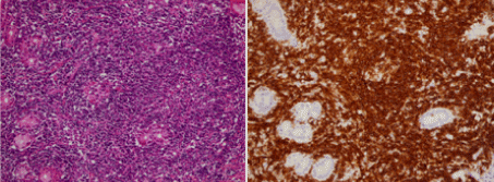
Figure 1: Initial histopathological examination: diffuse and abundant lymphoid infiltrate, realizing lymphoepithelial lesions: (Hematoxylin eosin x 200) and CD20 expression of tumor cells with immunohistochemistry (G x200).
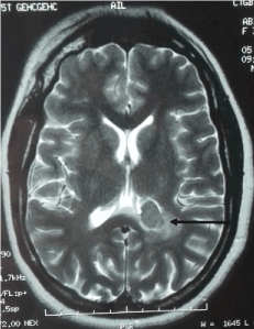
Figure 2: Initial MRI showing an expanding tissue tumor process of the left choroid plexus associated with a signal anomaly opposite: peri-lesional edema (arrow).
Lymphoma was classified stage IV E according to modified Ann Arbor Classification. She received chemotherapy (CHOP protocol). Endoscopic control showed a regression of the lymphoma with histopathological remission according to GELA score. Headache improved after treatment and control MRI (Figure 3) showed partial regression of the tumor after systemic treatment (after 3 months).
2021 Copyright OAT. All rights reserv
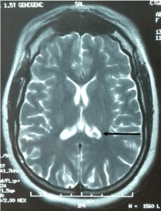
Figure 3: Control Computed tomography showing partial regression of the cerebral lesion (arrow).
Editorial Information
Editor-in-Chief
Prof. Yoshiaki Kikuchi
Tokyo Metropolitan University
Japan
Article Type
Image Article
Publication history
Received date: July 06, 2018
Accepted date: July 12, 2018
Published date: July 16, 2018
Copyright
© 2018 Meriam S. This is an open-access article distributed under the terms of the Creative Commons Attribution License, which permits unrestricted use, distribution, and reproduction in any medium, provided the original author and source are credited.
Citation
Meriam S, Norsaf B, Dorra B, Asma O, Raja J, et al. (2018) Cerebral localisation of a gastric malt lymphoma with partial response after chemotherapy. Neurol Neurosci Rep 1: DOI: 10.15761/NNR.1000108
Corresponding author
Dr Meriam Sabbah
Departement of pathology, Habib Thameur Hospital, Tunis, Tunisia.
E-mail : bhuvaneswari.bibleraaj@uhsm.nhs.uk
Figure 1: Initial histopathological examination: diffuse and abundant lymphoid infiltrate, realizing lymphoepithelial lesions: (Hematoxylin eosin x 200) and CD20 expression of tumor cells with immunohistochemistry (G x200).
Figure 2: Initial MRI showing an expanding tissue tumor process of the left choroid plexus associated with a signal anomaly opposite: peri-lesional edema (arrow).
Figure 3: Control Computed tomography showing partial regression of the cerebral lesion (arrow).