The Bladder exstrophy-epispadias complex is one of the most complex anomalies in Paediatric Urology. This complex anomaly comes with utmost distress to the patient and family due to the apparent physical anomaly and constant urine soiling which is socially unacceptable. The management of bladder exstrophy therefore requires a holistic approach to address all patient components namely, anatomic, functional, cosmetic, sexual, reproductive and psycho-social. The aim of this paper is to apply this holistic approach to highlight the challenges faced in our institution while managing a series of 34 cases with classic BEEC and reviewing literature on these challenges.
bladder exstrophy, holistic care, challenges in management
The management of Bladder exstrophy-epispadias complex (BEEC) is one of the greatest challenges to the pediatric urologist. The aim of a successful repair of bladder exstrophy is successful bladder closure and penile reconstruction in order to provide an adequate low-pressure and competent functioning reservoir as well as a good cosmetic appearance of the genitalia with unimpaired function while ultimately preserving renal function [1].
This complex anomaly comes with utmost distress to the patient and family due to the apparent physical anomaly and constant urine soiling which is socially unacceptable. The surgeon on the other hand is unable to give complete assurance due to the technicalities in surgical repair and the impending risk of poor outcome [1].
The management of bladder exstrophy therefore requires a systematic approach to address all patient components namely, anatomic, functional, cosmetic, sexual, reproductive and psycho-social. This article aims to highlight the need of holistic care in BEEC by featuring the various challenges faced in the management of a series of patients treated in a tertiary facility in Kenya and reviewing literature on these challenging factors.
This is a retrospective case series of patients managed for classic BEEC at Kenyatta National Hospital which is the largest teaching and referral hospital in Kenya, East Africa. Perioperative and follow-up data was collected from electronically retrieved medical records of patients treated between 2010 and 2017. The main variables in this series were categorized based on the clinical characteristics of patients, the outcome of surgery, the psychological assessment and the socio-economic status. The data was presented in text, tables and charts by use of frequencies and proportions based on the categories outlined below. Institutional approval to conduct the case series was sought and granted.
Categorization of results
Clinical characteristics of the patients that directly affect outcome were categorized under the patient factor. These include timing of diagnosis, age at presentation, presence of associated anomalies and the quality of the bladder plate. Timing of diagnosis was classified as either prenatal or postnatal. Isolated BEEC was defined as disorder occurring without the presence of associated systemic anomalies. Patients with poor quality bladder plates that were either fibrotic, polypoid or too small for primary repair were defined as unfavorable.
The surgical procedure done was the Modern Staged Repair of Exstrophy (MSRE) as described by Gearhart [2]. Outcome following surgery was categorized under the surgical factor. These include complications such as repair breakdown and incontinence after repair. Patients who were not able to complete treatment follow-up were also categorized in this section as lost to follow-up. In all patients who underwent urine diversion, the technique performed was uretero-sigmoidostomy with anti-reflux anastomosis as described by Leadbetter [3]. Patients who underwent urine diversion and developed complications are also classified in this category.
All patients and/or parents of children with BEEC in our hospital undergo psychological assessment as part of standard therapy in the management of BEEC. We retrieved data from reports of these assessments and recorded the variables under the psycho-pathologic factor.
The socio-economic factor was assessed based on the geographical origin on marginalization. Marginalized lands in Kenya are arid and semi-arid lands (ASAL) generally associated with low economy, social exclusion and inadequate infrastructure [4,5]. Patients residing in these areas were assessed for presence of medical insurance and treatment follow-up status as an indirect measure of socio-economic status.
The patient factor
A total of 34 patients are presented in this case series. The main challenges faced were lack of prenatal diagnosis, late age at presentation and poor-quality bladder plates which were unfavorable for primary repair. There were 4 patients with associated anomalies, of these 2 patients succumbed due to congenital heart disease. The other two have renal anomalies. Table 1 below summarizes the patient factor component of the participants.
Table 1. Table showing the patient factors affecting outcome
|
Variable
|
No.
n=34
|
%
n=100%
|
|
Sex
|
Male
|
21
|
62.0
|
|
female
|
13
|
38.0
|
|
Timing of Diagnosis
|
Prenatal
|
0
|
0.0
|
|
Postnatal
|
34
|
100.0
|
|
Age at presentation
|
Birth – 1 week
|
7
|
20.6
|
|
1 week – 12 weeks
|
6
|
17.6
|
|
More than 12 weeks
|
21
|
61.8
|
|
Associated anomalies
|
Isolated BEEC
|
30
|
88.2
|
|
Associated Anomalies
|
4
|
11.8
|
|
Quality of bladder plate
|
Favorable
|
23
|
67.6.
|
|
Unfavorable
|
11
|
32.4
|
Surgical factor
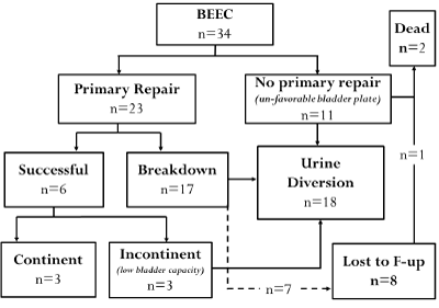
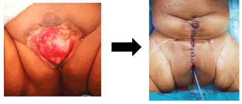
Figure 1 below summarizes the surgical journey of the 34 participants in this series. The mean age at surgery was 13 months. Eleven patients had unfavorable bladder plates and did not undergo primary repair. Three patients with favorable plates were continent after primary repair and all were female (Figure 2). The main challenges faced in those who had primary repair were; late timing of surgery, breakdown after surgery (failed repair) and incontinence after repair which was primarily due to small bladder capacity. Additionally, a total of 8 patients were lost to follow up after failed primary repair

Figure 1. Chart showing the surgical outcome after primary repair

Figure 2. Wide favorable bladder plate before and after repair
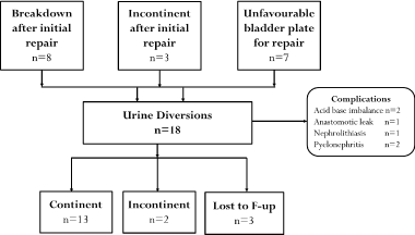
The indications for urine diversion in this series were; breakdown after primary repair, incontinence after repair and unfavorable bladder plates for repair. Figure 3 below summarizes the outcomes after urinary diversion. Continence rates of 72% were achieved after urine diversion. However, there were 2 patients with incontinence and 3 patients who were lost to follow-up. A total of 6 patients encountered complications related to urine diversion.

Figure 3. Chart showing surgical outcome after urine diversion
The psycho-pathologic factor
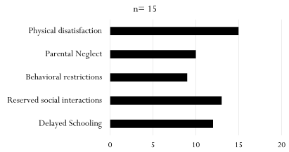
Data on 15 patients who underwent psychological assessment was retrieved. Most patients suffered from physical dissatisfaction and reserved social interactions. The results are summarized in Figure 4.

Figure 4. Graph showing the psychological impact of bladder exstrophy
The socio-economic factor
The socioeconomic factor is associated with geographic marginalization. The impact is seen more on patients residing from arid and semi-arid lands (ASAL). Patients from these areas are seen to present late to hospital, have no medical insurance and are often lost to follow up. Table 2 describes this association.
Table 2. Table showing the socioeconomic impact in bladder exstrophy
|
Variable
|
ASAL
n=19
|
Non-ASAL
n=15
|
Total
|
|
Presentation
|
Early
|
3
|
10
|
13
|
|
Late
|
16
|
5
|
21
|
|
Follow-up
|
Ongoing
|
9
|
13
|
22
|
|
Lost
|
10
|
2
|
12
|
|
Medical Insurance
|
Registered
|
5
|
11
|
16
|
|
Unregistered
|
14
|
4
|
18
|
In the quest to develop a model that integrates holistic care in BEEC, highlighting the challenges faced on various aspects that affect outcome is warranted. In this case series, we outline the journey of 34 patients with classic BEEC treated in a tertiary hospital in Kenya with particular emphasy on the various factors that require address during the management of this complex anomaly.
The patient factor
The patient related factors include, sex, age at presentation, associated anomalies and the quality of the bladder plate. Sex appears to play a role in the outcome after surgery. Interestingly, in an Indian experience 6 girls were noted to be completely dry immediately after primary bladder closure alone, unlike male counterparts who usually require additional procedures to achieve continence [6]. Consequently, in our series we note that all successful repairs with continence achieved were in female patients.
BEEC can be diagnosed prenatally as early as 15 weeks gestation via a prenatal ultrasound or a fetal MRI scan. Features of a prenatal scan suggestive of BEEC include absence of bladder filling, low-set umbilicus, pubic bone diastasis, diminutive genitalia and a lower abdominal mass that increases in size with progression of gestation [7]. The benefits of prenatal diagnosis include. Early prenatal parental counselling, plan for delivery in a center with expertise and early surgical repair. None of the patients in our series had a prenatal diagnosis of BEEC, a factor that led to late age of presentation.
Associated anomalies occur in approximately 30 – 60% of patients with BEEC. The spectrum and prevalence of associated anomalies include GUT 36%, cardiac 6%, GIT 7% and musculoskeletal anomalies 40%. In this German multicenter study, there is evidence to support increased incidence of congenital heart disease in patients with BEEC [8]. Four patients in our series had associated anomalies, two of which died as a result of complications related to congenital heart disease.
The quality of the bladder plate at the time of surgery presents a challenge to the surgeon. Histological studies carried out on the exstrophied mucosa shows various degrees of inflammation, fibrosis and polyposis [9-11]. Such degenerative changes are thought to occur primarily due to chronic environmental exposure. In our series we experienced difficulty in offering primary closure to patients with poor quality plates, most of these patients presented to hospital after 6 months of age and therefore suffer from chronic environmental insults on the bladder mucosa.
The surgical factor
The approaches to repair of BEEC include the modern staged repair (MSRE), the complete primary reconstruction of bladder exstrophy (CPRE) and the radical soft tissue mobilization (RSTM). In all these repairs the most important factor is the expertise of the surgeon performing the repair [2,12,13]. This is a challenge in our setup as there are only a few number of surgeons and centers with the ability to perform such repairs.
Timing of primary closure is a matter under controversy. Initial closure performed within 72 hours of life is thought to prevent environmental injury of the bladder mucosa, ensure early bladder cycling which is important to increase bladder capacity and bladder musculature development. This early closure also provides better pelvic approximation due to greater pliability of the neonatal pelvic bones [14,15]. However delayed approach has gained popularity as recent studies show that repair done within 6 -12 weeks of age is associated with better tolerance to anesthesia and analgesia; mature renal function and adequate time for mother-child bonding. Additionally, there is evidence citing no much histological changes on the bladder mucosa [1]. In our series timing of surgery was dependent on time of presentation and quality of bladder plate. Half of the patients who presented after the neonatal period had fibrotic and polypoid plates unamenable to primary repair. Gearhart and colleagues document a 58% rate of breakdown with bladder dehiscence and prolapse after initial repair [16]. In our series we experienced a high rate of repair breakdown which may be attributed to delayed presentation and lack of osteotomy procedures.
The role of osteotomy in management of BEEC is to enable a tension free symphyseal approximation and abdominal wall closure while allowing the bladder to sit deep into the pelvis [17]. However, its use remains controversial and there is no consensus regarding the necessity of this procedure with arguments raised on the recurrence of pelvic diastasis after bladder exstrophy surgery with or without osteotomy [18-20]. In our series none of the patients underwent osteotomy due to both technical and resource-based difficulties faced in our setup in offering this approach.
Incontinence after successful repair of BEEC occurs due to several factors which include wide bladder neck, small bladder capacity, urinary obstruction and failure to do adequate toilet training [1,21,22]. In this regard surgical technique with bladder neck reconstruction at an appropriate age usually after 4 years provides continence by allowing resistance whilst avoiding a tight repair which can cause urinary obstruction with overflow incontinence and risk of renal impairment [15]. In patients with small bladder capacities after initial repair, bladder augmentation with clean intermittent catheterization (CIC) is the preferred option for increasing bladder capacity and achieving continence, this however requires careful co-operation by the caregivers to ensure safe CIC practice [23]. Our setup does not favor this approach due to low socio-economic backgrounds, low levels of education and poor access to clean water. In this regard we offer urine diversion as an alternative approach to patients with small bladder capacities or those with poor bladder plates unamenable to repair.
Urine diversion offers the advantage of creation of a low-pressure reservoir with a continence mechanism that provides acceptable protection for the upper tracts [15]. Our indications for urinary diversion were breakdown after initial repair, incontinence after initial repair due to small bladder capacity and poor-quality bladder plates unamenable to initial repair. In our series urine diversion was offered to almost half of all patients with classic BEEC and the resultant outcomes were favorable. However, evident from this study, urine diversion is associated with complications such as acid base imbalances, stone disease, anastomotic leaks, infections and malignancy [24].
The psycho-pathologic factor
Children with BEEC suffer from a psychopathologic component which is more or less likely to be overlooked by the more apparent surgical aspects. The physical defect is obvious and may demand multiple surgeries. Thereafter the child is faced with the difficulties of mastering urinary control at atypical ages and growing up with an anomalous genital appearance and abnormal genito-urinary function, the greatest being urinary leak and soiling [25].
A systematic review revealed that 50% of the adolescents with bladder exstrophy and epispadias met the criteria for psychiatric diagnoses and 36% had scores implying significant psychosocial dysfunction [26]. In our series a psychological assessment concluded that patients with BEEC suffered from psychological disturbances including physical dissatisfaction, parental neglect, reserved social interactions and delayed schooling. This psychopathological factor needs to be incorporated in the holistic management of BEEC with considerations that the predictors of psychological function are parental warmth, the patients' genital appraisal, urinary continence function and future sexual abilities [26].
The socio-economic factor
The incidence of BEEC varies with socioeconomic status, geographical distribution and insurance status [27]. As a determinant of disease, this increased risk with low socioeconomic status may be related to lack of access to adequate prenatal care, maternal exposure to teratogens and increased maternal age and parity [28]. Furthermore, the challenges faced by such mothers after the child with BEEC is born includes delay in seeking health care, lack of access to health care, lack of medical insurance and lost to follow-up. All these factors are seen affecting patients in our series. In Kenya only 36% of the total population resides in arid and semi-arid lands (ASAL), therefore it is a significant finding that a total of 56% of patients with BEEC hail from ASALs. Consequently, most of patients affected with low socioeconomic status come from these areas which are marginalized lands supporting the environmental component in the etiopathogenesis of BEEC [4,5].
The long-term factor
The impact of the long-term factors including renal function, sexual function, reproductive function and malignancy could not be assessed in this series. Nevertheless, the holistic management protocol for BEEC requires incorporation of these factors. The main challenges faced in male patients include reduced fertility rates with erectile dysfunction and retrograde ejaculation. Although fertility potential is unaffected in females there is generally low orgasmic function and risk of obstetric complications during pregnancy and delivery [6].
Patients with BEEC are at risk of malignancy. In a paper by Smeulders et al. patients with exstrophy have an almost 700-fold greater incidence of carcinoma of the bladder than the age-matched general population, a greater risk appears to be in patients who had urine diversion with exposure to mixing of urine and faeces in a colorectal reservoir [29]. Prospective evaluation that includes assessment of renal function, fertility potential and screening for malignancy is required to determine long-term outcome [15].
In our setup this patient series highlights specific challenges that require employment of measures in holistic care including; Promotion of prenatal diagnosis & prompt initiation of specialized care; consortium based capacity building at designated high volume centers; promotion of universal health care in marginalized setting; psychological support programs for patient rehabilitation and targeted research on long term outcome to guide management
Bladder Exstrophy-Epispadias complex presents a challenge in the field of pediatric urology. There is need to develop a model that integrates holistic care in the management of this anomaly. The model must incorporate patient factors, surgical factors, psychopathological factors, socioeconomic factors and long-term factors.
University of Nairobi, Department of Surgery.
Kenyatta National Hospital, Department of specialized surgery.
None
None
- Promm M, Roesch WH (2019) Recent trends in the management of bladder exstrophy: the gordian knot has not yet been cut. Front Pediatr 7: 110. [Crossref]
- Gearhart JP, Jeffs RD (1989) State-of-the-art reconstructive surgery for bladder exstrophy at the Johns Hopkins Hospital. Am J Dis Child 143: 1475-1478. [Crossref]
- Leadbetter WF (1951) Consideration of problems incident to performance of Uretero-Enterostomy: report of a technique. J Urol 65: 818-830. [Crossref]
- CRA Kenya. Survey report on Marginalised areas/Counties in Kenya. CRA Working Paper No. 2012/03. Available online from: https://www.crakenya.org/wpcontent/uploads/2013/10/SURVEY-REPORT-ON-MARGINALISED-AREASCOUNTIES-IN-KENYA.pdf.
- Njoka JT, Yanda P, Maganga F, Liwenga E, Kateka A, et al. (2016) Kenya: Country situation Assessment. Working paper. Pathways to Resilience in Semi-arid Economies (PRISE) project. Available online from: https://www.prise.odi.org/wp-content/uploads/2016/01/Low-Res_Kenya-CSA.pdf.
- Mahajan JK, Rao KL (2012) Exstrophy epispadias complex- Issues beyond the initial repair. Indian J Urol 28: 382-387. [Crossref]
- Gearhart JP, Ben-Chaim J, Jeffs RD, Sanders RC (1995) Criteria for the prenatal diagnosis of classic bladder exstrophy. Obstet Gynecol 85: 961-964. [Crossref]
- Ebert AK, Zwink N, Jenetzky E, Stein R, Boemers TM, et al. (2018) Association between exstrophy-epispadias complex and congenital anomalies: a german multicenter study. Urology 123: 210-20. [Crossref]
- Rösch WH, Bertz S, Ebert AK, Hofstaedter F (2010) Mucosal changes in the exstropic bladder: is delayed timing of reconstruction associated with premalignant changes? J Pediatr Urol 6: 555.
- Novak TE, Lakshmanan Y, Frimberger D, Epstein JI, Gearhart JP (2005) Polyps in the exstrophic bladder. A cause for concern? J Urol 174: 1522-1526. [Crossref]
- Ferrara F, Dickson AP, Fishwick J, Vashisht R, Khan T, et al. (2014) Delayed exstrophy repair (DER) does not compromise initial bladder development. J Pediatr Urol 10: 506-510. [Crossref]
- Grady RW, Mitchell ME (1999) Complete primary repair of exstrophy. J Urol 162: 1415-142. [Crossref]
- Kelly JH (1995) Vesical exstrophy: repair using radical mobilization of soft tissues. Pediatr Surg Int 10: 298-304.
- Gearhart JP (2001) Chapter 32 The bladder exstrophy-epispadias-cloacal exstrophy complex. In: Gearhart JP, Rink RC, Mouriquand PDE, editor. Pediatric Urology. Philadelphia, PA: WB Saunders Co; p. 511-46.
- Ebert AK, Reutter H, Ludwig M, Rösch WH (2009) The exstrophy-epispadias complex. Orphanet J Rare Dis 4: 23. [Crossref]
- Gearhart JP, Baird A, Nelson CP (2007) Results of bladder neck reconstruction after newborn complete primary repair of exstrophy. J Urol 178: 1619. [Crossref]
- Wild AT, Sponseller PD, Stec AA, Gearhart JP (2011) The role of osteotomy in surgical repair of bladder exstrophy. Semin Pediatr Surg 20: 71-8. [Crossref]
- Borer JG (2014) Are osteotomies necessary for bladder exstrophy closure? J Urol 191: 13-14. [Crossref]
- Castagnetti M, Gigante C, Perrone G, Rigamonti W (2008) Comparison of musculoskeletal and urological functional outcomes in patients with bladder exstrophy undergoing repair with and without osteotomy. Pediatr Surg Int 24: 689-693. [Crossref]
- Kertai MA, Rösch WH, Brandl R, Hirschfelder H, Zwink N, et al. (2016) Morphological and functional hip long-term results after exstrophy repair. Eur J Pediatr Surg 26: 508-513. [Crossref]
- Baradaran N, Cervellione RM, Stec AA, Gearhart JP (2012) Delayed primary repair of bladder exstrophy: ultimate effect on growth. J Urol 188: 2336. [Crossref]
- Gargollo P, Hendren WH, Diamond DA, Pennison M, Grant R, et al. (2011) Bladder neck reconstruction is often necessary after complete primary repair of exstrophy. J Urol 185: 2563-2571. [Crossref]
- Bhatnagar V (2011) Bladder exstrophy: An overview of the surgical management. J Indian Assoc Pediatr Surg 16: 81-87. [Crossref]
- Mostafa K, Mansi MS (1999) Continent urinary undiversion to modified ureterosigmoidostomy in bladder extrophy patients. World J Surg 23: 207-213. [Crossref]
- Reiner WG (2011) A brief primer for pediatric urologists and surgeons on developmental psychopathology in the exstrophy-epispadias complex. Semin Pediatr Surg 20: 130-134. [Crossref]
- Diseth TH (1999) Mental health, psychosocial functioning, and quality of life in patients with bladder exstrophy and epispadias. An overview. World J Urol 17: 239-48. [Crossref]
- Nelson CP, Dunn RL, Wei JT (2005) Contemporary epidemiology of bladder exstrophy in the United States. J Urol 173: 1728-1731. [Crossref]
- Siffel C, Correa A, Amar E, Bakker MK, Bermejo-Sánchez E, et al. (2011) Bladder exstrophy: an epidemiologic study from the International Clearinghouse for Birth Defects Surveillance and Research, and an overview of the literature. Am J Med Genet C Semin Med Genet 157: 321-332. [Crossref]
- Smeulders N, Woodhouse CR (2001) Neoplasia in adult exstrophy patients. BJU Int 87: 623-628. [Crossref]


