Abstract
Objectives: to describe a Jordanian family three members of them diagnosed with cleidocranial dysostosis and the treatment approaches for each one of them.
Case description: First one; the older girl was a thirteen years old female treated with surgical teeth exposure and orthodontic treatment. The father; who was treated before by extraction of badly destructed teeth, construction removable partial dentures. Third one was the young girl, four years old, still under observation and follow up until the appropriate time to start dental treatment. Treatment of the first case was successfully positioned teeth into a proper alignment through two stages of treatment. while the second one gave good results with satisfactory results.
Conclusion: Early diagnosis is essential for initiating the appropriate treatment approach.
Key words
Cleidocranial dysostosis, orthodontic treatment, surgical exposure, delayed eruption.
Introduction
Cleidocranial dysostosis (CCD) also known as Marie and Sainten disease, mutational dysistosis [1] is a rare congenital disorder of bone with an autosomal hereditary dominant mode of inheritance [2,3].
Cleidocranial dysostosis has been mapped to a microdeletion of chromosome band 6P214. This disorder can be caused by mutation in the transcription factor, cone binding factor α-1 CBFA1 (RUNα-1) [4,5,6]. The CBFA1 gene controls differentiation of precursor cells into osteoblasts and is thus essential for membranous as well as endochondral bone formation which may be related to delayed ossification of the skull, teeth, pelvis and clavicles [5,7].
Affected person is characterized by short stature, clavicular hypoplasia or aplasia, imperfect or delayed ossification of the cranial sutures and fontanels, increased skull width and resulting hypertelorism usually appear associated with biparietal and frontal bossing. The skull base is dysplastic and reduced in growth [8].
Skeletal abnormality commonly found included Wormian bones, brachydactyly with hypoplastic distal phalanges, hypoplasia of the pelvis with widened symphysis pubis and bell-shaped thorax [9]. The face appears small in relation to the cranium with hypoplastic maxillary, lachrymal, nasal and zygomatic bones. Maxillary sinuses may be small or missing [10], the maxilla is under developed and the palate may be high [11]. Cone shaped epiphysis and premature closure of epiphyseal growth plates are frequently observed and lead to shortening of other bones. Terminal phalanges are poorly developed that give the appearance of tapered digit, hypoplastic or dysplastic nails [12].
Dentally; primary teeth erupted normally but sometimes there is some delay or even failure to erupt. Expholiation of the primary teeth is always delayed, also absence or paucity of cellular cementum on the roots of the teeth. Presence of supernumerary teeth has been hypothesized to result from incomplete or delayed resorption of primary teeth, the super numerary teeth usually resemble their normal counterpart functionally and morphologically [9,13,14]. The palate is offer high arch and cleft of soft and hard palate have been described. Surgical procedure to promote eruption of teeth include extraction of all deciduous teeth and the removal of bone overlying the crypts of the unerupted teeth. The situation is further complicated by the presence of multiple supernumerary teeth that displace the developing teeth and obstruct their eruption [12].
Early diagnosis of this syndrome is very essential in order to start the best treatment plan and time of treatment. The aim of this paper is to describe a family three members of them had CCD the treatment approach in one of them.
Materials and methods
Thirteen years old female patient was referred to the periodontal clinic after her deciduous teeth were extracted to improve her oral hygiene and then to be referred to prosthetic clinic in order to construct upper and lower removable partial denture. After extra and intra oral examination; the patient had the classical signs of CCD: frontal bossing and hypertolerism with low nasal bridge (depressed nasal bridge). Attempt to place her shoulder adjacent to the midline, relatively short stature (Figure 1). Chest X-ray revealed hypoplastic clavicle with small high lying scapula (Figure 2), anterioposterior skull view showed macrcephale with widened anterior fontanel and sagital suture (Figure 3).
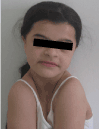
Figure 1. Older girl approximating her shoulders to the mid line

Figure 2. Chest X-ray for the older girl.

Figure 3. Anterioposterior skull view
Intra-oral examination (Figure 4) revealed mixed dentition in her oral cavity. Teeth were

Figure 4. Oral cavity after improving oral hygiene and before starting surgical exposure and orthodontic treatment.
6 E D / 1 D E 6
6 E D / 12 D E 6
The patient has poor oral hygiene, points of bleeding, but no pockets more than 3mm or tooth mobility.
The panoramic view (Figure 5) revealed the following impacted teeth 8 7 5 4 3 2 1 / 2 3 4 5 7 8
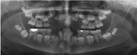
Figure 5. Panoramic view before starting treatment
8 7 6 4 3 2 / 3 4 7 8
No supernumerary teeth. The patient had skeletal class III malocclusion. The radiograph demonstrated that the upper anterior teeth were close to the gingival margin, and so the lower anterior teeth. After improving the oral hygiene and periodontal condition, consultation with the orthodontic department about the possibility of orthodontic treatment after surgical exposure of the impacted teeth. As the impacted teeth in more than one sites so surgical exposure was performed in more than one visit and orthodontic traction of the impacted teeth was started by powerful unity anchorage.
Because of the impaction that included more than one tooth, the stage of traction the impacted teeth took long time and required many visits and cooperation from the patient. All impacted teeth that were exposed surgically and activated orthodontically (Figure 6). Occlusal relationship had been established with class III but aesthetically acceptable (Figure 7).

Figure 6. During surgical exposure

Figure 7. During orthodontic treatment
The father presented to the clinic before with the same clinical (Figure 8) and radiographic signs of CCD. Anterio-posterior view for the skull revealed persistent sagital sutures with multiple wormian bone (Figure 9). Chest X-ray showed absent of the distal third of both clavicles (Figure 10) He was presented before with many impacted and supernumerary teeth and with multiple carious and badly destructed teeth. He was treated by extraction of the badly destructed teeth (Figure 11) and construction upper and lower removable partial denture (Figure 12). The father was happy and satisfied with this kind of treatment.

Figure 8. The father approximating his shoulders to the midline

Figure 9. Anterioposterior skull view
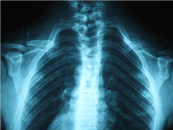
Figure 10. Chest X-ray showing abcent of the distal third of both clavicles

Figure 11. Panoramic view after dental treatment.
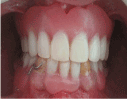
Figure 12. Oral cavity after dental treatment
The little girl presented to the clinic when she was two years old and presented with cardinal signs of CCD. Attempt to approximate her shoulder to the midline, face appears small in relation to the cranium, low nasal bridge, hypertelorism, frontal bossing (Figure 13). At the age four years radiographical investigations were performed for her; chest X-ray revealed hypoplastic clavicle (Figure 14). Lateral skull view showed widened anterior fontanele, multiple wormian bone, brachoiocyphalic with abnormal skull shape (Figure 15). Panoramic view; all deciduous teeth were erupted and the tooth buds of the permanent teeth, no super numerary teeth could be detected from the X-ray (Figure 16). Dental and paediatric follow up are recommended for the little girl in order to start the dental treatment in the suitable time and to have medical treatment if she needs.

Figure 13. The little girl at the age of two years

Figure 14. Chest X-ray of the little girl at the age two years
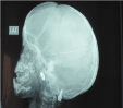
Figure 15. Lateral skull view
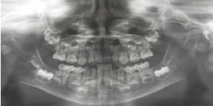
Figure 16. Panoramic view for the little girl at the age 4years.
Discussion
This Jordanian family consists of eight members, three of them were diagnosed as CCD. The father and two daughters, the older and the younger one. Up to the author knowledge no reported cases of CCD from Jordan. Although the treatment of the older daughter was not planned before, but surgical exposure and orthodontic treatment was the best choice and ended up good results. Orthodontic treatment is the best choice of treatment in order to direct the eruption of malposed and impacted teeth. Treatment of CCD needs teamwork, in order to find out the treatment plan and the best time to start treatment.
The older girl psychological status was significantly improved as she moved from removable partial denture in this young age to a full mouth permanent teeth aesthetically and functionally acceptable, in addition to improvement in the phonetics. It was noted that the mandibular prognathism was due to prominent lower third of the body of the mandible. This prognathism may be treated in the future either by composite facing or porcelain veneers. Although father treated by extraction badly destructed teeth and construction of removable upper and lower dentures, but the father was happy with this kind of treatment and functionally satisfied.
Farronato et al [15] reported a 28 years old case of CCD who were treated orthodontically and ended with class III and in order to improve the aesthetic a prosthetic treatment was performed to the patient after one year of orthodontic treatment.
Vicki et al 2004 [16] treated a case of CCD had several unsuccessful surgical interventions to expose the impacted teeth, then endo-ossous implants were inserted in the upper and lower arch with fixed prosthesis. This treatment option was effective and described one of the protocols that offers an effective treatment options, although a lack of evidence-based data that support the osseointegration in patients with CCD but that case was successful. da-Cuhna et al 2004 [17] reported a 25years old case of CCD treated orthodontically and because the teeth had enamel hypoplasia, microabrasion, dental bleaching and direct composite resin were performed for aesthetic purpose. Chaky L. et al 2015 [19] reported a 29years old case of CCD who was treated after orthodontic treatment with fixed prosthesis and dental implants.
The dental treatment in CCD varies and primarily depends on patient’s needs, age of diagnosis, a social and economic circumstances, but still the main objective of treatment remain the restoration of craniofacial and dental function together with aesthetic. Three known ortho-surgical regimes used for the dental treatment of CCD. The first one is Toronto-Melborne: it is based on timed serial extraction of deciduous teeth depends on the permanent teeth have developed. Removing of super numerary teeth to facilitate the spontaneous eruption of unerupted permanent teeth. The second one is Belfast -Hamburg: depends on extraction of deciduous and supernumerary teeth, exposure of unerupted permanent teeth, then orthodontic treatment. All that is in a single surgical operation under general anaesthesia. The third one is Jerusalem approach; almost like the second approach but in at least two surgical interventions, the first one to extract the primary and supernumerary teeth with exposure of anterior teeth. At the age of thirteen years, the residual primary teeth are extracted, unerupted canines and premolars are exposed, and the necessary orthodontic and surgical processes are completed [19,20]. Furrow et al 2018 [21] also concluded that orthodontically aided eruption should always attempted but stabilizing occlusion is very is very important, and that comes through orthodontic treatment, which also improve facial esthetic. But when orthodontic treatment is partially efficient prosthetic treatment
Patients with CCD require a team approach with good cooperation and communication from the patient. Timing of the intervention is critical, and many surgeries might be required. The collaboration of dentists with clinical geneticists to produce early diagnosis is very important.
Consent form
Written informed consent was obtained from the patient for the publication of this report and any accompanying images
Competing interests
The authors declare that they have no competing interest
Conflict of interest and source of funding statement
Authors declare that they have no conflict of interests.
Declaration of Helsinki
This article was undertaken with the understanding and written consent of subjects according to the Medical Association Declaration of Helsinki
References
- Kalliala E, Taskinen PJ (1962) Cleidocranial dysostosis. Report of six typical cases and one atypical case. Oral Surg Oral Med Oral Pathol 15: 808-822. [Crossref]
- Brueton LA, Reeve A, Ellis R, Husband P, Thompson EM, et al. (1992) Apparent cleidocranial dysplasia associated with abnormalities of 8q22 in three individuals. Am J Med Genet 43:612-18.2. [Crossref]
- Dard M (1993) Histology of alveolar bone and primary tooth roots in a case of cleidocranial dysplasia Bull Group Int Rech Sci Stomatol Odontol. 36: 101-107. [Crossref]
- Mundlos S, Mulliken JB, Abramson DL, Warman ML, Knoll JH, et al. (1995) Genetic mapping of cleidocranial dysplasia and evidence of a microdeletion in one family. Hum Mol Genet 4: 71-75. [Crossref]
- Mundlos S, Otto F, Mundlos C, Mulliken JB, Aylsworth AS, et al. (1997) Mutations involving the transcription factor CBFA1 cause cleidocranial dysplasia. Cell; 89:773-779. [Crossref]
- Machuca-Tzili L, Monroy-Jaramillo N, Gonza´ lez del Angel A, Kofman-Alfaro S (2002) New mutations in the CBFA1 gene in the two Mexican patients with cleidocranial dysplasia. Clin Genet 61:349–353. [Crossref]
- Yoshida T, Kanegane H, Osato M, Yanagida M, Miyawaki T, et al. (2002) Functional analysis of RUNX2 mutations in Japanese patients with cleidocranial dysplasia demonstrates novel genotype-phenotype correlations, Amer J Human Genet, 71: 724–738. [Crossref]
- Golan I, Baumert U, Hrala BP, Müssig D (2003) Dentomaxillofacial variability of cleidocranial dysplasia: clinicoradiological presentation and systematic review. Dentomaxillofac Radiol 32: 347-354. [Crossref]
- Tyndall DA (1983) Cleidocranial dysostosis: a nearly unrecognized case. Gen Dent 31: 390-393. [Crossref]
- Goaz PW, White SC. 5th ed. Elsevier publications. Oral Radiology: Principles and Interpretation, 2004.
- Jensen BL, Kreiborg S (1990) Development of the dentition in cleidocranial dysplasia. J Oral Pathol Med 19: 89-93. [Crossref]
- Mundlos S (1999) Cleidocranial dysplasia: clinical and molecular genetics. J Med Genet 36: 177-182. [Crossref]
- Hitchin AD (1975) Cementum and other root abnormalities of permanent teeth in cleidocranial dysostosis. Br Dent J 139: 313-318. [Crossref]
- Jensen BL, Kreiborg S (1992) Dental treatment strategies in cleidocranial dysplasia. Br Dent J 172: 243-247. [Crossref]
- Farronato G, Maspero C, Farronato D, Gioventù S (2009) Orthodontic treatment in a patient with cleidocranial dysostosis. Angle Orthod 79: 178-185. [Crossref]
- Petropoulos VC, Balshi TJ, Balshi SF, Wolfinger GJ (2004) Treatment of a Patient with Cleidocranial Dysplasia Using Osseointegrated Implants: A Patient Report. Int J Oral Maxillofac Impl 19:282–287. [Crossref]
- Leonardo Fernandes da Cunha, IsabelaMaria Caetano, Fernando Dalitz, Carla Castiglia Gonzaga, and JoséMondelli. Cleidocranial Dysplasia Case Report: Remodeling of Teeth as Aesthetic Restorative Treatment
- Lee C, Jung HS, Baek JA, Leem DH, Ko SO (2015) Manifestation and treatment in a cleidocranial dysplasia patient with a RUNX2 (T420I) mutation. Maxillofac Plast Reconstr Surg 37:41. [Crossref]
- Smylski PT, Woodside DG, Harnett BE (1974) Surgical and orthodontic treatment of cleidocranial dysostosis. Int J Oral Surg 3: 380-385. [Crossref]
- Hall RK, Hyland AL (1978) Combined surgical and orthodontic management of the oral abnormalities in children with cleidocranial dysplasia. Int J Oral Surg 7: 267-273. [Crossref]
- Farrow E, Nicot R, Wiss A, Laborde A, Ferri J (2018) Cleidocranial Dysplasia: A Review of Clinical, Radiological, Genetic Implications and a Guidelines Proposal. J Craniofacial Surg 29: 382–389. [Crossref]