Abstract
Liquid injectable silicone is occasionally used off label as a cosmetic filler in patients desiring breast augmentation. The appropriate identification of the sequelae of silicone breast injections in breast imaging is important to avoid unnecessary work up. The purpose of this case report is to review the breast imaging findings on mammography, breast ultrasound and breast MRI associated with the sequelae of silicone injections. A companion case is also presented to illustrate an example of early breast cancer obscured in a patient with silicone mastopathy.
Key words
2021 Copyright OAT. All rights reserv
radiology, breast imaging, silicone, breast oncology, breast surgery, breast cancer, mammography
Introduction
For over a century, different methods of breast augmentation have been performed to achieve desired cosmetic results, many of which are not regulated or even legal. Before today’s surgical implantation techniques many substances, including Paraffin and autologous fat, have been injected subcutaneously into the breast or into the pectoralis muscle often with undesirable consequences. Beginning after World War II, the leftover silicone liquid initially used to insulate electrical transformers was injected into the breasts of Asian sex workers who desired to have a more prototypical Western appearance [1]. The popularity of liquid silicone injections then spread throughout Asia and to the United States where it eventually was regulated by the FDA in 1965 and subsequently banned from injection into the breast. Termed “Cleopatra’s Needle,” silicone breast injections were largely performed in Hollywood and Las Vegas performers in the 1960s and 1970s as a “black market-type” procedure, often without medical supervision [2]. Though known complications ranging from local granulomatous reactions to systemic emboli can occur, nevertheless silicone injections remain a cheaper and faster alternative for breast augmentation in patients willing to travel to other countries or forego medical supervision [3].
Case presentation
A 37-year-old Hispanic female presented to the emergency department with a self-palpated painful mass in her right breast for one week. She was not currently breastfeeding and had not experienced any associated nipple discharge. At the time of presentation, she reported no history of surgery. The emergency physician palpated a 4 cm x 3 cm mass at the 11-12 o’clock region 2 cm from the nipple, without erythema or warmth, and she was discharged to the breast center for follow-up mammogram and ultrasound.
Bilateral MLO views demonstrated multiple bilateral circumscribed and partially calcified dense oval masses, the largest in the right breast corresponding to the patient’s area of palpable concern (Figure 1A). Ultrasound of the right breast at the site of concern demonstrated a large anechoic oval mass at with posterior acoustic enhancement at 12 o’clock with surrounding echogenic breast tissue (Figure 1B).

Figure 1: 37-year-old woman with a history of silicone injections, presenting for further evaluation of a palpable abnormality in the 12 o’clock position of the right breast. (A) Mediolateraloblique (MLO) mammography images demonstrate multiple circumscribed, dense partially calcified bilateral breast masses. (B) High resolution panoramic ultrasound of the right breast spanning the 12 o’clock to 2 o’clock positions obtained using a high frequency (7-12 MHz) transducer demonstrates an avascular anechoic oval mass (arrows) with circumscribed margins and posterior acoustic enhancement. There is surrounding granular, hyperechoic breast tissue which has lost its texture. This is consistent with liquid silicone droplets surrounded by fibrosing tissue.
Upon further questioning, the patient confirmed that she had silicone injections to the breast six months prior.
Discussion
Mammographic findings
Mammographic findings in patients who have been injected with liquid silicone include multiple cystic masses ranging from 0.2 cm to 2.0 cm in maximum diameter [1]. Often the masses demonstrate a rim of calcification. If a patient has undergone a larger volume injection, there may be more confluent opacities within the breast. Scaranelo et al. [4] evaluated 14 patients who had undergone liquid silicone breast injections. The study categorized mammographic findings into macronodules, defined as dense nodules greater than 1 cm in all quadrants of the breast with 4 times as many larger nodules when compared to smaller nodules and micronodules, defined as dense nodules less than 1 cm in all 4 quadrants. The macronodular pattern was seen in 7 patients with a mixed pattern seen in 6 patients. Architectural distortion was seen in 5 patients. Our case appears typical, with a mixed macro and micronodular mammographic pattern but without architectural distortion.
Ultrasound findings
Sonographic findings are more variable, with Scaranelo et al. [4] categorizing the macronodular pattern as greater than 1 cm anechoic nodules with smooth margins, posterior acoustic enhancement, and surrounding echogenic noise or “snowstorm” appearance. The micronodular pattern consisted of multiple hypoechoic or anechoic nodules less than 1 cm with variable posterior acoustic enhancement and surrounding echogenic noise or “snowstorm” appearance. The macronodular pattern was seen in 2 patients, micronodular pattern in 3 patients, and mixed pattern in 6 patients. 3 patients demonstrated a “snowstorm” appearance without nodules. Cole-Beuglet et al. [5] described a clinical scenario where ultrasound was inadequate in the evaluation of silicone injection granulomata due to scattering created by the interface of the multiple granulomata and breast parenchyma. The finding of echogenic noise is more specific for free silicone. Rosculet et al. [6] demonstrated homogenous low-level echogenicity in 17 of 19 cases with silicone implant ruptures, with 2 cases in which ruptures were diagnosed only by echogenic noise seen on ultrasound, as the mammograms were normal. Although our case involves silicone injections rather than implants, the study illustrates that free silicone is not always identified by mammogram. Our case again appears typical, exhibiting a mixed pattern of macro and micronodules with surrounding echogenic noise. The lymph nodes did not demonstrate free silicone migration.
MRI findings
Classically MRI findings of free silicone include low signal on fat suppressed T1 weighted images and high signal on water suppressed T2 weighted images [7]. Silicone selective pulse sequences may also be obtained to increase sensitivity [8]. Unfortunately, silicone granulomata can enhance in a pattern similar to breast carcinomas and may not be distinguished without tissue sampling [9]. While MRI is advantageous in detecting rupture of silicone implants, it is not definitely superior to mammography or ultrasound in detecting free silicone. In our case, we were confident based on mammographic and sonographic imaging findings in conjunction with the patient’s history that this was a clear-cut case of silicone injection granulomata. Therefore, MRI was not pursued.
Patients with a history of silicone breast injection may present with multiple painful masses due a granulomatous reaction that forms within the breast. This may occur on the order of months as in this case to years. Not only does it contribute to patient’s morbidity, but it can also potentially obscure detection of subtle underlying breast cancers.
A case of breast cancer in the augmented breast
We present a companion case of breast cancer developing in a background of silicone mastopathy in a patient with a history of silicone injections 17 years prior in Mexico. A 59-year-old woman presented for annual screening mammography. Her screening mammogram from two years prior demonstrated diffuse silicone droplets and parenchymal heterogeneity, obscuring a retrospectively seen asymmetry in the inferior right breast (Figure 2A). Her subsequent screening mammogram two years later demonstrated a global asymmetry in the right breast measuring 9 cm (Figure 2B). Breast ultrasound demonstrated a complex cystic and solid mass, indeterminate given her history of injection granulomata (Figure 2C). Subsequent biopsy revealed invasive mammary carcinoma, Nottingham histological grade 1, estrogen and progesterone receptor positive, HER-2 non-amplified, Ki-67 15-20%. A breast MRI obtained to determine extent of disease demonstrated a large enhancing, necrotic mass in the right breast measuring approximately 11 cm with multiple prominent bilateral axillary silicone laden lymph nodes, unable to be adequately assessed by MRI for staging purposes (Figure 2D, 2E). In this case, the early cancer was obscured by changes related to silicone mastopathy, and the malignancy was not identified until it became much more advanced.
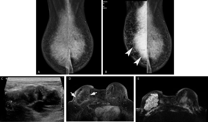
Figure 2: 59-year-old woman with a history of silicone injections, presenting for routine screening mammography. (A) Bilateral Mediolateraloblique (MLO) mammography images from her prior screening mammogram in 2016 demonstrate an asymmetry seen in retrospect in the inferior right breast, difficult to see given diffuse curvilinear calcified silicone droplets and parenchymal heterogeneity related to silicone mastopathy and fibrosis. (B) Bilateral Mediolateraloblique (MLO) mammography images from screening mammogram in 2018 demonstrate a global asymmetry in the lower outer quadrant of the right breast (arrow heads). There is diffuse skin thickening of the right breast compared to the left breast. Curvilinear calcified silicone droplets and parenchymal heterogeneity are again seen. (C) Transverse ultrasound image of the right breast using high frequency (7-12 MHz) transducer demonstrates an irregular, deeply situated mass measuring at least 3.9 cm with associated internal calcifications. Ultrasound guided needle biopsy was performed, revealing invasive ductal carcinoma, Nottingham histological grade 1, estrogen and progesterone receptor positive, HER-2 non-amplified, Ki-67 15-20%. Right axillary ultrasound (not shown) revealed a “snow-storm” appearance of silicone within multiple lymph nodes. (D) Bilateral breast MRI was performed to determine disease extent given the difficulty seeing the mass on mammography. Axial T1 weighted early post contrast imaging demonstrates a large necrotic mass measuring 11 cm in the right breast at 8 o’clock (arrows) with associated right breast skin thickening and enhancement. (E) Axial T2 weighted imaging at the same location demonstrates the T2 hyperintense mass. The chronic fibrotic silicone mastopathy medial to the cancer appears hypointense.
Conclusion
While liquid injectable silicone is still occasionally used off label as a cosmetic filler, its use as a breast augmenting agent is advised against as the large amount of liquid silicone required to provide a cosmetic result can lead to complications such as migration, cysts, and granulomata [10]. While its regulated use in the United States is limited, patients looking for a cheap and fast alternative for breast augmentation as well as patients from countries with fewer regulations on the use of silicone may present with silicone related granulomata. Additionally, these granulomata may make it difficult to detect early breast cancers by screening mammography. Mammographic findings include dense, circumscribed, partially calcified nodules. Sonographic findings include hypoechoic and anechoic nodules with echogenic noise. MRI findings include low T1 and high T2 signal, with silicone selective pulse sequences available, though there is not substantial evidence demonstrating the efficacy in diagnosing free silicone by MRI. Silicone breast injections, though rare and illegal in the United States, can cause typical findings on multiple imaging modalities which are important to recognize to avoid unnecessary intervention.
References
- Peters W, Fornasier V (2009) Complications from injectable materials used for breast augmentation. Can J Plast Surg 17: 89-96. [Crossref]
- Matelski E (2017) Reducing bodies. Mass culture and the female figure in postwar America. Basingstoke: Taylor & Francis Ltd.
- Lyapichev K, Chinea FM, Poveda J, Pereda J, Bejarano PA, et al. (2016) Pulmonary Empty Spaces: Silicone Embolism-A Decade of Increased Incidence and Its Histological Diagnosis. Case Reports in Pathology 2016: 1-5.
- Scaranelo AM, Maia MDFR (2006) Sonographic and mammographic findings of breast liquid silicone injection. J Clin Ultrasound 34: 273-277. [Crossref]
- Cole-Beuglet C, Schwartz G, Kurtz AB, Patchefsky AS, Goldberg BB (1983) Ultrasound mammography for the augmented breast. Radiology 146: 737-742. [Crossref]
- Rosculet KA, Ikeda DM, Forrest ME, Oneal RM, Rubin JM, et al. (1992) Ruptured gel-filled silicone breast implants: sonographic findings in 19 cases. AJR Am J Roentgenol 159: 711-716.
- Caskey CI, Berg WA, Hamper UM, Sheth S, Chang BW, et al. (1999) Imaging Spectrum of Extracapsular Silicone: Correlation of US, MR Imaging, Mammographic, and Histopathologic Findings. RadioGraphics 19: S39-S51. [Crossref]
- Turpin IM (1996) MR detection of leakage from silicone breast implants: Value of a silicone-selective pulse sequence. Plast Reconstr Surg 98: 380.
- Yang N, Muradali D (2011) The augmented breast: a pictorial review of the abnormal and unusual. AJR Am J Roentgenol 196: W451-460. [Crossref]
- Narins RS, Beer K (2006) Liquid Injectable Silicone: A Review of Its History, Immunology, Technical Considerations, Complications, and Potential. Plast Reconstr Surg 118: 77-83. [Crossref]