Myelodysplastic syndroma (MDS) is heterogeneous group of hematological malignancies characterized by ineffective hematopoiesis and peripheral cytopenia with a variable risk of developing acute leukemia [1]. The incidence rate is about 5 per 100000 persons per year in the general population, but increases to 20 to 50 cases per 100000 persons per year after age 60 years old [2,3]. The number of MDS patients is destined to increase in the next decades. Incidence rate of MDS in our region was underestimated and largely misdiagnosed. The demographic statistic of Moroccan population increase up to 5.8 million in 2030, this projection can explain why MDS in our region will be a real public health problem. Due to socioeconomic constraints and incomplete diagnosis tools, support care of MDS will be the most challenging issue in the future.
The purpose of the current study was to determine the profile of this disease in the principal hematology department in Morocco, to improve diagnosis and to select appropriate management of patients for better invest.
In this retrospective descriptive study, we included all patients newly diagnosed with primary MDS admitted in hematology-oncology pediatric department in Casablanca from January 2008 to December 2015. The diagnosis and classification were defined according on WHO classification. We excluded Patients with history of prior chemo radiotherapy, prior malignancy before diagnosis of MDS, and acute myeloid leukemia with MDS.
All clinical and biological data were collected to the medical record. Including features: age, sex, geographic origin, profession, biologic test (blood cell and reticulocyte count, ferritin, dosage of folate and cobalamin, renal and liver function and serology). Blood and bone marrow smears were examined by using May-Grunewald-Giemsa and iron staining. The morphology analysis was revised by two hematologist cytologists in hematology laboratory department IBN Rochd in Casablanca using WHO 2008 classification of myeloid neoplasm recommendation, which changed in 2016. Cytogenetic of bone marrow was done in a single private laboratory for at least 20 metaphases and karyo typing was performed according to the international system for human cytogenetic nomenclature (ISCN 2005). The definition of group risk related to the characteristics of MDS was based on the international prognostic scoring system (IPSS). There was no strategy about treatment; supportive care consisting on symptomatic transfusion of erythrocytes and platelet, iron chelating, erythropoietin stimulating. Depending on categories group, economic situation and availability of treatment in hospital patient can received low chemotherapy (cytarabine), hypomethylation treatment (azacytidine, thalidomide) and allogenic stem cell transplant. Statistical analysis was performed using SPSS 21.0 software, the overall survival of the entire cohort was assessed using the Kaplan Meier method.
One hundred seventy-eight patients of primary MDS from 8623 hematological malignancies, (2% of MDS) were included in the study. Geographical distribution noted that 40% of patients were from Casablanca region, 26% from central region, 24% from Rabat and north region and 10% from south region. 78% of patients were from the rural regions and 75% had farmer profession. Distribution of patients during eight years showed a median at 22 patients per year.
The median age was 59 years (range: 16–90 years) with sex ratio at 1.2, three patients were under 15 years old. The Median delay from first symptoms and diagnosis was 7 months (range 0,5-48 months).
The commonest complaint of patients was anemia syndrome in 70 % of patients. Lymphadenopathy and splenomegaly were found in 19% of cases, dermatosis and systemic autoimmune disease affected 5.6% of patients (Table 1).
Table 1. Clinical and Biological Characteristics of 178 patients with primary MDS
Parameter |
N |
% |
Clinical: |
No symptoms |
20 |
11 |
Anemic syndrome |
125 |
70 |
Hemorragic syndroma |
47 |
26 |
Infection |
31 |
17 |
Tumoral syndroma |
34 |
19 |
Biological: |
Hemoglobin ≤ 10g/dl |
122 |
68 |
Neutrophil ≤ 1,5 10 9 /l |
62 |
34,8 |
Platelet ≤ 100 10 9/l |
83 |
46,6 |
Number of cytopenia
1
2
3 |
-
76
55
36 |
-
42
30
20
|
The most frequent laboratory finding on peripheral blood examination was anemia (73%) and thrombopenia (46%). In all patients bone marrow aspiration was done, 54% of cases performed blood smears; 20% confirmed diagnosis with bone marrow biopsy and 46% of total patient had cytogenetic analysis (Figure 1).
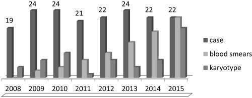
Figure 1. Evolution of diagnosis by blood smears and cytogenetic analysis according to years
The assessment of dysplasia on peripheral blood and bone marrow smears was the mainstay for the diagnosis of MDS, all patients were included in analysis, and were classified by WHO classification in 7 subtypes of MDS, the most frequently one is MDS with multilineage dysplasia (MDS-MLD) in 67% of cases. The results of cytogenetic analysis were available in 83 patients, among whom 48% had a normal Karyotype. About 80 patients who had cytogenetic results (3 failed), the IPSS score was calculated and classified in four categories groups, and 69.9% were in low risk (Table 2).
Table 2. Characteristic and classification of patients
Parameter |
N |
% |
WHO 2008 Classification :
RCMD
AREB1
AREB2
CRUD
RARS
MDS with isolated del 5q
MDS unclassified |
178
119
20
17
7
6
6
3 |
67
11.2
9.5
4
3.3
3.3
1.7 |
Cytogenetic: Normal karyotype
Clonal abnormalities
Del 5 q
Del Y
Del 11q
Del 20q
+ Chr 8
+ Chr 21
Other abnormalities
Complex abnormalities
Failed |
83
40
6
3
2
1
3
1
13
11
3 |
48
7
3.6
2.4
1.2
3.6
1.2
15.6
13
3.6 |
Group risk*:
High risk
Intermediate 2
Intermediate 1
Low risk |
80
13
11
45.5
11 |
16.2
13.7
56.2
13.7 |
*group risk according IPSS score
Without consensus of treatment in unit, according risk factors, and availability of medications, some therapeutic option was proposed: Symptomatic transfusion in 66% of patients and 11% of them had iron chelation therapy. Patients in low risk had erythropoietin injection in 4,5% of cases. About patients in high risk, low chemotherapy with cytarabine injection was done in 2,2% of cases, 2.2% patients received azacytidine, 1.6% of patients was treated by thalidomide and only one patient was transferred to France for allogenic stem cell transplant.
The MDS has heterogeneous features such as morphological clinical, biological and cytogenetic profile, only few patients are reported in national register. Actually, this disease represented a real problem of public health and here diagnosis need a complete cytogenetic analysis. This study consisted of analysis 178 patients with primary myelodysplasia; the mean age was 59 years with 53% of males. The common of patients presented anemia syndrome in 70% of cases, RCMD in 67%, normal chromosomal karyotype in 48% of cases and 56.2% of patients was in group risk intermediate1.
Epidemiological studies on MDS are rare worldwide, enjoying continuous success and developing in molecular diagnosis, classification and treatment, because the incidence is in increasing progress [4]. Recent studies suggest that many MDS cases are undetected by cancer registries, so the true incidence of MDS might be higher [5]. Demographic projection of Moroccan population older than 60 years in 2030 will increase to 6 million habitants (8% in 2004 to 15.4%) [6], this could be attributed to the prolongation of life expectancy and the amelioration of diagnosis. The first study of Moroccan hematology society which attempts to describe data of 3461 cases has found 3.5% of MDS [7], particularly in our unit the MDS was found in 2% of all hemopathy, these results remain underestimated. Epidemiologic data of the study don’t represent all country, but Casablanca unit is one of the reference center in Morocco. MDS is a disease of the elderly with median age varies between 65 and 74 years old [8], but in our result the population was younger at 59 years old. the sex ratio found a male predominance and 70% of patients presented anemia, this clinical profile is in comparable to literature [5-8]. Autoimmune abnormalities, although uncommon, may complicate the course of MDS because of his immune therapy and genetic predisposition, Clinical analysis of our results found 5,6% of cases less than literature 23% [9].
In this study cases were classified by WHO 2008 changed in 2016 [10-11], and a dominant finding was MDS-MLD, which is compatible to results of Maghrebian countries like Tunisia Algeria [12-14] and others countries (Table 3).
Table 3. Comparison of distribution of MDS cases in literature classified by WHO classification
WHO 2008 |
Our study (178) |
Tunisia (88) |
China (435) |
France (907) |
Canada (66) |
CRDU/ AR (%) |
4 |
10 |
4.3 |
18.6 |
18 |
ARSC (%) |
3.3 |
12 |
1.2 |
17.3 |
18 |
RCMD (%) |
67 |
51 |
70 |
15.4 |
41 |
AREB1(%) |
11.2 |
15 |
13 |
20 |
7 |
AREB2(%) |
9.5 |
10 |
11 |
18 |
14 |
5q- (%) |
3.3 |
2 |
0.5 |
4 |
2 |
Cytogenetic analysis has a major role to determine clonality in patients with suspected MDS, chromosomal abnormalities were presented in 50% to 60% of patients with MDS [12] and it’s very important for diagnosis to identify non random chromosomal abnormalities and the diagnosis of MDS may be difficult in patients with a normal karyotype or noninformative cytogenetics who do not have robust morphologic markers such as ring sideroblasts or excess of myeloblasts.
The most frequent single cytogenetic abnormalities include del(5q), monosomy 7 or del(7q), trisomy 8, and del(20q) [12]. In our results 48% of patients had normal chromosomal analysis and good cytogenetic prognosis with del 5q (7%), delY (3.6%), del11q (2.4%) and 20q in 1.2%. The common of patient are in lower risk groups of prognosis which are similar with others series [12-14] (Table 4).
Table 4. Comparison of distribution of MDS cases classified by cytogenetic prognosis and scoring IPSS
WHO 2008 |
Our study |
Tunisia |
France |
British |
Group risk% |
Low R |
62 |
82 |
72 |
82 |
Inter R |
25 |
11 |
14 |
12 |
High R |
13 |
7 |
13 |
6 |
IPSSscore% |
Low R |
13.7 |
27 |
34.8 |
44 |
Inter 1 |
56.2 |
62 |
32.5 |
38 |
Inter 2 |
13.7 |
8 |
16.3 |
15 |
High R |
16.2 |
3 |
16.3 |
3 |
Unfortunately, the Karyotype is not done systematically, in France 82.5% [12] of patients had been performed this exam and in our unit 46% of patients realized this analysis. But we noted an improvement of diagnosis procedures during last years. In 2015 all patients had blood smears and 86% had a cytogenetic analysis.
The assessment of dysplasia on peripheral blood and bone marrow smears is the mainstay for the diagnosis of MDS. It’s should be performed in all patients with suspected MDS in whom bone marrow examination is indicated. For evaluation of morphology and dysplasia in blood and bone marrow, WHO classification of myeloid neoplasms is recommended [7] and some criteria are important to realized, like percentage of dysplasia, percentage of blast, presence of Auer rods, iron staining [8,9]. During this study, we revised all bone marrow smears in hematology laboratory of IBN ROCHD hospital using the international recommendation of quantification and morphology diagnosis, because we detected many problems and incorrect classification of diagnosis according WHO classification. This incomplete diagnosis is explained by the low socio economic resources of local population, and the limited option of treatment in Morocco.
The outcome of MDS is very variable with some patients transforming into acute leukemia while others remain asymptomatic for years, so there are many risk factors predicting the evolution of disease and several scoring systems have been developed. The IPSS is the most commonly used in the world and included both disease and patient-related variables (performance status, age, platelet count, hemoglobin, bone marrow blasts, white blood cell count, and karyotype) [13]. The results of our study showed that 56.2% of patients are in intermediate1 risk group and 16.2% in low risk, which is comparable to results of countries in Maghreb [10] but in developed country like Europe [14,15] the group of low risk is more frequent.
In fact, there is limited possibility for allogenic stem cell transplantation and limited means for hypomethylation and iron chelation agents available in our condition. 30% of patients with a poor prognosis are candidates for up-front intensive treatments 66% of them received chemotherapy and hypomethylation agents. From patients with low-risk disease only 14% of patients were treated by erythropoietin stimulation. A high proportion of MDS patients are not eligible for potentially curative treatment because of advanced age and/or clinically relevant co morbidities and poor performance status so they received symptomatic transfusion in 66% of cases and only 11% had chelation.
The assessment of individual risk can define the group who needs invest to better therapeutic options but this stratification for management require a complete biological diagnosis including molecular and genetic analysis [16,17], it’s important to have a same consensus of procedures diagnosis and national recommendations such us program transfusion and chelation, for younger patients with good prognosis. So the Moroccan society of hematology has developes a national recommendation, and particularly in the department of study with specific consultation to better support and management of patients.
Myelodysplasia will be the disease of national public health because here Incidence will increase significantly. The present study describes profile of patients with MDS in Morocco, and show the lack of means in diagnosis and treatment. This results have improved the quality of diagnosis and therapeutic care in our department.
The authors declare no competing financial interests.
We expressed a deep gratitude to our patients who participated in this study.
- Heaney ML, Golde DW (1999) Myelodysplasia. N Engl J Med 340: 1649-1660. [Crossref]
- Radlund A, Thiede T, Hansen S, Carlsson M, Engquist L (1995) Incidence of myelodysplastic syndromes in a Swedish population. Eur J Haematol 54: 153-156.
- Neukirchen J, Schoonen WM, Strupp C, Gattermann N, Aul C, et al. (2011) Incidence and prevalence of myelodysplastic syndromes: data from the Düsseldorf MDS-registry. Leuk Res 35: 1591-1596. [Crossref]
- Avgerinou C, Alamanos Y, Zikos P, Lampropoulou P, Melachrinou M, et al. (2013) The incidence of myelodysplastic syndromes in Western Greece is increasing. Ann Hematol 92: 877-887. [Crossref]
- Troussard X, duchenet v (2009) Epidemiologie des syndromes myelodysplasiques et des syndromes myelodysplasiques /myeloproliferatif. RFL 431: 25-29.
- Maroc 2030 (2011) quelle demographie? haut commissariat au plan. Prospective.
- Quessar A, Bennani Othmani M (2005) Hemopathies malignes au Maroc: Enquete realisée par la societe marocaine d’hematologie pp. 204-218.
- Maynadie M, Verret C, Moskovtchenko P, Mugneret F, Petrell T, et al. (1996) Epidemiological characteristics of myelodysplastic syndromes in a well-defined French population. Br J Cancer 74 :288-90.
- Anderson LA, Pfeiffer RM, Landgren O, Gadalla S, Berndt SI, et al. (2009) Risks of myeloid malignancies in patients with autoimmune conditions. Br J Cancer 100: 822-828. [Crossref]
- Malcovati L, Lindberg E, Bowen D (2013) Diagnosis and treatment of primary myelodysplastic syndromes in adults: recommendations from the European LeukemiaNet. Blood 122: 2943-2964.
- Bennett JM, Catovsky D, Daniel MT, Flandrin G, Galton DA, et al. (1982) Proposals for the classification of the myelodysplastic syndromes. Br J Haematol 51: 189-199. [Crossref]
- Sendi HS, Hichri H, Elghezal H, Gribaa M, Laatiri A, et al. (2002) Cytogenetic survey of 117 Tunisian patients with de novo myelodysplastic syndrome. Ann Genet 45: 131-135. [Crossref]
- Mufti GJ, Bennett JM, Goasguen J (2008) International Working Group on Morphology of Myelodysplastic Syndrome. Diagnosis and classification of myelodysplastic syndrome: International Working Group on Morphology of myelodysplastic syndrome (IWGM-MDS) consensus proposals for the definition and enumeration of myeloblasts and ring sideroblasts. Haematologica 93: 1712-1717.
- Kelaidi C, stamatoullas A, beyneeauzy O (2010) Daily practice management of myelodysplastic syndromes in France: data from 907 patients in a one week cross- sectional study by the GFM. Haematologica 955: 892-899.
- Haase D, Germing U, Schanz J, Pfeilstöcker M, Nösslinger T, et al. (2007) New insights into the prognostic impact of the karyotype in MDS and correlation with subtypes: evidence from a core dataset of 2124 patients. Blood 110: 4385-4395. [Crossref]
- Greenberg P, Cox C, LeBeau MM (1997) International scoring system for evaluating prognosis in myelodysplastic syndromes. Blood 89: 2079-2088.
- Solé F, Luño E, Sanzo C, Espinet B, Sanz GF, et al. (2005) Identification of novel cytogenetic markers with prognostic significance in a series of 968 patients with primary myelodysplastic syndromes. Haematologica 90: 1168-1178.