Lumbar radiculopathy is a common and debilitating condition with 30% of people affected experiencing pain and disability beyond 12 months. Physiotherapy is recommended as well as pain management as first line management in the absence of red flags, but due to the complex nature of the condition and the variety of causative factors and symptom presentations it can be difficult to treat. This case study describes in detail the clinical reasoning utilised to treat a common presentation of lumbar radiculopathy highlighting the complex and systematic thinking and decision making required to provide effective individualised treatment, reducing the likelihood of persistent pain and disability.
Lumbar radiculopathy is a common and disabling condition that often resolves within several weeks. A substantial group (30%) however still have pain and disability beyond 12 months (Coster, De Brujin &Tavy, 2010). Sound clinical reasoning is important in modern physiotherapy practice, particularly in back pain and radiculopathy, with symptoms influenced by a wide array of intrinsic and extrinsic risk factors [1]. This case study explores the clinical reasoning involved in the management of a middle aged woman, under the pseudonym Kate, suffering from discogenic radiculopathy. Using a biopsychosocial approach, several risk factors were identified as contributing to the complex nature of the case. Management was directed at addressing these factors, using a combination of manual therapy, education and exercise to achieve a successful outcome.
Kate, a fifty-two year old female presented with a six week history of right sided lower lumbar pain with associated referral down the right lateral thigh and calf (body chart Figure 1).
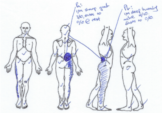
Figure 1. Body chart
Pain started in the right lower lumbar region (Pa), triggered on lifting a box (5kg). Over several hours she developed leg pain (Pb), subsequently presenting to a general practitioner (GP). The GP ordered an MRI (Figure 2), prescribed NSAIDs for one week and recommended walking. This referral was against current guidelines, MRI to be considered in patients for radiculopathy symptoms who do not respond to conservative management (pain control, medical management and physiotherapy) after 4-6 weeks [2]. Since commencing NASIDs her symptoms had eased somewhat. Her symptom presentation on initial physiotherapy consultation is shown in Table 1, the remainder of subjective information is shown in Table 2.
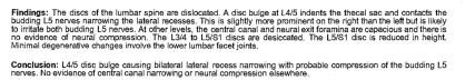
Figure 2. MRI report (2/52 post injury)
Table 1. Symptom presentation (initial appointment)
|
Pa |
Pb |
Symptoms |
I/M sharp grab with aggravating activity (3/10 NRS), easing to no pain at rest |
I/M deep burning ache with aggravating activity (4/10), easing to no pain at rest |
Aggs |
- Lumbar flexion (bending to put on socks)
- Sit to stand
|
- Sitting >20 mins worse in car and couch
- Lumbar flexion (bending to put on socks)
|
Irritability |
Settles immediately |
Up to 3 hours to settle |
Eases |
- Laying prone
- Walking
- Prednisolone
|
24hrs |
|
- Evenings worse (workdays)
- Nill WIN past 2 weeks
|
Relationship |
- Pb worsens with continued aggravation of Pa
- Pb can be present without presence/aggravation of Pa
|
Table 2. Subjective examination
Past history |
|
Medical history |
- 10kg overweight (longstanding)
- Voltaren 25mg (ceased 4/52)
- Postmenopausal (2 years)
|
Family history |
|
Social history |
- IT consultant (37.5 hrs/week)
- 4 weeks sick leave used, past 2 weeks working through pain (decreased concentration and performance)
- Works from home (laptop on kitchen table) 2 days
- Works at office (desktop with sit/stand desk) 2 days
- Clinical Pilates x1 weekly (Ceased since injury)
- Run/walk 5km x2-3 weekly (Ceased since injury)
- Lives independently
|
Yellow flags |
- Frustrated not being able to do regular exercise regime
- anxious that not exercising will cause weight gain
- Work affected due to pain causing significant stress – supportive boss
|
Red flags |
|
Patient goals |
- Return to pre-injury exercise regime without symptoms
- Weight loss
- Work symptom free requiring:
- Sit for 3 hours minimum
- Sit on bus ~ 40 mins (office days)
|
Kate’s main concerns were; 1.) affected work performance secondary to pain and; 2.) weight gain secondary to reduced exercise. Subsequently the most important findings for re-assessment were sitting tolerance and exercise capacity as these were directly linked with her goals and concerns.
Using the hypothesis framework presented by Ford, Hynes, et al. (2018) [3], it was hypothesised the source of symptoms were most likely from the L4/5 disc (Pa) and the right L5 nerve root (Pb). Factors consistent with this hypothesis were:
- Location and nature of symptoms
- Pa; nociceptive quality, in region for L4/5 disc pathology [4]
- Pb; neuropathic quality, in distribution of the right L5 nerve root
- Injury in flexion whilst lifting weight common in discogenic injury [5]
- MRI findings
- Aggravating and easing factors
- Pa aggravated with annulus fibrosis loading
- Pb worse in positions of relative nerve root compression (sitting and lumbar flexion) and eased with relative decompression (laying prone) [6]
A patho-anatomic explanation also supported the hypothesis. Outer layers of the annulus fibrosis are richly innervated containing nociceptive neurotransmitters capable of initiating the production of cytokines and provoking nociceptive input from the disc [7]. Annular tears and extravasation of the pro-inflammatory nucleus material may provoke nerve ending sensitisation and discogenic pain (Pa). Inflammatory mediators released in close proximity to the budding nerve root can also cause neural irritation and the development of radicular symptoms, supporting the relationship between Pa and Pb [8]. A compression component was also reasoned, Pb aggravated in positions of potential nerve root compression without a related worsening of Pa. Mechanical compression of the nerve root elevates the intraneural pressure, reduces blood flow causing ischemia, triggering neuropathic symptoms [9].
Kate showed several risk factors for developing a discogenic radiculopathy. Intrinsic risk factors included:
- Age; discogenic radiculopathy common in those 30-55 years of age [10].
- Weight; overweight individuals have an increased risk of back pain [11].
- Post-menopausal; hormonal changes increase risk of disc injury [12].
Extrinsic risk factors identified were:
- Mechanism; forward lumbar flexion under load is commonly associated with lumbar disc herniation [13].
- Work requirements; prolonged sitting exposes lumbar discs to high compressive forces [14].
- Posture: use of a laptop for work not being ergonomically setup can significantly increase lumbar disc load [15].
Precautions/contraindications
Whilst symptoms had been improving, Pb was still highly irritable. During physical examination, care was taken in aggravating positions and movements, such as the SLUMP test, to limit any post-assessment flare up that may cloud the effectiveness of treatment [16].
Prognosis
The natural course of radiculopathy varies in the available literature however a high quality study with long term follow up showed that 70% of patients similar to Kate, attending to hospital for radiculopathy symptoms, still had some symptoms 13 years later [17]. Kate showed several positive prognostic factors her low fear avoidance and working full time reflecting improved chance of recovery, however her age is associated with poorer prognosis [17].
Physical examination
Physical examination results shown in Table 3 were consistent with the suggested hypothesis.
Table 3. Key physical examination findings
Examination |
Result |
Functional tests |
- Sitting posture: slumped
- Standing posture: nad
- Gait: nad
- Sit to stand: Pa (2/10 NRS)
|
Range of motion (ROM) |
- Pa; (2/10 NRS) hands ½ thigh
- Pb; P1 hands ¼ thigh, P2 hands to knee
- Standing extension: 20° easing (Pb+Pa)
- Standing lateral flexion:
- R: hand to knee nad,
- L: hand ½ thigh P1 (Pb), ¾ thigh P1 (Pa) + P2 (Pb)
|
Neurological |
- R -30° knee extension P1 (Pb), -20° P2 (Pb)
- L clear
- Regular: R 60° lim Pb, L 80° nad
- + ADF: R 55° lim Pb
- + APF and inversion: R 60° lim Pb
- Myotomes: clear
- Dermatomes: clear
- Reflexes: LL clear
- Valsalva: clear
|
Palpation |
- Central PA L4; P1@50% (Pa 2/10 NRS), P’R2 @90%
- Central PA L5; P1@40% (Pa 3/10 NRS), P’R2 @ 80%
- Right PA L4/5; P1@60% (Pa 1/10, NRS Pb awareness) P’R2@65%
- Right PA L5/S1; P1@20% (Pa 1/10, Pb 2/10 NRS) P’R2 @ 40%
|
Functional tasks were examined first, meaningful to the patient they can also highlight several impairments such as pain provocation, motor control strategies, confidence and protective behaviours. Kate was relatively slumped in sitting, increasing load through her lumbar spine, potentially exacerbating her pain at work with prolonged sitting [18].
Neurological screening was included as stipulated by guideline recommendations in the presence of radicular signs [19]. Kate’s neurological findings were consistent with mild right sided L5 nerve root involvement, neural tensioning tests (SLUMP and PSLR) reproducing Pb.
Range of motion (ROM) testing confirmed a flexion compressive pain pattern consistent with discogenic radiculopathy, symptoms worse with lumbar flexion and contralateral flexion, easing in extension [1].
Although lumbar palpation has poor inter-rater reliability and diagnostic accuracy as a sole clinical test, palpation methods provoking a reproduction of symptoms in combination with clinical reasoning can be useful in diagnosis and as a re-assessment marker [20]. Palpation of Kate identified several segmental abnormalities through the lower lumbar spine correlating to the hypothesised location of pathology and reproducing her symptoms.
Due to time constraints, initial management consisted of ergonomic education to encourage postures at work that could reduce posterior annulus compression. Kate was provided with a lumbar roll, evidence supporting its capacity to facilitate this change [21]. Reassurance was also provided in an effort to allay her anxiety surrounding work and exercise limitations, reducing fear important in optimising patient outcomes [22].
On follow up two days later, Kate reported improved comfort at work (sitting tolerance 45 minutes). Treatment of central mobilisation of L4 significantly improved lumbar flexion, reassessment showing cessation of Pa. Mobilisation of L5 and repeated extension in laying (REIL) improved Pb in flexion and PSLR. These techniques were chosen as physical examination identified extension as easing Kate’s symptoms, each technique used to introduce relative extension to the area. Grade three mobilisations were used as the limiting factor at both levels was resistance and higher grades may have aggravated her symptoms having no prior history of her response to manual therapy. Kate was given repeated extension in laying (Figure 3) for home exercise, as it led to in session improvement. Session 2 treatment is shown in Table 4.
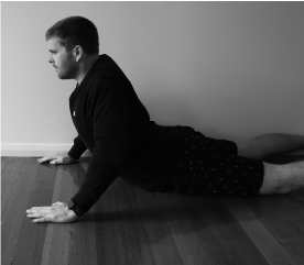
Figure 3. Repeated extension in laying
Table 4. Treatment and reassessment session 2
Treatment 1 |
Test: |
- Pa (2/10 NRS) hands ½ thigh
- Pb; P1 hands ¼ thigh, P2 hands to knee
|
Treatment: |
Central PA L4 GIII 3x 60 secs |
Re-test: |
|
Treatment 2 |
Test: |
As above |
Treatment: |
Central PA L5 GIII 3x 60 secs |
Re-test: |
- Pb improved; P1 hands to knee, P2 ½ shin
- PSLR + ADF: improved; 70° lim Pb
|
Treatment 3 |
Test: |
As above |
Treatment: |
REIL x10 |
Re-test: |
- Pb improved; P1 hands to knee, P2 3/4 shin
- PSLR + ADF: improved; 75° lim Pb
|
Kate reported a week later that her sitting tolerance improved (>60 minutes) and she had not experienced Pa since last treatment. Shown in Table 5, central mobilisation was repeated through L5 as it yielded greatest improvement in the previous session but increased (GIV) due to the limiting factor being end of range stiffness only. Right sided unilateral PA mobilisation was trialled through L4-S1 as they were still restricted on palpation triggering Pb, L5/S1 mobilisation leading to reduced neural symptoms and restriction on key markers post intervention. The patient was keen on re-starting her exercise routine and was recommended to re-commence run/walking building by 1km per week dependent on symptoms and clinical Pilates limiting lumbar flexion initially.
Table 5. Treatment and reassessment session 3
Treatment 1 |
Test: |
- Central PA L5; R1@50% P’R2 @ 80%
- Right PA L4/5; P1@70% (Pb awareness) P’R2 @ 80%
- Right PA L5/S1; P1@20% (Pb 2/10 NRS) P’R2 @ 40%
- Standing flexion: Pb; P1 ½ shin, P2 ¾ shin
- PSLR + ADF: 70° lim Pb
- SLUMP; Pb P2 -10° knee extension
|
Treatment: |
Central PA L5 GIV 3x 60 secs |
Re-test: |
- Central PA L5; R1@80%
- Standing flexion: NC
- PSLR + ADF: NC
- SLUMP: NC
|
Treatment 2 |
Test: |
As above |
Treatment: |
Right PA L4/L5 GIII 3x 60 secs |
Re-test: |
- Right PA L4/5; P’R2 @ 90%
- Standing flexion: Pb; P1 ¾ shin, limited bilateral hamstring tightness
- PSLR + ADF: 75° limited by hamstring tightness, mild pb awareness
- SLUMP: Pb; -10° knee extension
|
Treatment 3: |
Test: |
As above |
Treatment: |
Right PA L5/S1 GIII 3x 60 secs |
Re-test: |
- Right PA L5/S1; P’R2 @ 70%
- Standing flexion: hands to ankle limited by hamstring tightness R=L
- PSLR + ADF: 80° limited by hamstring tightness R=L
- SLUMP: negative, -5° knee extension R=L
|
A week after the third session Kate reported that running had helped to ‘loosen her up’ and her sitting was not limited. She was happy, able to work pain free and get back into her exercise regime. Assessment highlighted all impairments from the original assessment were normal except for awareness of Pb with neural tension tasks (SLUMP and PSLR) and some palpatory stiffness of the lumbar spine. Trialling neurodynamic mobilisation exercise, shown in Figure 4, significantly improved re-test results (SLUMP and PSLR). This exercise was included as a home exercise, 3 sets of 30 seconds slow gliding daily.
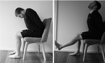
Figure 4. Neurodynamic mobilisation exercise
Over the next month Kate was reviewed twice more, monitoring her key signs. Lower back mobilisation yielded no further improvement in palpatory stiffness and her exercise returned to pre-injury levels. Fourteen weeks after initial injury she was discharged to self-management, encouraged to maintain her exercises. A follow up call 2 months later found her fit and healthy, having lost 5 kg through exercise.
Back pain is complex and multifactorial, requiring a detailed subjective examination to increase confidence in physical tests that are often of low sensitivity. Clinical accuracy is ascertained through a combination of subjective and objective findings coupled with the patient response to intervention which can be changed as necessary if diagnosis changes. Treatment needs to target contributing factors and patient beliefs to optimise outcomes. In this case a combination of several targeted interventions through sound clinical reasoning and structured assessment processes ensured a successful outcome.
- Alyami H, Albarrati AM (2016) Comparison of Spinal Angles in a Typing Task on a Laptop and a Desktop Computer: A Preliminary Study. Am J Occup Ther 70: 7006350020p1-8.
- Billy GG, Lemieux SK, Chow MX (2014) Changes in lumbar disk morphology associated with prolonged sitting assessed by magnetic resonance imaging. PM&R 6: 790-795.
- Dankaerts W, O'Sullivan P, Burnett A, Straker L (2006) Differences in sitting postures are associated with nonspecific chronic low back pain disorders when patients are subclassified. Spine 31: 698-704.
- Delitto A, George SZ, Van Dillen LR, Whitman JM, Sowa G, et al. (2012) Orthopaedic section of the American Physical Therapy Association: low back pain. J Orthop Sports Phys Ther 42: A1-57.
- Ford J (2018) Spinal physiotherapy part a (lumbar). St Vincents Private Hospital East Melbourne, Australian Physiotherapy Association.
- García‐Cosamalón J, Del Valle ME, Calavia MG, García‐Suárez O, López‐Muñiz A, et al. (2010) Intervertebral disc, sensory nerves and neurotrophins: who is who in discogenic pain? J Anat 217: 1-5.
- Jones MA (2014) ch. 2, Clinical reasoning: from the Maitland concept and beyond. Maitland’s vertebral manipulation. London, Elsevier.
- Lin JH, Chiang YH, Chen CC (2014) Lumbar radiculopathy and its neurobiological basis. World J Anesthesiology 3: 162-173.
- Lou C, Chen H, Mei L, Yu W, Zhu K, et al. (2017) Association between menopause and lumbar disc degeneration: an MRI study of 1,566 women and 1,382 men. Menopause 24: 1136-1144.
- Makhsous M, Lin F, Bankard J, Hendrix RW, Hepler M, et al. (2009) Biomechanical effects of sitting with adjustable ischial and lumbar support on occupational low back pain: evaluation of sitting load and back muscle activity. BMC Musculoskelet Disord 10: 17.
- Nachemson AL (1981) "Disc pressure measurements." Spine (Phila Pa 1976) 6: 93-97.
- Nykvist F, Hurme M, Alaranta H, Kaitsaari M (1995) Severe sciatica: a 13-year follow-up of 342 patients. Eur Spine J 4: 335-338.
- O’Neill CW, Kurgansky ME, Derby R, Ryan DP (2002) Disc stimulation and patterns of referred pain. Spine 27: 2776-2781.
- Oliveira CB, Maher CG, Pinto RZ, Traeger AC, Lin CW, et al. (2018) Clinical practice guidelines for the management of non-specific low back pain in primary care: an updated overview. Eur Spine J 27: 2791-2803.
- Rao D, Scuderi G, Scuderi C, Grewal R, Sandhu SJ (2018) The Use of Imaging in Management of Patients with Low Back Pain. J Clin Imaging Sci 8.
- Rea W, Kapur S, Mutagi H (2012) Intervertebral disc as a source of pain. Continuing Education in Anaesthesia, Critical Care & Pain 12: 279-282.
- Schneider M, Erhard R, Brach J, Tellin W, Imbarlina F, et al. (2008) Spinal palpation for lumbar segmental mobility and pain provocation: an interexaminer reliability study. J Manipulative Physiol Ther 31: 465-473.
- Schoenfeld AJ, Laughlin M, Bader JO, Bono CM (2012) Characterization of the incidence and risk factors for the development of lumbar radiculopathy. Clin Spine Surg 25: 163-167.
- Seidler A, Bolm-Audorff U, Siol T, Henkel N, Fuchs C, et al. (2003) Occupational risk factors for symptomatic lumbar disc herniation; a case-control study. Occup Environ Med 60: 821-830.
- Sheng B, Feng C, Zhang D, Spitler H, Shi L (2017) Associations between obesity and spinal diseases: a medical expenditure panel study analysis. Int J Environ Res Public Health 14: 183.
- Traeger AC, Moseley GL, Hübscher M, Lee H, Skinner IW, et al. (2014) Pain education to prevent chronic low back pain: a study protocol for a randomised controlled trial. BMJ open 4: e005505.
- Van Der Windt DA, Simons E, Riphagen II, Ammendolia C, Verhagen AP, et al. (2010) Physical examination for lumbar radiculopathy due to disc herniation in patients with low‐back pain. Cochrane Database Syst Rev 2010: Cd007431.