Introduction: Cardiac output and index, measured during right heart catheterization are used to define vasoreactivity and predict mortality. The Fick and thermo-dilution methods are routinely used, but have not been compared following vasoreactivity.
Methods: We retrospectively reviewed the RHC data of 50 patients; that met inclusion criteria, of a right heart catheterization with inhaled nitric oxide challenge and had both methods of CO and CI calculations before and after the challenge. Intra-class correlation was used to demonstrate correlation.
Results: There was a significant correlation between the CO/CI calculated by TD before and after iNO with an ICC of 0.9 (0.83170, 0.94147) for CO and ICC of 0.89 (0.81457, 0.93583.) for CI. There was a weak yet statistically significant correlation between CO/CI calculated by FM before and after iNO with an ICC 0.45 (0.20323, 0.64301) for CO and 0.43 (0.17657 0.62996) for CI. The average pre iNO pulmonary artery (PA) saturation was 67.3% and the average PA saturation post iNO was 79.8%. There was a significant difference between PA saturations of 12.5% (p<0.001).
Conclusion: We found that the CO and CI pre- and post iNO had a stronger correlation by TD method than by FM. The FM is dependent on hemoglobin saturations which is higher in patients with PAH post iNO challenge. The TD is independent of the PA hemoglobin saturations and therefore is not augmented post iNO challenge and should be the preferred method of calculating CO and CI in patients undergoing RHC with iNO challenge.
pulmonary hypertension, right heart catheterization, cardiac output, Fick calculation, Thermo-dilution, Vaso-reactivity test, inhaled Nitric Oxide
Pulmonary Arterial Hypertension (PAH) is characterized by remodeling of small pulmonary arteries, resulting in increasing pulmonary vascular resistance and ultimately leading to right ventricular failure [1]. Despite all advances in therapy, PAH continues to be a devastating disease and has a poor prognosis if it is not diagnosed and treated early. Unfortunately, most patients are still diagnosed in the late course of the disease [2]. This disorder affects about 15 individuals per million [3]. The diagnosis of PAH is based on hemodynamics obtained during a Right Heart Catheterization (RHC). The current accepted hemodynamic definition of PAH is a mean Pulmonary Artery Pressure (mPAP) equal or greater than 25 mm Hg or a Pulmonary Vascular Resistance (PVR) greater than 3 Wood units along with Left Ventricular End-Diastolic Pressure (LVEDP) or the surrogate measurement of pulmonary capillary wedge pressure (PCWP) when it is less than or equal to 15 mmHg; [4]. The Cardiac Output (CO) and index (CI) are also important measurements during the RHC, they are strong predictors of mortality in patients with PAH and they are used to define “response” to the Vasoreactivity Challenge (VC) [5]. The VC is recommended only for patients with Idiopathic PAH (IPAH) [4]. It involves intravenous or inhaled administration of a rapid acting specific pulmonary vasodilator [6]. Inhaled Nitric Oxide (iNO) is known to vasodilate the pulmonary arteries and thus decreases the pulmonary vascular resistance PVR) [5,7]. A positive VC is defined as a decrease in mPAP by 20% (about 10 mmHg) and the mPAP should be less than 40 mmHg or into the normal range. The CO must be preserved [5]. The importance of determining acute response is both diagnostic and prognostic. Acute responders typically have better long-term survival and are candidates for initial calcium-channel blocker therapy [8-10].
To calculate the CO and CI we used two accepted methods; the Fick Method (FM) and the Thermodilution method (TD) [11]. Several studies have compared different technologies to measure CO, but only a few studies have tried to answer whether the FM or TD is better in patients with PAH [12]. Recent studies show that there are significant discrepancies between the CO measured by FM or TD [13,14]. We have recently studied and found a good correlation of TD and FM in mild, moderate and severe PAH pre iNO [15]. Rich et al showed that, a Non-Invasive Output Monitor (NICOM) was precise and reliably measuring CO at rest and after vasodilator challenge, and was comparable with FM and TD to detect such changes [16].
In this manuscript, we retrospectively review the methods of calculating CO and CI via FM and TD and analyze whether these correlated better before and after iNO challenge.
We conducted a retrospective chart review of patients who underwent RHC for diagnosis or follow up of PAH at a university center and a tertiary referral hospital. All procedures were performed in a cardiac catheterization laboratory guided by fluoroscopy. To be included in the study, patients had to meet the definition for pulmonary arterial hypertension [4], and were non-responders to VC. All patients included in the study underwent a VC with iNO at 40ppm with 100% oxygen for 5 minutes. Mean Pulmonary Artery Pressure (mPAP), PCWP, CO and CI measurements by both FM and TD were measured pre- and post iNO challenge. Saturations were measured in the inferior vena cava, superior vena cava, right atrium and pulmonary artery. All variables were summarized by means and Standard Deviations (SD). Demographic data and co-morbidities were recorded for each patient. An Intra-class Correlation Coefficient (ICC) was used to demonstrate strength of correlation between CO and CI calculated by FM before and after iNO challenge and similarly by TD before and after iNO challenge. We used student’s “t” test to compare the difference between the mean saturations before and after iNO challenge.
Methods to measure cardiac output
Thermodilution method: The TD method is based on the principles of dilution uses a room temperature (cold) bolus (10 mL) of sterile solution that is injected into the proximal port of a pulmonary artery catheter. This ends up in the right atrium. Here, the cold saline mixes with the blood and passes through the tricuspid valve. A thermistor within the catheter senses the change in blood temperature as the blood passes the catheter tip located in the pulmonary artery. A curve that shows the change in temperature over time is generated by a computer and converted into a measurement of CO/CI given that CO/CI is inversely proportional to the area under the curve [17]. In other words, the higher the CO/CI the less time the bolus has to warm to body temperature.
Fick method: The FM states that uptake of oxygen by any organ is the product of the arteriovenous (A-V) concentration difference of oxygen and the blood flow to that organ. As the blood moves, more oxygen is taken out of circulation and the distal saturations drops. CO is calculated based on the following formula; CO = [O2 consumption (ml/min)]/A-Mixed Venous O2 difference (ml O2/ 100ml blood) x 10. The arteriovenous oxygen (A- Mixed Venous O2) difference is calculated from arterial x mixed venous (PA) O2 content, where O2 content = saturation% x 1.36 x hemoglobin.
The used Resting O2 Consumption was 125ml O2 per square meter of body surface area per minute [18].
One hundred and eighty patient’s charts were reviewed; out of them, 50 (27%) met inclusion criteria matching the diagnosis of PAH and were non-responders to VC. Sixty two percent were females. The most common comorbidities were obstructive lung disease (28%), obstructive sleep apnea (18%) and autoimmune/connective tissue disorder (14%). The hemodynamic parameters before and after iNO are summarized in Table 1. There was a statistically significant and strong correlation between the CO calculated by TD before and after iNO with an ICC of 0.9 (95% CI 0.83, 0.94) (Figure 1). There was also a statistically significant and strong relationship observed for CI calculated by TD before and after iNO with an ICC of 0.89 (95% 0.81, 0.94.)
Table 1. Hemodynamic Parameters before and after Inhaled Nitric Oxide
|
PRE – iNO (sd) |
POST - iNO (sd) |
Difference |
p |
mPAP (mmHg) |
39.6 (10.7) |
34.9 (10.9) |
4.7 |
0.55 |
PCWP (mmHg) |
11.1 (4.0) |
13.0 (5.0) |
-1.9 |
0.93 |
PVR(Woods Units) |
6.09 (4.7) |
|
|
|
Fick CO (L/min) |
5.77 (2.3) |
7.43 (3.8) |
-1.66 |
0.99 |
Fick CI (L/min/m2) |
2.94 (1.0) |
3.69 (1.5) |
-0.75 |
0.99 |
TD CO (L/min) |
5.58 (2.4) |
5.61 (2.4) |
-0.03 |
0.5 |
TD CI (L/min/m2) |
2.80 (1.1) |
2.85 (0.9) |
-0.05 |
0.08 |
PA Saturation (%) |
67.3 (8.3) |
79.9 (10.1) |
12.6 |
p<0.001 |
iNO: inhaled Nitric Oxide, mPAP: mean Pulmonary Arterial Pressure, PCWP: Pulmonary Capillary Wedge Pressure, PVR: Pulmonary Vascular Resistance, Fick CO: Cardiac Output by Fick method, Fick CI; Cardiac Index by Fick method, TD CO: Cardiac Output by Thermodilution Method, TD CI: Cardiac Index by Thermodilution Method, PA: Pulmonary Artery
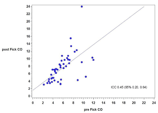
Figure 1. Correlation between cardiac output by Fick method before and after Inhaled Nitric Oxide.
There was a statistically significant weak correlation between CO calculated by FM before and after iNO with an ICC 0.45 (95% 0.20, 0.64) (Figure 2). There was the same statistically significant weak relationship observed for CI calculated by FM before and after iNO with an ICC of 0.43 (95% 0.18, 0.63). The COs by FM and TD before iNO had a difference of the means of 0.19 L/min, with an ICC 0.7 (95% 0.53, 0.82). The average COs by FM and TD after iNO had a difference of means of 1.92 L/min with a statistically significant weak correlation with ICC of 0.49 (95% 0.25, 0.67).
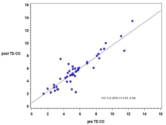
Figure 2. Correlation between cardiac output by thermo-dilution method before and after Inhaled Nitric Oxide.
The average pre iNO PA saturation was 67.3% (sd; 8.3%) and the average PA post iNO was 79.9% (sd; 10.1%). (There was a statistically significant difference between saturations of 12.6% (sd; 6.3%) (p<0.001)
The FM first described by Adolph Fick in 1870, uses the quotient of the oxygen consumption and the difference of the arterial and mixed venous oxygen content [19].
TD method has been widely used since its inception in 1954 [20]. It involves injecting a known volume of saline with known temperature into the PAC’s right atrial opening and measuring the drop-in temperature at the distal opening 20 cm away. The change in temperature overtime detected at the distal end of the catheter (the thermistor) is used to calculate CO and CI [21]. The results of this study indicate that the CO and CI calculated by the TD, before and after iNO, have a strong correlation in patients undergoing RHC with ICC of 0.9. This strong correlation observed in CO and CI calculated by TD may be due to the lack of dependence on the arterial and mPAP hemoglobin saturation. The FM does depend on these hemoglobin saturations. Inhaled nitric oxide improves ventilation-perfusion matching in the lung by increasing blood flow to well-ventilated areas of the lung [22]. This transient increase in ventilation perfusion allows for the oxygen tension in the mPAP to increase. In addition, the 40ppm of iNO was given with a FiO2 close to 100%, which also increases arterial oxygen saturation. As the mPAP saturation increases, calculation of the CO and CI by the FM is then falsely elevated. The CO by the FM may not detect an otherwise dropping CO nullifying an otherwise positive vasoreactivity challenge. This can lead to a trial of calcium channel blockers instead of other potentially beneficial agents and may ultimately be deleterious.
2021 Copyright OAT. All rights reserv
There have been several studies suggesting that the TD method is inaccurate in patients with Tricuspid Regurgitation (TR) pre iNO [23,24]. Some studies suggest that TD is valid even in patients with severe TR [25]. In a more recent study, Fares et al recommend that both of these measurements be taken during the RHC in patients with PAH [13]. We have previously documented that pre iNO, the TD correlates with the FM in mild, moderate, and severe PAH [15]. The 5th World Symposium on PAH recommended TD over FM in the pre vasoreactive phase [4]. This decision is based on data from Hoeper et al [4,21] who compared the TD to the gold standard (direct FM) and found a positive correlation. The FM has less reliability because it uses estimated values of oxygen uptake derived from tables.
In conclusion, the CO and CI in patients with PAH predict both prognosis and whether the patients have a positive vasodilation challenge. The weak correlation of the CO and CI by FM after iNO can be explained by the augmentation of the PA saturation. During the iNO challenge, there is a stronger correlation between CO and CI obtained by TD than by FM. It can be explained by the TD’s independence on the arterial or mPAP hemoglobin saturations. Most of the patients studied were non-responders and no change in the CO and CI were expected. Therefore, for iNO vasoreactivity testing in patients with PAH, the TD measurement of the CO and CI should be used rather than the FM, as TD has a much stronger correlation before and after the iNO challenge.
This study was retrospective study and some patients with chronic hypoxemic respiratory failure were given oxygen before and during the procedure. This may have altered the pre iNO pulmonary artery saturations. A prospective randomized trial would be needed to validate our study.
All authors listed on this manuscript m made a significant contribution on the Conception and design of the project, data collection analysis and interpretation, rafting of the manuscript and final approval of the version to be published.made substantive intellectual contributions to a published studysubstantial contributions to conception and design, or acquisition of data, or analysis and interpretation of data; 2) drafting the article or revising it critically for important intellectual content; 3) final approval of the version to be published; and 4) agreement to act as guarantor of the work (ensuring that questions related to any part of the work are appropriately investigated and resolved
- Farber H (2004) Mechanism of disease pulmonary arterial hypertension. NEJM 39: 133-136.
- Barst R, Badesh D (2010) Pulmonary arterial hypertension Baseline Characteristics from the REVEAL Registry Web site.
- Humbert M, Sitbon O, Chaouat A, Bertocchi M, Habib G, et al. (2006) Pulmonary arterial hypertension in france: Results from a national registry. Am J Respir Crit Care Med 173: 1023-1030.
- Hoeper MM, Bogaard HJ, Condliffe R, Frantz R, Khanna D, et al. (2013) Definitions and diagnosis of pulmonary hypertension. J Am Coll Cardiol 62 (25 Suppl): D42-D50. [Crossref]
- McLaughlin VV, Archer SL, Badesch DB, Barst RJ, Farber HW, et al. (2009) ACCF/AHA 2009 expert consensus document on pulmonary hypertension a report of the american college of cardiology foundation task force on expert consensus documents and the american heart association developed in collaboration with the american college of chest physicians; american thoracic society, inc.; and the pulmonary hypertension association. J Am Coll Cardiol 53: 1573-1619. [Crossref]
- Rumbak M, Cox J, Dray D, Weston M, Solomon D. The right heart catheterization for the pulmonologist: The diagnosis, management, and prognosis of patients with pulmonary arterial hypertension. Clinical Pulmonary Medicine 17: 239-243.
- Norbert F. Voelkel, MD; Robert A. Quaife, MD; Leslie A. Leinwand, et al. (2006) Right ventricular function and failure: report of a National Heart, Lung, and Blood Institute working group on cellular and molecular mechanisms of right heart failure. Circulation 114: 1883-1891. [Crossref]
- Sitbon O, Humbert M, Jais X, Ioos V, Hamid AM, et al. (2005) Long-term response to calcium channel blockers in idiopathic pulmonary arterial hypertension. Circulation 111: 3105-3111. [Crossref]
- Morales-Blanhir J, Santos S, de Jover L, et al. (2004) Clinical value of vasodilator test with inhaled nitric oxide for predicting long-term response to oral vasodilators in pulmonary hypertension. Respir Med 98: 225-234. [Crossref]
- Malhotra R, Hess D, Lewis GD, Bloch KD, Waxman AB, et al. (2011) Vasoreactivity to inhaled nitric oxide with oxygen predicts long-term survival in pulmonary arterial hypertension. Pul Circ 1: 250-258. [Crossref]
- Strang V (2008) The nuts and bolts of right heart catheterization and PA catheter placement. In: Cardiovascular hemodynamics for the clinician. Blackwell Publishing Ltd. PP: 17-36.
- Reuter DA, Huang C, Edrich T, Shernan SK, Eltzschig HK (2010) Cardiac output monitoring using indicator-dilution techniques: Basics, limits, and perspectives. Anesth Analg 110: 799-811. [Crossref]
- Fares WH, Blanchard SK, Stouffer GA, Chang PP, Roseamond WD et al. (2012) Thermodilution and fick cardiac outputs differ: Impact on pulmonary hypertension evaluation. Can Respir J 19: 261-266. [Crossref]
- Engoren M, Barbee D (2005) Comparison of cardiac output determined by bioimpedance, thermodilution, and the Fick method. Am J Crit Care 14: 40-45. [Crossref]
- Geck R, Rumbak M, Seleznick M, (2012) The correlation of fick and thermodilution cardiac indices in patients with pulmonary hypertension as A function of the severity of disease. Clinical Evaluation of Pulmonary Hypertension. Am J Respir Crit Care Med 185: A1894.
- Rich JD, Archer SL, Rich S (2013) Noninvasive cardiac output measurements in patients with pulmonary hypertension. Eur Respir J 42: 125-133. [Crossref]
- Gawlinski A (2004) Measuring cardiac output: Intermittent bolus thermodilution method. Crit Care Nurse 24:74-78. [Crossref]
- Hoeper MM, Maier R, Tongers J, Niedermeyer J, Hohlfeld JM, et al. (1999) Determination of cardiac output by the fick method, thermodilution, and acetylene rebreathing in pulmonary hypertension. Am J Respir Crit Care Med 160: 535-541. [Crossref]
- Zuntz N (1892) Ernahrung des herzens und ihre beziehung zu seiner arbeitsleistung. Leipzig G. Thieme.
- Fegler G (1954) Measurement of cardiac output in anesthetized animals by a thermo-dilution method. Q J Exp Physiol Cogn Med Sci 39: 153-164. [Crossref]
- Hoeper MM, Maier R, Tongers J, Niedermeyer J, Hohlfeld JM, et al. (1999) Determination of cardiac output by the Fick method, thermodilution, and acetylene rebreathing in pulmonary hypertension. Am J Respir Crit Care Med 160: 535-541. [Crossref]
- Ichinose F, Roberts JD, Zapol WM (2004) Inhaled nitric oxide: A selective pulmonary vasodilator: Current uses and therapeutic potential. Circulation 109: 3106-3111. [Crossref]
- Cigarroa RG, Lange RA, Williams RH, Bedotto JB, Hillis LD (1989) Underestimation of cardiac output by thermodilution in patients with tricuspid regurgitation. Am J Med 86: 417-420. [Crossref]
- Heerdt PM, Pond CG, Blessios GA, Rosenbloom M (1992) Inaccuracy of cardiac output by thermodilution during acute tricuspid regurgitation. Ann Thorac Surg 53: 706-708. [Crossref]
- Kadota LT (1985) Theory and application of thermodilution cardiac output measurement: a review. Heart Lung 14: 605-616. [Crossref]