Evolution in dentistry has bridged the gap between impossible and possible. Componeer is one such evolution. A thin composite shell which can be placed on anterior teeth with minimal preparation to bring out change in aesthetics and morphology has an edge over porcelain veneers. It has notably minimised the chair side time and there is no lab work needed. This article discusses esthetic restoration of fractured anterior tooth, diastema closure and masking of fluorosis stains with componeers.
Composite veneers, aesthetics, anterior teeth
Componeers as the name suggests are polymerized, prefabricated, enamel shaded composite laminates. The veneer coatings as thin as 0.3 mm cervically and 0.6-1.0 mm incisally, conserves tooth structure during preparation. Componeers are manufactured to follow the M proportion and combine the favorable properties of both the direct and indirect veneering systems with some inherent properties that no other veneering system offers. Veneering of anterior teeth was introduced to Dentistry by Dr. Pincus in 1937 [1].
Prefabricated composite veneers emerged in mid-1970’s but were abandoned due to former technical limitations. This concept has been renewed and reintroduced with a new shade guide comprising enamel shells, along with bonding agent of different colours to give desired effects in esthetics. The componeer is an affordable and less time-consuming alternative for patients who are not financially prepared for porcelain veneers. These combine the advantages of direct composite restoration with those of laboratory made veneers.
Componeers have bridged the gap between the direct bonded composite veneers and porcelain veneers not only in terms of the benefits but also the cost. A “natural layering concept” is followed, and a guide has been developed to allow the combination of all dentin and enamel shades [2]. This concept is based on a two-layer incremental technique, mimicking the anatomy of natural teeth [3].
- Restorations [4]
- Discoloration
- Tooth fracture
- Malformation
- To correct midline diastema, attrition, abrasion, erosion
- Cosmetic correction [4,5]
Severe bruxism and active caries.
This paper demonstrates the use of componeers in three different clinical situations-esthetic restoration of fractured anterior tooth, diastema closure and masking of fluorosis stains.
A 16-year-old boy reported to a private clinic with moderate fluorosis and midline diastema (Figure 1). He wanted a conservative treatment without any considerable reduction of the tooth structure and had already explored the conservative option of tooth bleaching/whitening but was unsatisfied with the result. A treatment plan for componeers was discussed with the patient and planned accordingly.
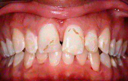
Figure 1. Midline diastema and mild fluorosis stains are visible.
Teeth were isolated with rubber dam and the minimal tooth preparation was done. Enamel shell guide was superimposed over the dentin core to determine the shade. The size of the componeers were selected using the contour guides provided in the componeer kit. Selected sizes were customized to suit the length and gingival contour of the patient. Standard procedure of etching and bonding was done on the buccal surface and on the lingual surface near the incisal edge of the prepared teeth. The bonding agent was light cured on the teeth with mylar strips in proximal spaces to separate the teeth. A layer of selected shade of composite resin was applied on the buccal surface of the tooth and left without light curing. The componeer was clamped on the componeer holder (provided in the componeer kit) and bonding agent was applied on the inner concave surface of the componeer and selected shade of composite was added. Without light curing the bonding agent and composite on the componeer, the componeer was placed on the tooth and was adapted to the tooth using the componeer placer (also a part of the componeer kit). The flash of composite was removed using the sharp MBC instrument provided in the kit and after checking the final position of the componeer on the tooth, the componeer was light cured for 30 seconds lingual and 30 seconds buccally. All the componeers were placed using the same technique. Final check of marginal adaptation and voids was done using a probe and the componeers were finished using the finishing cups and points provided in the kit. The interproximal areas were polished using the polishing strips and final polishing was done with polishing brushes. Incisal shaping of the componeers was done using the Swissflex polishing discs mounted on the mandrel provided in componeer kit. Composer masked the stains efficiently (Figure 2).
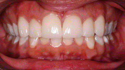
Figure 2. Componeers placed have masked the fluorosis stains and diastema.
This was a case of 15-year-old boy who reported to a private clinic with an Ellis Class II fracture of 21. The tooth was asymptomatic and after preventive therapy for the exposed dentin, componeer was placed using the standard protocol as discussed in Case 1. The results are as shown in Figures 3 and 4.
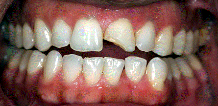
Figure 3. Ellis Class II fracture of 21.
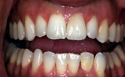
Figure 4. Componeer placed has masked the fracture and shade has blended well with the adjacent tooth.
This unique veneering system mimicks the properties of lab made Porcelain veneers (like identical size, shape, shade bilaterally, highly polished and glossed surface) and properties of direct composite veneers (like conservative, single visit procedure with polishability and repairablity). These veneers make the restoration as a monoblock unit,as they are made of hybrid composite resin and are cemented with the same.
Various studies have observed, the quality of fluorosed enamel to have an adverse effect on bonding,and a good bond is difficult to achieve [6]. This is due to the hypermineralization of tooth, and etchants are less effective in treating fluorosed tooth surfaces [7-9]. Restoring with the use of a dentinal bonding agent, dental composite, and prefabricated composite veneers is a viable option for treating severely stained and fluorosed teeth [10].
Composite veneers are less expensive, which makes this practice a feasible option, mostly to people with a lower income and satisfy the patient’s restorative needs and aesthetic desires [11].
The extremely thin and shiny veneer coatings of 0.3 mm conserves tooth structure during preparation and gives glossy, long lasting and natural looking aesthetics. Though porcelain is superior to componeers; it has higher chance of chipping off than componeers [12].
Componeers on the other hand, even if chipped can be repaired in minimal time and no lab work involved. The proximal line angles can be rounded in componeers compared to porcelain where they have to be parallel and at right angles.
It is a new breakthrough for restorative procedure as dependence on lab is highly reduced. Componeers do not require a mock up on the model; rather the final result can be demonstrated on the patients’ teeth and can be modified chair side to custom suit the patient’s profile. No impression needed for veneering. It can be repolished anytime to restore the aesthetics.
Componeers have bridged the gap between the direct bonded composite veneers and porcelain veneers not only in terms of the benefits but also the cost. The dependence on dental laboratory, in a single visit along with a vast flexibility of use in different clinical situations make it a necessary tool in clinical practice.
- Pincus CL (1937) Building mouth personality. California State Dental Association, California.
- Faunce FR, Myers DR (1976) Laminate veneer restoration of permanent incisors. J Am Dent Assoc 93: 790-792. [crossref]
- Helpin LM, Fleming JE (1982) Laboratory tech2021 Copyright OAT. All rights reservtion. Pediatr Dent 4: 48-50.
- Gomes G, Perdigão J (2014) Prefabricated composite resin veneers: a clinical review. J Esthet Restor Dent 26: 302-313. [crossref]
- Dietschi D (2008) Optimizing smile composition and esthetics with resin composites and other conservative esthetic procedures. Eur J Esth Dent 3: 14-29.
- Miller RA (1995) Bonding fluorosed teeth: new materials for old problems. J Clin Orthod 29: 424-427. [crossref]
- Denbesten PK, Thariani H (1992) Biological mechanisms of fluorosis and level and timing of systemic exposure to fluoride with respect to fluorosis. J Dent Res 71: 1238-1243.
- Hoffman S, Rovelstad R, McEwan WS, Drew CM (1969) Demineralization studies of fluoride treated enamel using scanning electron microscopy. J Dent Res 48: 1296-1302.
- Kochavi D, Gedalia I, Anaise J (1975) Effects of conditioning with fluoride and phosphoric acid on enamel surfaces as evaluated by scanning electron microscopy and fluoride incorporation. J Dent Res 54: 304-309.
- Toit JD, Patel N, Montalli V, Jain S (2012) Aesthetic treatment of severely fluorosed teeth with prefabricated composite veneers: a case report. Int Dent Sou Afr 2: 44-50.
- Pontons-Melo JC, Furuse AY, Mondelli J (2011) A direct composite resin stratification technique for restoration of the smile. Quintessence Int. 42: 205-211.
- Peumans M, Van Meerbeek B, Lambrechts P, Vanherle G (2000) Porcelain veneers: a review of the literature. J Dent 28: 163-177. [crossref]