Abstract
Congenital anomalies of gallbladder are extremely rare, nonetheless do occur and may have important clinical predilection. Anomalies of the gallbladder can hinder biliary drainage or may predispose cholecystitis or cholelithiasis. We present a case of congenital bilobed gallbladder in an adult female, who presented to the ED for right upper quadrant pain and a right upper quadrant ultrasound consistent with acute cholecystitis. Following cholecystectomy, a gallbladder specimen with two distinct lumens and a common cystic duct was observed.
The aim of this report is to review the literature of previous cases to contrast with the present one. An online search of available databases, including MEDLINE/PubMed, was conducted. Terms used included bilobed gallbladder, congenital gallbladder, anomalies of the gallbladder, and congenital anomalies of the gallbladder. Inclusion criteria comprised all cases identified in alive patients, and articles written in English.
Introduction
Gallbladder is a pearl-shaped intraperitoneal organ which is situated beneath the right lobe of the liver. It functions to concentrate, store, and release bile into the gastrointestinal tract. Congenital anomalies of the gallbladder are rare; however, some reported anomalies include agenesis of the vesical fellea, duplication, bilobed, gallbladder diverticulum, and absence of gallbladder [1]. Bilobed gallbladder anomaly is relatively rare in humans and occurs at a rate of 1 case per 3000 to 4000 patients [2]. This condition is not typically diagnosed preoperatively and maybe missed during surgery. However, preoperative diagnosis is extremely crucial for proper surgical planning and minimizing the risk of complications. In one reported case, there was a need for reoperation following a laparoscopic cholecystectomy of a duplicated gallbladder, where the accessory lobe was not retracted and later became inflamed and infected.
The current study presents a unique case of bilobed gallbladder in a 53-year-old female, who presented with nausea, food intolerance, and right upper quadrant pain with radiation to the back. Her preoperative and intraoperative diagnoses were consistent with acute cholecystitis. We received the specimen in the Pathology Department and found a 10.5 x 4.2 x 3.5 cm intact gallbladder with an accessory lobe measuring 8 x 2.5 x 1 cm on the mesenteric aspect of the gallbladder. The lumen of the accessory lobe had a different gross and microscopic morphology. To the best of our knowledge a similar case has not been reported in the literature.
Case presentation
A 53-year old female who was recently diagnosed with cholelithiasis on ultrasound imaging after being seen at an outside hospital. She came to the Emergency Department (ED) at our institution due to worsening of right upper quadrant and epigastric pain. The patient was seen in our ED the night before with similar complains and was discharged with plans for an elective cholecystectomy the following week. The patient endorsed worsening pain, nausea, and vomiting. She denied fever, chills, or recent antibiotics use. A stat ultrasound, which was consistent with acute cholecystitis was obtained and the patient received antibiotics, analgesics, and was prepped for a laparoscopic cholecystectomy. (Table 1 and 2)
WBC |
12.8 H |
Hemoglobin |
12.8 |
Hematocrit |
38.9 |
RBC count |
4.56 |
MCV |
85.6 |
Neutrophils |
89 H |
Absolute Neutrophil |
11.2 H |
Bilirubin Total |
1.4 H |
Table 1. Admitting labs
Urine PH and Specific Gravity |
7.0 and 1.017 |
Color |
Cloudy |
Urine Ketones |
1+ |
Urine Occult Blood |
3+ |
Urine HCG |
Negative |
Table 2. Urinalysis
Operative report
On the operative report, the patient underwent a robotic assisted laparoscopic cholecystectomy under general anaesthesia. Post-operative diagnosis was consistent with an acute cholecystitis with extensive inflammation and edema. On post-op day 1, the patient reported feeling better and her pain as controlled. She had tolerated diet and was stable for discharge.
Surgical pathology report
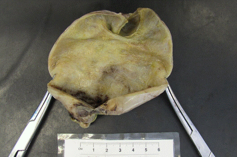
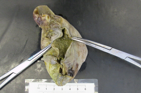
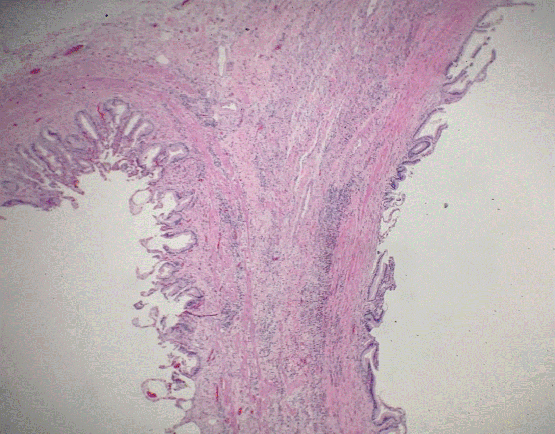
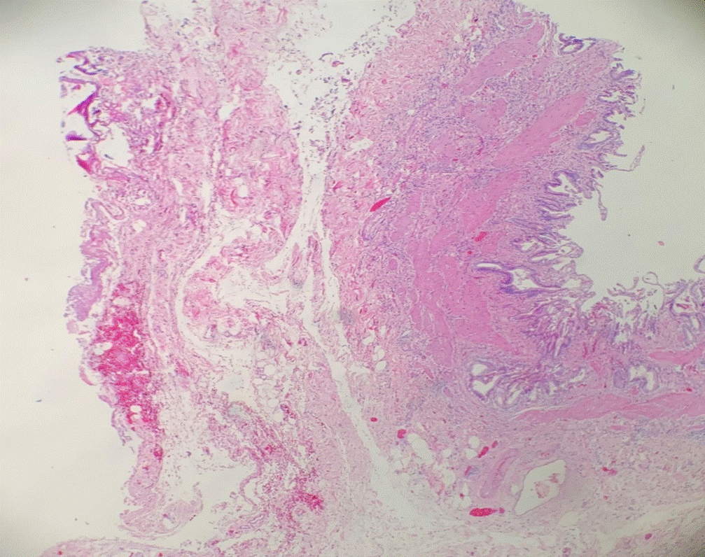
We received the specimen at the Pathology Department in a formalin fixed specimen container and designated as “gallbladder”. The specimen is a 10.5 x 4.2 x 3.5 cm bilobed gallbladder with a single cystic duct. The accessory lobe measures 8 x 2.5 x 1 cm. The specimen contains a copious amount of yellow, opaque bile with numerous (>25) yellow, multifaced choleliths ranging from 0.2 to 1.5 cm in size, in both lumens but predominantly within the accessory lobe. A 0.4 cm in thickness septum completely separates the two lobes of the gallbladder. The mucosa of the main lobe appears tan-pink and smooth; however, the mucosa of the accessory lobe is bile-stained green, with diffuse yellow stippling (Figures 1 and 2). Representative sections were obtained and the pathology report was consistent with a congenital bilobed gallbladder with acute cholecystitis superimposed on chronic cholecystitis and cholelithiasis (Figures 3 and 4).

Figure 1. Main gallbladder lobe: opened from cystic duct to fundus

Figure 2. Accessory gallbladder lobe: opened from cystic duct to fundus

Figure 3. Magnified septal epithelium (10X) showing infiltration of inflammatory cells into the lamina propria and muscularis propria

Figure 4. Magnified fundic epithelium (20X) showing acute inflammation, and two distinct epithelial morphologies
Discussion
Gallbladder is an endodermally derived organ, which develops from the hepatic diverticulum in the 4th week of intrauterine life. Rarely, congenital anomalies of the gallbladder occur where the gallbladder becomes atrophic, doubles itself, develops a septum, acquires a diverticulum, and or is completely absent [2,3]. Duplication of gallbladder is the result of splitting of the cystic primordium which typically occurs during the 5th or 6th weeks of embryogenesis. Additionally, an accessory lobe can be formed if there is budding of the biliary primordium to give rise to an accessory gallbladder [4]. The rate of occurrence of a congenital bilobed gallbladder is approximately 1 in 3000 to 4000 patients [5]. This case report is being presented due to the extremely rare occurrence of a bilobed gallbladder as well as the presence of such pathology with two distinct gross and microscopic mucosal morphologies. In previously reported cases of bilobed gallbladder, the main and accessory lobes have a similar histology [6]; however, in this case, the main gallbladder lobe grossly presents as tan and smooth while the accessory lobe is bile-stained with diffuse yellow stippling consistent with cholestasis.
Double lumen gallbladders are more likely to form choleliths due to common bile duct or cystic duct obstructions, as well as gallbladder motility disruptions. Additionally, patients with these anomalies are at an increased risk of infection and biliary stasis, as the result of ductal obstruction [6]. Furthermore, patient’s with gallbladder anomalies are at increased risk of surgical complications, especially for patients with H shaped double lumen subtypes, where there is an increased risk of injury to the bile duct or the common hepatic duct [7]. However, in general, it is safe and feasible to perform laparoscopic cholecystectomies in patients with double lumen gallbladder and so far, there is only one occurrence in which conversion to an open procedure following a laparoscopic cholecystectomy was necessitated [8].
In conclusion congenital anomalies of the gallbladder are extremely rare; however, if present can have significant clinical predilections. A better understanding of the biliary anatomy during the preoperative phase is essential for improved surgical and symptomatologic management.
References
- Heffernon EW (1960) Congenital Absence of the Gallbladder. Jama 174: 894.
- Boyden EA (1926) The accessory gall-bladder- an embryological and comparative study of aberrant biliary vesicles occurring in man and the domestic mammals. Am J Anatomy 38: 177-231.
- Anderson RE (1958) Congenital Bilobed Gallbladder. Arch Surg 76: 7.
- https://www-ncbi-nlm-nih gov.ezproxy.med.nyu.edu/pubmed/1259249?dopt=Abstract
- Kannan N (2014) Congenital Bilobed Gallbladder with Phrygian Cap Presenting as Calculus Cholecystitis. J Clin Diagn Res 8: NDO5-ND06. [Crossref]
- Yu W, Yuan H, Cheng S, Xing Y, Yan W (2016) A double gallbladder with a common bile duct stone treated by laparoscopy accompanied by choledochoscopy via the cystic duct: A case report. Exp Ther Med 12: 3521-3526. [Crossref]
- https://www.researchgate.net/publication/233426109_Double_Gallbladder
- Silvis R, Wieringen AJMV, Van Der Werken CHR (1996) Reoperation for a symptomatic double gallbladder. Surg Endosc 10: 336-337. [Crossref]


