Due to weakened immune system, the oncological patients are highly at risk with regard to the COVID-19 pandemic. The aim of this article is to contribute with a supplement for the database on symptomatology and chest imaging appearance in patients infected with COVID-19.
During the course of radiotherapy (RT), in four oncological patients we observed a variety of atypical clinical and laboratory expression: cough without fever, uroinfection with hematuria, gastrointestinal symptoms, leukopenia, which is difficult to associate with COVID-19 infection. In three out of seven patients, diffuse bilaterally ground glass opacities (GGO) are presented on the chest CT.
In oncological patients infected with COVID-19, a less common symptomatology such as gastrointestinal discomfort, dry cough without fever, which is difficult to associate with this viral infection, are observed. The features of pneumonia are accompanied by leukopenia, lymphopenia and accelerated ESR. Hematuria, proteinuria, elevated transaminases and Gamma-glutamyl transferase (GGT) levels are result of vasculitis which affects parenchymal organs like kidneys and liver. The radiological features of COVID-19 pneumonia are not specific. The ground glass opacities (GGO) are bilateral, asymmetrical, peripheral and subpleural in location, with predilection for the lower lobes. CT detection is very useful in time course of the disease with checking severity and prognosis of the disease.
cancer patients, risk, COVID-19, pneumonia, vasculitis, radiological findings
Due to weakened immune system, oncological patients are highly at risk with regard to the COVID-19 pandemic. The most typical symptoms are fever, sore throat, cough and shortness of breath in a recently exposed person [1]. New data suggests that the disease can present differently and it does not necessarily to affect the respiratory system [2]. This viral illness does not only present with respiratory symptoms. There has been increasing reports of patients presenting with gastrointestinal symptoms such as abdominal pain, diarrhoea, nausea and vomiting [3]. In this article, we present the diverse symptomatology and imaging of pulmonary pathological features in five oncological patients and two patients with different comorbidity infected with COVID-19.
The 1st case is an 81-year-old woman diagnosed with uterine carcinosarcoma G3-4, status after a radical total laparohisterectomy with bilateral adnexectomy. Admited in good general condition, afebrile and with normal laboratory values of peripheral blood count. After implementing percutaneous radiation treatment (RT) in the area of the small pelvis ( tumor bed and regional lymph nodes) to total dose (TD) 40 Gy, on 24/Jun/20 shows leukopenia 1,04 T/l (range 3,5-10,5 T/l); lymphopenia 0.37 T/l (range 0.77-4.62 T/l); 0.38 T/l (range 1.4-7.35 T/l) and thrombocytopenia 88.0 T/l (range 140-440 T/l) and normal ESR 12 mm/h (range 1-30 mm/h). The patient complained of general fatigue, with a one-day temperature increase up to 380 C, without coughing and sore throat. Three-digit values of liver enzymes AST-138 U/l (range 1-40 U/l) are reported; ALT-152 U/l (range 1-40 U/l) and GGT-120 U/l (range 5-35 U/l). The radiography and the native CT scan of the lung of 26/Jun/20 are presented on Figure 1 and Figure 2. She was admitted to the Lung Hospital, where after a PCR test, she tested positive on COVID-19. The rapid test for COVID-19 (IgG/Ig M antibodies): IgG antibodies-negative; Ig M antibodies- negative.
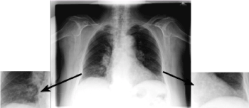
Figure 1. Chest X-ray/ 26.06.20- bilateral basal infiltrative shadows due to inflammatory changes. Conclusion: Bilateral pneumonia
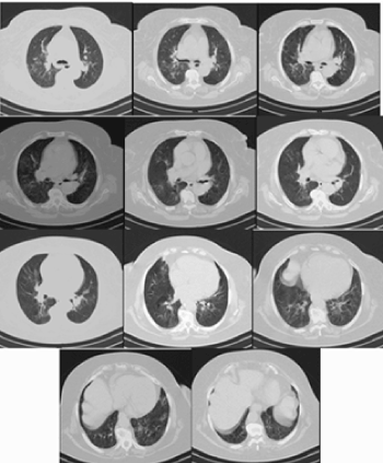
Figure 2. Native CT of the chest – Hazy patchy ground glass opacities with bilateral more peripheral and basal location
Treatment with Ceftriaxone 2 g i.v./daily and oral Azithromycin 500 mg daily was conducted. Chest X-ray on 01/Jul/20 (Figure 3A) and established high value D-dimer 0,98 mg/l (range 0.0-0.5 U/l mg/l). Peripheral blood count of 08/Jul/20: Leuco 3,8 T/l (range 3,5-10,5 T/l); neutrophilia/Neu 85% (range 39.3% -74.9%); lymphopenia/ Lym 0.4 T/l (range 0.94-2.99 T/l); mild thrombocytopenia Tr 124 T/l (range 140-440 T/l), at normalised D-dimer values of 0,25 mg/l and significantly reduced AST-43 U/l (range 1-40 U/l); ALT-47 U/l (range 1-40 U/l) and GGT-45 U/l (range 5-35 U/l). She was discharged from the Pulmonary Hospital with a negative PCR Covid-19 test and referred for home treatment and put in quarantine.
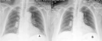
Figure 3. Chest X-ray-А/ 01/Jul/20 and В/ 08/Jul/20 – Moderate resorption of inflammatory changes in the right basal
The 2nd case is a 78-year-old woman with invasive ductal carcinoma Grade 3 of a right mammary gland, diagnosed in 2012. A radical right-sided mastectomy, 6 courses of adjuvant chemotherapy (Сh) and postoperative RT to TD 50Gy in the area of right chest wall and regional lymph nodes has been performed. In 2020 locally advanced invasive ductal carcinoma Grade 3 of left breast was proven. After 6 courses of neoadjuvant Ch, a left-sided radical mastectomy with axillary lymph dissection was performed and classified ypT4N0M0. The patient is reffered for postoperative RT of a left chest wall with the regional lymph nodes. Figure 4 presents a localization CT for RT planning on a left chest wall, which shows, that there are no pathological bilateral lung changes.
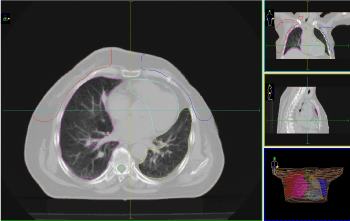
Figure 4. Localization CT for RT planning on a left chest wall
During RT, the patient complains of abdominal discomfort. From the ultrasound of the abdominal organs: Liver-homogeneous structure, without outbreak lesions. Pancreas and kidneys – without pathological changes. Gallbladder- stretary walls, concrement measuring 4 mm. Conclusion Cholelithiasis. So far it has implemented to TD 48Gy in the area of right chest wall and regional lymph nodes. The patient is afebrile, with a dry cough of 2-3 days. Targeted for radiography of the lung and CT scan of a chest cell (Figure 5 and 6).
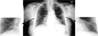
Figure 5. Chest X-ray- bilateral basal patchy iniltrative shadows due to inflammatory changes. Conclusion: Bilateral pneumonia
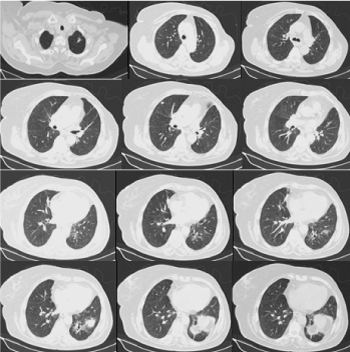
Figure 6. Native CT of the chest - Bilateral mammectomy. Subsegmental focal fibrosis apically and basally on the left. Bilateral peripheral lobular subsegmental consolidations most likely inflammatory type
After history for contact with COVID-19 positive patient (1st presented clinical case) and imaging data for pneumonia, on 28.06.20 the patient was admitted to the Pulmonary Hospital, where PCR test proved positive on COVID-19.
The rapid test for COVID-19 (IgG/Ig M antibodies): IgG antibodies-negative; Ig M antibodies positive. From the peripheral blood count, leukopenia/ Leuco 2,6 T/l (range 3,5-10,5 T/l); lymphopenia/Lym 0.5 T/l (range 0.94-2.99 T/l); neutrophilia/Neu 75.8% (range 39.3% -74.9%); increased ESR 54 mm/h (range 1-30 mm/h) and elevated AST-64 U/l (range 1-40 U/l) and GGT-40 U/l (range 5-35 U/l) is detected. From the blood-gas analysis: PH 7.4 (range 7,36-7.44); p CO2 32 mmHg (range 35-45 mmHg); pO2 51 mmHg (range 81-100 mmHg) %SO2C 88,9% (range 92-98 %); SBC 25,8 mmol/l (range 21-25 mmol/l).
In the Pulmonary Hospital, treatment with Ceftriaxone 2g i.v./daily, Amikacin 1000 mg i.v./daily was conducted for 10 days. On 02/Jul/20 and 09/Jul/20, control chest X-ray (Figure 7) were performed. From the peripheral blood count on 09.07.20: Leuco 3,7 T/l (range 3,5-10,5 T/l) is established; Neu 86% (range 39.3% -74.9%); lymphopenia/Lym 0.3 T/l (range 0.94-2.99 T/l); at normal values of D-dimer 0.16 mg/l (range 0.0-0.5 mg/l). On 10.07.20 g the patient was discharged from the Pulmonary Hospital with a negative PCR for COVID-19 and reffered for home treatment in quarantine.
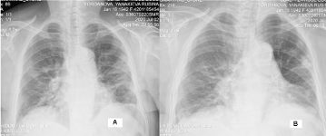
Figure 7. Chest X-ray- А / 02/Jul/20г. Bilateral non-homogeneous shading in lower and part in medium lung fields. In some places, these shadows confluate in the left middle lung field, where a denser shade of triangular shape is formed, with a tip directed to the hill/bilateral pneumonia- COVID-19; B/ 09/Jul/20 - Partial resorption of inflammatory changes bilaterally, slightly decreased, but still available
The 3rd is a 76-year-old woman diagnosed with invasive low-grade squamous cell carcinoma of the cervix in 2018. Radical hysterectomy was performed with bilateral adnexectomy and pelvic lymph dissection classified in T2N0M0 without postoperative RT. After a biopsy of the vagina due to genital bleeding in 2020, recurrence was proven. Admitted to the Department of RT for definitive irradiation of recurrence with the pelvic and inguinal regional lymph nodes bilaterally. Local status: In the left corner of the arc vagina exophyte with solutions 6/3 cm. From localization CT- no infiltration to the bladder and rectum. Pathologically enlarged lymph nodes bilaterally pelvic and inguinal are not reported.
In the course of RT, the patient complained of general fatigue, diarrhea and visible clear blood in the urine. Urinalysis shows erythrocyturia, leucocituria and high level proteinuria. From ultrasound of abdominal organs with conclusion: Diffuse parenchymal process in the right kidney and cyst in the upper pole of the left kidney with diam. 12 mm. Klebsiella has been isolated from urine culture. The patient was afebrile, with normal values of peripheral blood count and biochemistry - urea, creatinine, total protein, total bilirubin, AST, ALT, GGT.
Drug therapy was performed with Ceftriaxone and solvents. Chest X-ray on 29/Jun/20 (Figure 8). Due to contact with the above-mentioned COVID-19-positive patients, the patient was admitted to the Infectious Diseases Hospital with positive test for COVID-19 and discharged after 14 days with a negative PCR test.
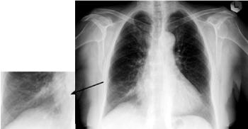
Figure 8. Chest X-ray – enhanced vascular interstitial pattern with some lobular patchy consolidations in the right lung base, most likely inflammatory changes
The 4th case is a 56-year-old woman diagnosed with undifferentiated medullary carcinoma Grade 3 of the left breast. After a left-sided quadrantectomy with axillary lymph nodes dissection and 8 courses of adjuvant chemotherapy, she was reffered to the Department of RT for postoperative RT. Two weeks after hospitalization, the patient was afebrile, with a complaint of dry cough and sore throat. Consulted with ENT specialist - Clinical data on acute pharyngitis. From radiography of the lungs no evidence of inflammatory changes. The complaints subside.
Two weeks later COVID-19 infection was proven due to history for contact with the 3-rd clinical case. Control chest X-ray were performed (Figure 9) no pathological lung changes are reported. The coronavirus (SARS coronavirus 2 RNA) test is positive. When TD 36Gy in the area of the left breast is realized, RT is terminated and considering the good general condition and the lack of symptoms, the patient is referred for home treatment.
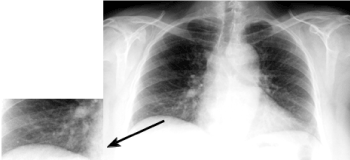
Figure 9. Chest X-ray - normal picture of the lungs
The other three patients, one of whom has oncological disease, were diagnosed at University Hospital "St. Marina"- Varna, Bulgaria. With them, we observed the typical signs and symptoms of COVID-19 infection: dry cough with fever and difficulty breathing.
The 5th case is an 81-year-old woman with cardiovascular comorbidity is hospitalized on 12/Jul/2020 and died the next day from COVID-19 (positive RTPCR test). Figure 10 presents a chest X-ray in lay down position, that reveals small pleural effusion on the right, multiple bilateral peripheral subsegmental consolidations, enhanced interstitial vascular markings, dilated hilar shadows. Conclusion: Bilateral pneumonia.
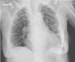
Figure 10. Chest X-ray -Small pleural effusion on the right, multiple bilateral peripheral subsegmental consolidations, enhanced interstitial vascular markings, dilated hilar shadows. Conclusion: Bilateral pneumonia
The 6th case is a 70-year-old woman with cardiovascular comorbidity, who was hospitalized on 4/Apr/2020 and signed discharged on 17/Apr/2020. Figure 11 presents a chest x-ray of the patient. Bilateral multiple subpleural confluent zones of consolidation with some little retraction, prominent dilated hilar shadows, bilateral pneumonia.
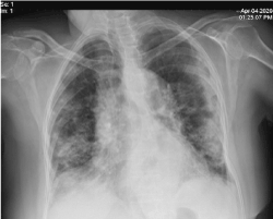
Figure 11. Chest x-ray- Bilateral multiple subpleural confluent zones of consolidation with some little retraction, prominent dilated hilar shadows. Conclusion: Bilateral pneumonia
The 7th patient is a 61-year-old woman with known multiple myeloma, who was hospitalized on 1/Jun/2020 and moved on 12/Jun/2020 into Infectious disease clinic. During the treatment in the hematology department the woman showed some clinical symptoms and was verified with positive test for COVID-19. On the chest CT many GGО zones are seen, with ALSKO zone soft crazy paving and subpleural fibrotic bands of consolidation with some retraction, prominent and dilated vessels in these zones and some distortion of the bronchi architecture (Figures 12A and 12B). Chest x-ray changes compatible with COVID-19 disease (Figure12C).
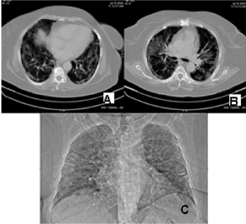
Figure 12. A, B/ Native CT of the chest – many GGO zones are seen, with ALSKO zone soft crazy paving and subpleural fibrotic bands of consolidation with some retraction, prominent and dilated vessels in these zones and some distortion of the bronchi architecture. C/ Chest x-ray changes compatible with COVID-19 disease
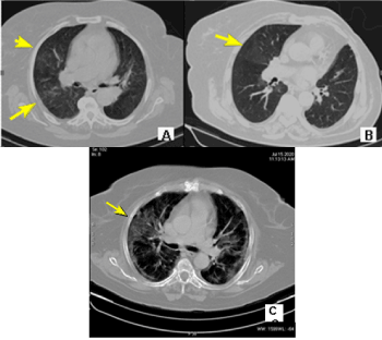
Figure 13. Native CT of the chest – Bilateral ground glass opacities zones unevenly distributed in the lungs, in cancer patients with positive PCR tests for COVID-19
It is well known that patients with oncological diseases have immunosupression related to antitumor treatment /chemotherapy, radiotherapy, targeted therapy or combined chemo-radiotherapy/. In all seven patients, nasal and pharyngeal swabs were collected and tested for SARS-CoV-2 RNA with RT-PCR assay [4]. Patients positive for COVID-19 with nonmalignant comorbidity express similar features of the disease. The most common symptoms at onset of illness were fever (90%-98.6%), fatigue (69.6%), dry cough [59.4%-75%]), myalgia (34.8%), and dyspnea (31.2%-50%) [5,6]. After radiotherapy (RT) to total dose 40 Gy in the area of small pelvis, the first clinical manifestation of infection in the patient with uterine carcinosarcoma were a general fatigue followed by leukopenia, lymphopenia, neutropenia and thrombocytopenia with a one-day temperature increase up to 380 C. Transaminases AST and ALT were significantly increased, as well as GGT. In patients with oncological disease infected with COVID-19 leucopenia and lymphopenia are a common clinical manifestation [7]. In Singapore, Fan et al. also found that patients requiring ICU support had significantly lower lymphocyte levels (p<0.001) at baseline [8].
The patient with bilateral mastectomy has understood aggressive complex treatment / 2012 - 6 courses chemotherapy (Ch), RT to total dose 50 Gy; 2020 g -6 courses 2nd line Ch. During the month-long period of RT, with a normal peripheral blood count, gastrointestinal symptomatology is manifested. From the ultrasound of abdominal organs, cholelithiasis is established. Despite the massive inflammatory changes bilaterally in the lung, the patient is non febrile, and the only manifestation is dry cough. A significant portion of coronavirus patients experiences abdominal discomfort before the onset of respiratory symptoms [3]. In both oncological patients, the development of bilateral COVID-19 pneumonia, is manifested by leucopenia, lymphopenia, accelerated ESR, increased values of AST, ALT and GGT. Increased values of liver transaminases are a consequence of pathological viral changes of liver cells. The presence of abnormal liver tests and liver injury were associated with the progression to severe pneumonia [9].
The 3rd patient with cervical cancer had a complaint of abdominal discomfort, and then general fatigue, diarrhea and hematuria with proteinuria. Ultrasound of abdominal organs reported a diffuse parenchymal process in the right kidney, and microbiological urine testing isolated Кlebsiella infection. We believe that the high dose regimen with Ceftriaхone prevents other consequences related to the illness. In patients infected with COVID-19 less common symptoms were headache, dizziness, abdominal pain, diarrhea, nausea, and vomiting. A small but significant subset has gastrointestinal symptoms [6]. 10.1% of patients with subsequent development of pneumonia initially presented with diarrhea and nausea 1 to 2 days prior to development of fever and dyspnea [5]. Besides, a prospective cohort study of 333 COVID-19 patients has identified kidney involvement in the form of acute kidney injury or abnormal urine dipstick within 75.4% of patients (65.8% presented with proteinuria, and 41.7% had haematuria) [10]. In addition, if this virus causes vasculitis, different reported clinical presentations can be eventually explained such as haematuria, haemoptysis, abdominal pain and the thrombo-embolic events [11].
Chest X-ray findings of COVID-19
The radiological signs of COVID-19 pneumonia are not specific. Common sign is ground glass opacities (GGO), which means hazy increased attenuation in parts of the lungs with preservation of bronchial and vascular markings [7,12]. These GGO are typically bilateral, asymmetrical, peripheral and subpleural in location, with predilection for the lower lobes (Figures 2, Figure 6, Figure 12). It could be a consequence of broad spectrum of pathology, including infection, acute alveolar injury and chronic interstitial disease. The CT GGO are typical for the early stages of the disease [13]. These lung changes cannot be seen on X-ray picture (Figure 1, Figure 3 and Figure 5). That’s why in the early stage of the disease first -line imaging modality must be high-resolution computed tomography (HRCT) [14]. In the three oncological patients, radiographic data on bilateral bronchopneumonia are established, and from CT scans of the lungs -bilateral GGO opacities zones (Figure 13). Despite the high dose antibiotic therapy, in both patients we observed slow resorption of pulmonary inflammatory changes. On control radiographs after 10 days of treatment, no significant reverse dynamics of inflammatory changes are taken into account (Figure 3, Figure 7).
The initial finding could be solitary round peripheral zone of GGO, sometimes with “reversed halo” sign - complete or partial consolidation, surrounding the GGO zone [15]. At later stage of the disease another CT sign is crazy paving pattern, which is a combination of GGO with thickened interlobular and intralobular septal lines [16] (Figure 12). Another common CT features are consolidations with traction bronchiectasis, cavitaions (Figure 6), fibrotic subpleural bands with distortion and dilated vessels in the affected region of the lung (Figure 12) [17,18]. In more severe cases the X-ray picture reveals massive confluent consolidations with small pleural effusions and sometimes could look like ”white lung” (Figures 10, and 11) [19].
The role of radiology in diagnosing COVID-19 disease is crucial in the early stage of the disease, when PCR test could be false negative. Fang Y. et al. showed prevalence of initial CT detection rate with 98% (50/51) over first RT-PCR detection rate 71% (31/57) patients [20]. CT scan is very useful in time course of the COVID-19 pneumonia by evaluating severity and prognosis of the disease. In the first four days after the presentation of the complaints, the CT in almost 50% of patients could be normal. Chest CT lacks complete sensitivity and cannot alone reliably fully exclude this disease, particularly early in the infection [21].
In oncological patients infected with COVID-19, a less common symptomatology such as gastrointestinal discomfort, dry cough without fever, which is difficult to associate with this viral infection, is observed. Due to depressed immunity, pneumonia is accompanied by leukopenia, lymphopenia and accelerated ESR. Hematuria, proteinuria, elevated transaminases and GGT level are due to pathological vasculitis changes in the parenchymal organs /kidneys and liver/. This viral pathology accompanies inflammatory changes in the lungs. The radiological signs of COVID-19 pneumonia are not specific. The ground glass opacities (GGO) are typically bilateral, asymmetrical, with peripheral and subpleural location, with predilection for lower lobes. CT detection is very useful in time course of the disease with checking severity of the disease, prediction of worsening or improvement.
- Zhai P, Ding Y, Wu X, Long J, Zhong Y, et al. (2020) The epidemiology, diagnosis and treatment of COVID-19. Int J Antimicrob Agents 55: 105955. [Crossref]
- Pedro J, Rodriguez C, Chicharro P, De Argila D, MunozHernandez P, et al. (2020) Reply to "Acute urticaria with pyrexia as the first manifestations of a COVID-19 infection": Urticaria-like lesions in COVID-19 patients are not really urticaria. A case with clinicopathologic correlation. J Eur Acad Dermatol Venereol.
- Cipriano M, Ruberti E, Giacalone A (2020) Gastrointestinal infection could be new focus for coronavirus diagnosis. Cureus 12: e7422.
- Huang C, Wang Y, Li X, Ren L, Zhao J, et al. (2020) Clinical features of patients infected with 2019 novel coronavirus in Wuhan, China. Lancet 395: 497-506.
- Wang D, Hu B, Hu C, Zhu F, Liu X, et al. Clinical characteristics of 138 hospitalized patients with 2019 novel coronavirus-infected pneumonia in Wuhan, China. JAMA 323: 1061-1069. [Crossref]
- Jiang F, Deng L, Zhang L, Cai Y, Cheung CW, et al. (2020) Review of the clinical characteristics of coronavirus disease 2019 (COVID-19). J Gen Intern Med.
- Zhang L, Zhu F, Xie L, Wang C, Wang J, et al. (2020) Clinical characteristics of COVID-19-infected cancer patients: a retrospective case study in three hospitals within Wuhan, China. Ann Oncol 31: 894-901. [Crossref]
- Fan BE, Chong VCL, Chan SSW, Lim GH, Eric Lim KG, et al. (2020) Hematologic parameters in patients with COVID-19 infection. Am J Hematol 95: E131-E134. [Crossref]
- Cai Q, Huang D, Yu H, Zhu Z, Xia Z, et al. (2020) COVID-19: Abnormal liver function tests. J Hepatol. [Crossref]
- Pei G, Zhang Z, Peng J, Liu L, Zhang C, et al. (2020) Renal involvement and early prognosis in patients with COVID-19 pneumonia. J Am Soc Nephrol 31: 1157-1165. [Crossref]
- Almashat SA (2020) Vasculitis in COVID-19: A literature review. J Vasc 6: 1.
- Franquet T (2011) Imaging of pulmonary viral pneumonia. Radiology 260: 18-39.
- Zu ZY, Jiang MD, Xu PP, Chen W, Qian Qian Ni, et al. (2020) Coronavirus disease 2019 (COVID-19): A perspective from China. Radiology 296: E15-E25.
- Ng M, Lee EYP, Yang J (2020) Imaging profile of the COVID-19 infection: Radiologic findings and literature review. Radiology: Cardio-Thoracic Imaging 2: e200034.
- Kong W, Agarwal PP (2020) Chest imaging appearance of COVID-19 infection. Radiology: Cardiothoracic Imaging 2: e 200028.
- Zhou S, Wang Y, Zhu T, Xia L (2020) CT features of coronavirus disease 2019 (COVID-19) pneumonia in 62 patients in Wuhan, China. AJR 214: 1-8.
- Ng M, Lee EYP, Yang J (2020) Imaging profile of the COVID-19 infection: Radiologic findings and literature review. Radiology: Cardio-Thoracic Imaging 2: e200034.
- Salehi S, Abedi S, Balakrishnan S, Gholamrezanezhad A (2020) Coronavirus disease 2019 (COVID-19): A systematic review of imaging findings in 919 patients. AJR 215: 1-7.
- Chinese society of radiology (2020) Radiological diagnosis of new coronavirus infected pneumonitis: Expert recommendation from the Chinese society of radiology (First edition). Chin J Radiol 54: E001-E001.
- Fang Y, Zhang H, Xie J, Lin M, Ying L, et al. (2020) Sensitivity of Chest CT for COVID19: Comparison to RT-PCR. Radiology.
- Chung M, Bernheim A, Mei X, Zhang N, Huang M, et al. (2020) Radiology 2020. CT Imaging Features of 2019 Novel Coronavirus (2019-nCoV). Radiology 295: 202-207. [Crossref]