Mesenteric panniculitis is a rare, benign condition. It is characterized by nonspecific inflammation of the mesenteric fatty tissue. Abdominal Computed Tomography is the most sensitive imaging modality for detecting mesenteric panniculitis. In this pictorial essay we present a 60-year-old case of mesenteric panniculitis, including the CT/MR images and discussed the imaging findings.
Although Computed Tomography is the most commonly used modality for the diagnosis of Mesenteric panniculitis, MR examination provides an effective differential diagnosis.
mesenteric panniculitis, MRI, CT
Mesenteric panniculitis (MP) is a rare, benign condition characterized by acute nonspecific inflammation of the mesenteric fatty tissue. It is generally localized at the root of the mesentery [1-5]. In 1924, the first case was reported [1]. In the English literature, there are several synonyms of MP, including mesenteric lipodystrophy and sclerosing/retractile mesenteritis. Each synonym is used according to histological findings, based on the relative amounts of inflammation, fat necrosis and fibrosis in mesenteric fatty tissue [6].
The etiology and pathophysiology of MP are yet unclear [1,7,8]. It may occur unaccompanied or in association with autoimmune disorders, infections, vasculitis, trauma (including surgery), granulomatous diseases, pancreatitis, or ischemia of the mesentery [1,8]. MP most commonly affects the mesentery of the small bowel, especially its root, and occasionally the mesocolon. However, MP can occur at any other site within the abdomen (e.g. pelvis, peripancreatic area, omentum) [2].
Computed tomography (CT) and magnetic resonance imaging (MRI) play important roles in the accurate diagnosis of MP [6]. Abdominal CT is the most sensitive imaging modality for detecting MP [5], and the diagnosis can often be made by using CT alone [1]. Here we present a pictorial essay of mesenteric panniculitis, including the CT/MR images, accompanied by a discussion of the imaging findings.
The patient was a 60-year-old woman admitted to our hospital complaining of epigastric abdominal pain. The patient exhibited no other systemic symptoms. Physical examination was normal. The laboratory examination results, including CBC, liver function and kidney function tests, amylase and tumor marker levels were in the normal ranges. C-reactive protein level and the erythrocyte sedimentation rate were slightly increased.
An abdominal ultrasonography examination revealed a heterogeneous mass between the small bowel loops. Abdominal CT examination was performed with contrast medium. CT examination revealed an inhomogeneous fatty mass localized at the small bowel mesentery on the midline. The attenuation values of the mass were higher than subcutaneous /retroperitoneal fat tissue. There was no evidence of small bowel and colon wall invasion. The density of the mass was -58 HU, which was higher than the retroperitoneal/subcutaneous fat tissue (Figure 1). 18 F-FDG PET/CT findings were in the normal range and did not show any positive results.
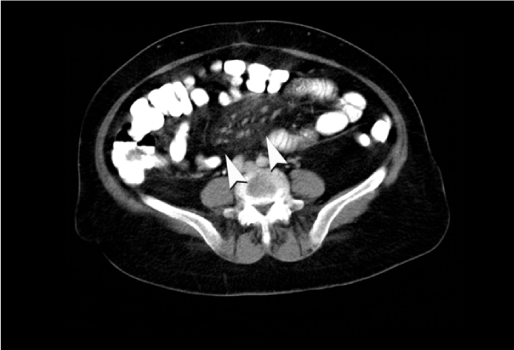
Figure 1. Axial contrast–enhanced CT image showed a hyperattenuated fatty mass with pseudocapsule (arrow) at the mesenteric root.
For further evaluation of the abdominal mass, MRI examination was performed. MRI showed a slightly inhomogeneous mass that predicted signal intensity changes on T1 and T2 weighted(W) sequences (Figures 2-5). All laboratory and radiologic examination results were compatible with MP. Oral steroid therapy was started. After 4 months, control MRI was performed done and presented that the MR imaging findings of MP were resolved.
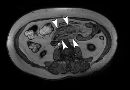
Figure 2. MRI examination-Axial dual echo FSPGR BH asset.

Figure 3. Axial Fiesta FATSAT.

Figure 4. Axial T2 SSFSE BH ASSET. slightly inhomogeneous mass that predicted signal intensity changes.
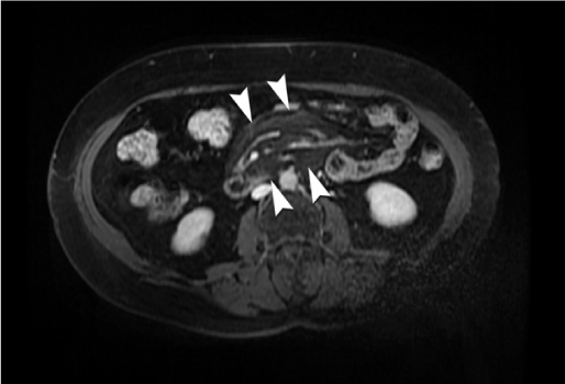
Figure 5. On axial contrast–enhanced MR image, multiple mesenteric lymph nodes and preservation of fat around vessels and lymph nodes (fat ring sign, arrow) were seen in the hyperintense fatty mass.
MP is a rare disease and its reported prevalence rates are inconsistent. In the literature, prevalence rates between 0.16 to 7.80% have been reported [8]. However, MP is thought to be occurred more frequently than reported [1].
Most studies and case reports about MP have reported this disease in patients in middle–late adulthood (median age: 65 years) with a slight male predominance (female/male: 1.3/1.8) [1,6,8].
MP has three stages, and the symptoms generally correspond to the histopathologic findings:
- At the fat necrosis/fatty tissue degeneration stage (mesenteric lipodystrophy), the disease is usually asymptomatic.
- At the inflammatory reaction stage (mesenteric panniculitis), abdominal pain, fever, and malaise are the most common symptoms.
- At the fibrosis and retraction stage (retractile mesenteritis), formation of an abdominal mass and possibly obstructive symptoms occur [2].
Patients may be asymptomatic or present with gastrointestinal complaints [8] such as abdominal pain, fever, constipation, diarrhea, weight loss and abdominal mass [8,9]. Abdominal pain is reported in 34.6% of all MP cases and abdominal mass is reported in 30.8% of cases [6]. In our case, the main symptom was abdominal pain, which was intermittent and vague. Diagnosis of the presented case was based largely on the imaging results.
CT is the most commonly chosen modality for the diagnosis of MP, Amor et al. [9] reported that abdominal CT was the most sensitive imaging modality for determining MP, although its specificity was limited.
Common CT findings suggesting MP are summarized below: [1,2,10,11]
- Encapsulated, solitary, inhomogeneous fatty mass,
- Increased attenuation values of mesenteric fat tissue,
- No vascular involvement, the caliber of the vessels is unchanged,
- No findings of bowel wall invasion,
- Well-defined mass, surrounded by a fatty halo, scattered soft tissue nodules of <5 mm,
- Lymph nodules <10 mm in diameter
In our patient, increased attenuation of mesenteric fat tissue, encapsulated solitary inhomogeneous fatty mass, no vascular involvement and well defined soft tissue nodules were seen on CT examination. In the literature, the main diagnostic criteria on the CT for MP is the increased density of the mesenteric fat tissue secondary to inflammation (approx. -40 up to -60 HU). We measured a fatty mass density of -58 HU in our patient. Generally definite diagnosis is accomplished by comparing it to the normal values of the retroperitoneal /subcutaneous fat tissue density (Normal Values: -100 to -160 HU).
In our study, MRI was performed to confirm the findings and MRI findings would be the main base for the follow-up. Furthermore, MRI examination provided the differential diagnosis. MRI revealed an inhomogeneous fatty mass with changing signal intensity on T1 and T2W sequences. Like CT; Encapsulated, solitary, inhomogeneous fatty mass, without any vascular involvement, with no bowel wall invasion, and lymph nodes were the main MRI findings. MRI was more suitable for differential diagnosis and follow-up rather than primary diagnosis [12]. Ghanem et al. described the role of MRI in the diagnosis of the disease [12].
In our case, all radiologic findings were compatible with MP; therefore, biopsy was not considered.
Wat et al. [2] suggested that, in the absence of a known malign tumor disease, the diagnosis of MP could be reached by using CT findings without biopsy. However, if there was a suspicious finding for MP, biopsy should be planned for differential diagnosis. For example, mesenteric lymph nodes larger than 10 mm were suspicious for MP and biopsy should be considered to exclude malignancy. During differential diagnosis; Carcinomatosis, carcinoid tumor, lymphoma, peritoneal mesothelioma, amyloidosis, lipoma, liposarcoma, infectious diseases and desmoid tumor should be considered [6].
MRI is a useful imag2021 Copyright OAT. All rights reservBy using MRI examination findings differential diagnosis can be done more accurately [2]. However, sometimes differential diagnosis cannot be completed by using radiologic methods alone. In some cases, histopathologic examinations are required [2,6]; however, in clinical practice, histopathological confirmation is rarely required [4]. In our case there were no suspicious imaging findings, so we did not perform a biopsy.
Although CT is the most commonly used modality for the diagnosis of MP, MRI examination has no risk of radiation intake and this examination provides an effective differential diagnosis, so we think that it will be appropriate to use MRI in the visualization of MP patients.
- Talwar A, Rayner H (2009) The medical mystery of the fatty mesentery. BMJ Case Rep. 2012; 27. 2. Jeon EJ, Song CM. Idiopathic isolated omental panniculitis confirmed by percutaneous CT-guided biopsy. Gut Liver 3: 321-324.
- Jerraya H, Khalfallah M1, Nouira R1, Dziri C1 (2015) Mesenteric Panniculitis: An Unusual Cause of Epigastric Pain. J Clin Diagn Res 9: PJ01. [Crossref]
- Alhazzani W, Al-Shamsi HO, Greenwald E, Radhi J, Tse F (2015) Chronic abdominal pain secondary to mesenteric panniculitis treated successfully with endoscopic ultrasonography-guided celiac plexus block: A case report. World J Gastrointest Endosc 7(5): 563-566. [Crossref]
- Gunduz Y, Tatli AP, Kara RO (2012) Mesenteric panniculitis: a case report and review of the literature. Maedica (Buchar) 7: 344-347. [Crossref]
- Daumas A, Agostini S, Villeret J, Ah-Soune P, Emungania O, et al. (2012) Spontaneous resolution of severe, symptomatic mesocolic panniculitis: a case report. BMC Gastroenterol 6: 59. [Crossref]
- Mitchell A, Caty V, Bendavid Y (2015) Massive mesenteric panniculitis due to fibromuscular dysplasia of the inferior mesenteric artery: a case report. BMC Gastroenterol 23: 71. [Crossref]
- van Putte-Katier N, van Bommel EF, Elgersma OE, Hendriksz TR (2014) Mesenteric panniculitis: prevalence, clinicoradiological presentation and 5-year follow-up. Br J Radiol 87: 20140451. [Crossref]
- Amor F, Farsad M, Polato R, Pernter P, Widmann J, et al. (2011) Mesenteric panniculitis presenting with acute non-occlusive colonic ischemia. Int Arch Med 22: 22. [Crossref]
- Shin NY, Kim MJ, Chung JJ, Chung YE, Choi JY, et al. (2010) The differential imaging features of fat-containing tumors in the peritoneal cavity and retroperitoneum: the radiologic-pathologic correlation. Korean J Radiol 11: 333-345. [Crossref]
- Badea R, Chiorean L, Damian D, Molnar G, Anton O (2013) Ultrasound aspect of mesenteric panniculitis. Case report. Med Ultrason 15: 247-249. [Crossref]
- Chawla S, Yalamarthi S, Shaikh IA, Tagore V, Skaife P (2009) An unusual presentation of sclerosing mesenteritis as pneumoperitoneum: case report with a review of the literature. World J Gastroenterol 15: 117-120. [Crossref]