Abstract
Dehiscence of the radiopaque guiding catheter tip during percutaneous coronary intervention is a rare case but a tendency to a potentially serious complication. The present case report embolization of the dehisced radiopaque EBU 3.5 6F guiding catheter tip and subsequent surgical removal of the entrapped radiopaque guiding catheter tip from distal ulnar artery.
Introduction
Guiding catheter facilitate coronary artery engagement. However, it also has a risk of complication. There are several case reports that vascular complication caused by broken or dehiscence of guiding catheter [1-6]. We should to report uncommon case that guiding catheter tip chipped off to the distal ulnar artery.
Case series
A 52-year old female patient underwent coronary angiography and intravascular ultrasound examination through her right radial artery at our Cath lab because of stable angina and positive treadmill test. Her cardiovascular risk factor is hypertension.
Diagnostic coronary angiography was performed through the right radial artery. This revealed tubular eccentric 50-70% stenosis in the middle segment of left anterior descending artery (Figure 1) and tubular eccentric 50-70% stenosis in posterolateral branch of right coronary artery (Figure 2). We decided to proceed intravascular ultrasound examination with EBU 3.5 6F guiding catheter and runthrough wire. Intravascular ultrasound finding revealed eccentric plaque at the middle segment of left anterior descending artery. We decided to recommend medical treatment and retrieved the guiding catheter. After retrieval of the guiding catheter, we noted that the tip of guiding catheter was being missed and retained in the body. We searched immediately for dehisced guiding catheter tip all over the body by fluoroscopy. Finally, we found the dehisced radiopaque guiding catheter tip at the distal part of right ulnar artery (Figure 3). Limited space of distal ulnar artery is difficult for access of non-surgical retrieval system. We considered surgical removal of entrapped guiding catheter tip at distal part of ulnar artery and consulted with the vascular surgeon. He agreed for surgical removal and successfully removed the entrapped guiding catheter tip from the distal part of right ulnar artery without serious complication on the next day (Figure 4, 5).
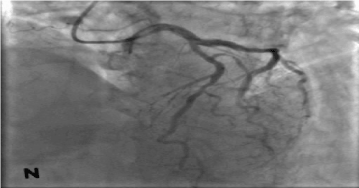
Figure 1. Middle segment of left anterior descending artery showing 50-70%stenosis
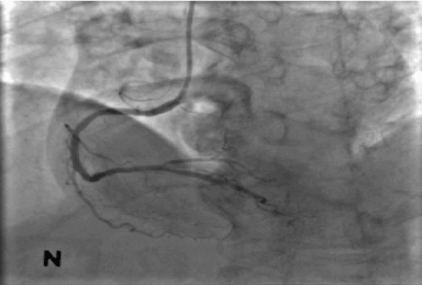
Figure 2. Posterolateral branch of right coronary artery showing 50-70% stenosis
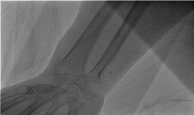
Figure 3. Fluroscopy showing entrapped dehiscence of radiopaque guiding Catheter tip at distal part of ulnar artery

Figure 4 and Figure 5. Showing surgical removal of entrapped dehiscence of guiding catheter tip
Discussion
Dehiscence of radiopaque guiding catheter tip is very uncommon. In our case, the guiding catheter tip may be damaged during difficult engagement to left main coronary ostium and complete dehiscence of guiding catheter tip may occur during passing the narrow and limited space of axillary artery or brachial artery at the time of retrieval. Embolization of dehisced guiding catheter tip may lead to a serious complication. This retained guiding catheter tip may carry the risk of any arterial occlusion, distal embolization of clot, vessel perforation, infection and ischaemic complications. Systemic embolization may lead to cerebrovascular accident or myocardial infarction. Removal of intravascular foreign bodies should be done immediately to avoid the serious complications. In our case, the dehisced guiding catheter tip fortunately embolized to distal part of right ulnar artery. Emergency removal of the dehisced guiding catheter tip is unnecessary because of collateral blood supply from radial artery prevent it from immediate distal ischaemic complication. Removal of an intravascular foreign body may require surgical intervention or non-surgical intervention. Snares, forceps and baskets are available for non-surgical removal of retained intravascular foreign bodies.
In this case, non-surgical retrieval technique may be difficult because of limited space of distal ulnar artery for retrieval instrument access and manipulation, and difficult to catch up for very small dehiscent guiding catheter tip. On the other hand, distal part of ulnar artery is favorable for surgical access. So, we decided to remove the guiding catheter tip by surgical intervention. Finally, vascular surgeons successfully removed the entrapped guiding catheter tip without serious complications.
In the intervention era, broken and complete separation of guide wire, diagnostic catheter, guiding catheter, advanced invasive diagnostic and therapeutic instruments, dislodgement of mounted stent from its delivery balloon are going to increase.
Conclusion
Intervention cardiologists and cardiovascular surgeons around the world are going to encounter more of these complications. The angioplasty operator and cardiovascular surgeon should be familiar with various techniques of basket, snare, forceps and optimal surgical management in cases of unsuccessful non-surgical retrieval or inaccessible site for non-surgical retrieval instruments.
References
- Chen Y, Fang CC, Yu CL, Jao YT, Wang SP (2002) Intracoronary retrieval of the dehisced radiopaque ring of a guiding catheter: an unusual complication of coronary angioplasty. Catheter CardiovascInterv 55: 262-264.
- Kern MJ (2013) The interventional cardiac catheterization handbook. 3rd ed. Philadelphia: Saunders.
- Kharge J, Bharatha A, Ramegowda RT (2012) Intracoronary dehiscence of radiopaque ring of a guiding catheter and its retrieval. J Invasive Cardiol 24: E182-E184. [Crossref]
- Nanjappa MC, Bhat P, Panneerselvam A (2011) Revascularization after removal of broken catheter from left circumflex coronary artery. J Cardiovasc Med 14: 222-224.
- Papayannis AC, Michael TT, Brilakis ES, (2012) Challenges associated with use of the guideliner catheter in percutaneous coronary interventions. J Invasive Cardiol 24: 370-371. [Crossref]
- Lee YP, Tan HC, Lee CH (2008) Complete fracture of an Ikari guiding catheter in the axillary artery during transradial coronary intervention. Int J Angiol 17: 40-42. [Crossref]