Introduction: Full thickness skin grafts have long been an indispensable and frequently practiced standard procedure in plastic surgery. The defatting and thinning of the grafts with scissors or scalpel to a thickness of 0,8 – 1,2 mm is very time-consuming even for experienced surgeons in the case of large transplants. Devices for defatting full thickness skin graft are not available on the market. For this reason, we developed a relatively simple device that enables complete and safe defatting and thinning of the graft in the shortest possible time.
Materials and methods: The machine has a dimension of 38cm width, 24cm length, 23cm height. The cutting thickness can be set to 0.6mm, 0.8mm and 1.0mm. Central part is a crimper run manually with a crank to guide the specimen through a cutting blade. The machine is made of stainless steel, can be completely disassembled and is easy to sterilize.
Results: With this device small and very large skin grafts up to 15 cm width can get completely degreased within a few seconds.
Discussion: The time for defatting is reduced by many times. Precious operating time is spared. It can be assumed that the healing of the graft is improved due to a more complete degreasing, a much shorter manipulating time of the skin and a more even cut surface compared to the manual procedure. The risk of defects in the graft and of injury of the surgeon is minimized compared to manual preparation.
Key words
technical innovations, surgical procedure, full thickness skin grafts, reconstruction, burn
Introduction
Full thickness skin transplants have long been an indispensable and frequently practiced standard procedure in plastic surgery. Important indications for a full thickness skin transplant are primary coverage of deep skin defects after trauma or tumor excision and secondary plastic corrections of scars after deep 3rd and 4th degree burns. A prerequisite for the healing of the full thickness skin graft is the thinning of the skin to about 0.75-1.2 mm, because until new capillaries grow in, the graft can only be treated by diffusion [1-4]. In the case of small transplants, the skin can be removed thinly, but even after this further thinning is usually necessary. Large transplants, such as those required in a sternomental contracture, are usually removed in the form of a spindle with a wedge-shaped subcutaneous fat body attached, so that a low-tension closure of the removal defect is possible. The defatting or thinning of the grafts is carried outstretched out over the finger with scissors or scalpel [5]. This work must be carried out very thoroughly but also carefully in order to obtain a sufficiently thin graft on the one hand and not to cut a defect into the graft on the other. Therefore, this procedure is very time-consuming and takes easily more than 30 minutes even for experienced surgeons in the case of large transplants. This time-consuming, rather anachronistic work has prompted the authors to think about alternative ways of full-skin degreasing.
For this reason, we developed a relatively simple device that enables complete and safe defatting or thinning of the graft in the shortest possible time. The basis for this degreasing unit are machines for deriding animal skins, which have been in use in the meat processing industry for a long time and have proven themselves over many years. In cooperation with Mr. Dietmar Anti (MAJA / Kehl-Goldscheuer), we have developed a device from such a Skinner, with which full thickness skin grafts can be degreased within seconds in such a way that an ideal graft is created.
Material and methods
In the period 2017-2020, series of experiments were carried out on both animal and human skin. In 2017, several preliminary tests with animal skins were carried out and a first prototype was developed, which was modified several times up to its final version. The prototype was then used to carry out a total of 10 test series with human skin, which accumulated during plastic surgery.
The machine is made of stainless steel, can be completely disassembled into 10 numbered parts and is therefore easy to sterilize. The core of the LOMA is a crimper which is driven by a hand crank. The fat is separated by a sharp fixed blade. The cutting thickness can be adjusted in 3 heights: 0,6 mm, 0,8 mm and 1 mm.
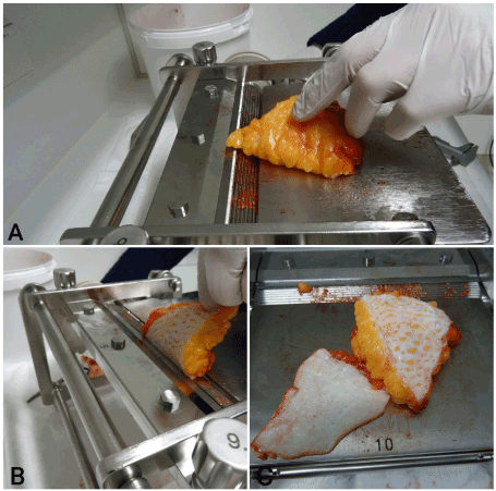
Process: The epidermal side of the graft to be degreased is placed on the carrier table and one corner of the graft is brought up to the crimper (A). By manually turning the crimper, the epidermal side of the graft is captured by the corrugation of the crimper and guided through the cutting edge (B). The process takes about 5 seconds. The result is a completely degreased graft, with more or less dermis, depending on the skin's thickness and position (C) (Figure 1).
Figure 1. The upper subcutaneous tissue should be guided straight in the direction of movement at an angle of approx. 45° during cranking with very minimal uniform tension (A-C)
Results
Preliminary tests with professional skinning machine and piglet skin
A TEM (table skinning machine) was used as the machine. The electric drive was switched off and a hand crank attached to the gearbox. By manually turning the crimper, the machine could be operated without any problems. The experiment was carried out from a butcher with fresh suckling pig skin, as this is thinner than the skin of older pigs and more similar to human skin. The site of removal was in the area of the lower abdomen, as the skin is very soft there. The skin was removed together with subcutaneous fat in the form of a 120mm x 50mm skin spindle. The rind was manually guided to the blade, by turning the crimper the epidermis was captured and the piece was pulled through the blade. However, the epidermis showed superficial cuts due to the sharp-edged ribs of the roller. An initial prototype of the machine has now been designed to meet the requirements for defatting full thickness skin transplants in the operating theatre: The device should be able to be operated by hand. It should also be possible to degrease large full thickness skin grafts, e.g. from the lower abdomen with a size of 20 x 20 cm. The surface of the crimper should be such that it safely grasps the epidermis without injuring it.
Experiments with first functional model and piglet skin
After completion of the first functional model, experiments with fresh piglet skin were initially carried out. The preparation was completely and evenly degreased. Three different crimpers had been produced, each with a different corrugation on the surface. It was found that completely rounded edges of the corrugation did not sufficiently capture the preparation, while sharp edges injured the epidermis. The ideal condition of the crimper corrugation was determined during the tests. This corrugation with rounded edges was used in the future.
Experiments with first functional model and human skin
The very large pieces of skin after abdominal wall plasty were cut with scissors in such a way that skin spindles were created typical for full thickness skin transplants of any size. The skins were completely degreased. The impressions from the crimper on the epidermal side were unproblematic and disappeared completely after 2 days. Series were carried out with different settings of the knife holder, with identical, reproducible results. The grafts can be degreased to almost any size. The cut surface is completely even and completely degreased. The cut surface of the separated fat shows that whitish dermis remains can be seen on it, i.e. the depth of cut was in the area of the dermis. Corrections were made to the first prototype: It turned out that the resistance when turning the crank was too high, so a gear transmission was necessary.
Experiments with improved second functional model and human skin
The second functional model has a cutting width of 20 cm. This can be used to degrease transplants of any length with a maximum width of 15 cm. According to the authors' experience, this corresponds to the maximum size of removable full thickness skin grafts, e.g. in the lower abdomen from spina to spina. The crimper drive is equipped with a 2:1 ratio. This means that even wide transplants can be easily degreased by one person. The height of the frame has been optimized so that the crank can still be operated comfortably.
Experiments with improved third functional model and human skin
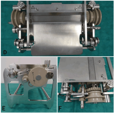
The final device has a dimension of 38 cm width (without crank), 24 cm length, 23 cm height. The maximum cutting width is 20 cm (D-F). The crimper has a corrugation that is suitable for gripping the graft securely without damaging it. The impressions from the crimper on the epidermal side are unproblematic and disappeare completely after 2 days. In order to allow easy turning, the crank force was increased by a transmission (E). The cutting thickness can be set to 0.6 mm, 0.8 mm and 1.0 mm. According to our measurements, the transplants actually have a slightly greater thickness. This is exactly within the required range of 0.8-1.2 mm (Figure 2).
Figure 2. Final device (D), lateral view (E), crimper from the machine (F)
Discussion
The thorough degreasing of a full thickness skin graft, which leads to the necessary reduction of the diffusion distance from the graft base to the epidermis, is the most important prerequisite for successful full thickness skin grafting, in addition to good fixation of the graft and prevention of graft infection. This important but simple work is carried out worldwide with scissors, whereby the graft is usually stretched over a finger. The procedure is time-consuming and requires a great deal of attention so that neither the surgeon's finger is injured, nor defects are cut into the graft.
Devices for full thickness skin graft defatting are not available on the market. For this reason, we have developed the transplant degreaser described above. These are the advantages of this new device:
The defatting process is shortened by many times compared to manual preparation.
Precious operating time is spared.
Patient is shorter under anesthesia.
Defatting takes place independently of the thoroughness and patience of the degreaser, so defatting will usually be more complete.
The cut surface is not irregular, as with manual defatting, but flat and even, as you can easily see in the illustrations.
The risk of a graft infection could be lower because the time of manipulation of the graft, a few seconds, is very short compared to manual defatting over many minutes.
The last three points might lead to reduced infection rates.
The risk of defects in the graft is minimized compared to manual preparation. Manual defatting frequently leads to perforations of the preparation, especially in the production of very thin grafts. When defatting with the device, we were only able to provoke a defect in the graft if the subcutaneous fat was pulled strongly during the cutting process when the subcutaneous fat was held up. This should therefore be avoided at all costs.
The risk of injury to the surgeon's fingers is also minimized compared to manual defatting. If the sharp blade is handled properly - just like a dermatome blade - injury to the surgeon and operating personnel should be ruled out.
The weight of the device can be mentioned as a disadvantage. This size resulted from the design, which was repeatedly modified by experiments, as the device was also to degrease large pieces of full thickness skin and the necessary stability made a smaller version not advisable.
Conclusion
The defatting process with this new device is shortened by many times compared to manual preparation and the operating time is spared. Defatting takes place independently of the thoroughness and patience of the degreaser, so defatting will usually be more complete. Moreover, the risks of defects and injury of the surgeon`s fingers are minimized. Summarized the machine is a new invention to improve the process of a full skin graft transplantation.
Funding
None.
Conflicts of interest
We have no conflict of interest to declare. Also there is no commercial interest in the device from all Authors. This statement is to certify that all Authors have seen and approved the manuscript being submitted. We warrant that the article is the Authors' original work. We warrant that the article has not received prior publication and is not under consideration for publication elsewhere. On behalf of all Co-Authors, the corresponding Author shall bear full responsibility for the submission.
Ethical approval
Not required.
References
Pommer A, Schumacher G (2020) Clinic Guide Surgery, Urban und Fischer, 6th edition, p.300.
Kaufmann R, Podda M (2011) Dermatological operations: Colour Atlas and Textbook of Skin Surgery, Thieme, 4th ed. p.60.
Katsch J, Krausse s, Müller R (1994) Skin transplants, S 55 ff in: Mahrle G, Schulze H-J, War T: Wound Healing Wound Closure, Springer.
Schiestl C, Stark g, Lenz Y, Neuhaus K (2017) Plastic Surgery for Children and Adolescents, Springer, pp, 185.
Ratner D (1998) Skin grafting. Dermatologic Clinics 16: 75-82.
Editorial Information
Editor-in-Chief
Article Type
Research Article
Publication history
Received date: August 12, 2020
Accepted date: August 19, 2020
Published date: August 25, 2020
Figure 1. The upper subcutaneous tissue should be guided straight in the direction of movement at an angle of approx. 45° during cranking with very minimal uniform tension (A-C)
Figure 2. Final device (D), lateral view (E), crimper from the machine (F)