Abstract
This study analyses the factors influencing the graft stability after a suture less and glue-free conjunctival autograft in pterygium surgery.
Patients and method
A prospective study, all patients underwent pterygium surgery with suture less glue less conjunctival autografting
The parameters studied were:
Intraoperative, evaluation of graft stability at the end of the procedure. Postoperative, the graft position on day 1 after surgery.
Results
Sixty-four grafts remained stable at the end of the intervention, i.e. 83.12% while 16.88% were unstable. In the postoperative follow-up, 79.22% of grafts were well-positioned including 76.62% of grafts flattened in place and 2.60% of grafts retracted. Grafts displacements were observed in 20.78% of cases including 11.69% of minor displacements and 9.09% of major displacements.
One patient had an excessive bleeding, which stopped on day one postoperative. Unstable grafts (84.62%) developed secondary displacements compared to stable grafts (7.81%). The difference was statistically significant, P=002. Out of the 7 cases with major displacement, 5 cases had unstable grafts (6.49%), and 2 cases had stable grafts (2.59%).
Conclusion
It is an effective and safe technique with good graft position and stability despite intraoperative surgical adjustments as formerly described in the literature. The absence of postoperative irritation and suture related complications makes it a useful method for graft fixation in pterygium surgery. However, still some improvements are needed for better graft stability.
Key words
pterygium surgery, conjunctival autograft
Introduction
Conjunctival autografting is giving good results in pterygium surgery [1,2]. The most commonly used means of fixating conjunctival autografts is by sutures or fibrin glue [3,4]. More recently, the use of autologous blood has been reported to be advantageous in minimizing the risk of postoperative infection, irritation and a reducing in the cost of surgery.
However, the absence of suture or biological glue poses problem of graft fixation. Indeed, this graft fixation is potentialized by fibrin glue at the junction of the conjunctival autograft and the conjunctival bed [5]. In a review of the literature conducted in India, major displacement rates ranged between 0 to 12% while Boucher in Canada noted a 20% rate of graft loss [6,7]. Several studies have shown that on the first postoperative day the graft is already firmly fixed [6,8]. Most graft loss occurs before this time.
This study presents some technical aspects aimed at improving the graft stability on the first postoperative day.
Patients and method
Patients
Our study was conducted as part of a decentralized surgical activity in a transportable hospital. The primary target was patients with cataracts, but pterygium also was operated on. Thus 77 eyes of 77 patients were operated from 09-07-2018 to 08-08-2018
Inclusion criteria
- Nasal pterygium
- Undergoing a pterygium surgery with conjunctival autografting by autologous blood Non-inclusion criteria
- Patient with any coagulation disorder
- temporal or bipolar pterygium
Method
This is a prospective study, which took place in the department of Pool located in south of Congo Brazzaville. The selected patients were seen by an anaesthesiologist who also assessed blood haemostasis parameters. The patients were then operated on under peribulbar anaesthesia. They underwent a pterygium surgery with suture less and glue-free conjunctival autografting.
Surgical technique
The procedure begins with the excision of the corneal part of the pterygium with a crescent knife, then its conjunctival area with scissors, keeping the adjacent tenon capsule and the sheath of the medial rectus muscle. Only bleeding vessels were cauterized. The size of the graft was determined by measuring the area of exposed sclera. This is taken from the supero-temporal bulbar conjunctiva of the same eye, starting from the posterior part. The conjunctiva was gently isolated; this is better verified by returning the graft forward of the limbic anterior border not yet resected (Figure 1). Resection of the limbic margin is done, with the epithelial face against the cornea, trying to collect the maximum limbic cells.
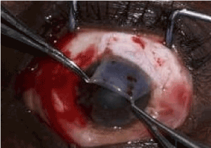
Figure 1. Harvesting of the graft in the superior temporal region
The graft is then transferred to the pterygium bed, and then returned to the tenonian bed on the conjunctival bed of the pterygium, the limbic margin facing the cornea. If a clot had formed on the operating bed, while the graft was removed, it was removed. Spreading-compression of the graft is performed with a foam instrument, as if ironing the graft. Graft stability is confirmed when the graft is no longer displaced despite moderate pressure of a stream of saline using a Rycroft cannula mounted on a syringe, and the absence of displacement of the graft following gentle opening and closing movements of the eyelids. The physiological saline infiltrating under the graft causes its expansion and strengthens its fixation (Figure 2).
Figure 2. Conjunctival autograft fixation autologous
At the end of the procedure, a shell is put in place before the dressing to avoid direct contact of the compress with the graft or via a secondarily formed clot; knowing that grafts and clots adhere more firmly to the compress than the pterygial bed.
The patient was seen again 24 hours after the operation.
OUTCOME OF THE GRAFT AT THE END OF INTERVENTION
At the end of the procedure before placing the dressing, graft fixation was assessed according to the following criteria:
- The absence of displacement of the graft following irrigation of the cornea and the graft,
- The absence of displacement of the graft following the opening and closing movements of the eyelids.
Evaluation of the stability of the graft at day 1 post-operative
We found either:
- A graft in place
- A retraction of the graft, conjunctival defect of approximately 1 mm
- A minor displacement of the graft, extending over the corneal surface.
- A major displacement with sometimes loss of the graft.
Results analysis: The analysis of the results was done with epi-info 7. We used the K2 test for data comparison.
Results
Mean age was 52.04+/-11.76 years (25 to 80 years)
Our sample consisted of 43 women and 34 men, a sex ratio of 0.79
The right eye was concerned in 62.34% (48 patients) cases versus 37.36% (29 patients) cases for the left eye. Cornand’s classification was used in the Table 1 [9]. The mean operation time was 7.49 minutes+/-2.1 (4 to 15 minutes) (Table 2).
Table 1. Distribution of patients according to the stage of Pterygium
Stage of Pterygium |
cases |
% |
Stage I |
6 |
7.79 |
Stage II |
23 |
29.87 |
Stage III |
48 |
62.34 |
Total |
77 |
100 |
Table 2. Graft outcome
Intraoperative outcome |
cases |
% |
Stable graft |
13 |
16.88 |
Unstable graft |
64 |
83.12 |
Total |
77 |
100 |
In the postoperative follow-up, 79.22% of grafts were well-positioned including 76.62% of grafts flattened in place and 2.60% of grafts retracted; 20.78% of transplants were displaced, with 11.69% of minor displacements, and 9.09% of major displacements (Table 3).
Table 3. Immediate postoperative graft outcome
Graft outcome at day one postoperative |
cases |
% |
Graft in Place |
59 |
76.62 |
Graft retraction with non-displacement |
2 |
2.60 |
Minor graft displacement |
9 |
11.69 |
Major graft displacement |
7 |
9.09 |
Total |
77 |
100 |
1 patient complained of continuous bleeding sensation, with the first postoperative day a completely wet compress, and bleeding already stopped.
Unstable grafts (84.62%) developed secondary displacements compared to stable grafts (7.81%). The difference is statistically significant, P=002. Out of the 7 cases with major displacement, 5 cases had unstable grafts (6.49%), and 2 cases had stable grafts (2.59%) (Table 4).
Table 4. Relationship between intraoperative and postoperative graft outcome, Chi-square - Mantel-Haenszel
Intraoperative graft outcome |
Well-positioned graft |
Graft displacement |
Total |
Unstable graft |
2 (15.38%) |
11 (84.62%) |
13 (100%) |
Stable graft |
59 (92.19%) |
5 (7.81%) |
64 (100%) |
Total |
61 |
16 |
77 |
Discussion
We included 77 patients including 43 women and 34 man with a sex ratio of 0.79. Men are the most professionally exposed to Pterygium. This inverted sex ratio, already found by Kwon, can be explained in our series, by the access to free healthcare to the most vulnerable, especially women [10-12]. The average age was 52.04+/-11.76 years old (25 to 80). This average age like the one in the literature, with 45 years old for Kammoun in Tunisia, and 40 years old for Moukouri [3,13,14] in Cameroon. Zarrouki in a comparative study, had mean ages of 51.5 and 54 years old. Stage III pterygium was the most frequent with 60% of cases. Kammoun found almost the same distribution of pterygium per stage with 68% of stage III pterygium, whereas Zarrouki in Morocco had 50% of stage III pterygium [3,14]. We had to choose between several cases, and we selected patients with the most advanced stages of pterygium.
We used physiological saline rather than a buffer or a compress in case of bleeding, to prevent an accidental contact of the graft with the buffer or the compress resulting to adhesion between these two, which could cause the loss of graft orientation. It is important to avoid any contact of the compress with the graft even when dressing by using a sterile draping. The draping, however, prevents compressive haemostasis provided by a bandage on the non-cauterized excision area. Therefore, it reduces the risk of postoperative bleeding.
The clots adhering to the graft should be removed while keeping the graft in place. Intraoperatively, the grafts were either stable or unstable, with, as stability criteria, an absence of graft displacement the following the irrigation of the cornea and of the graft or the eye opening and closure movements after surgery. Thirteen grafts were unstable (16.88%), compared to 83.12% of stable grafts.
The stability of the graft is a related to the shape and size of the graft. Tapered grafts were the most unstable with their ends peeling off and wrapping following an irrigation. When grafts were too wide compared to the pterygium exeresis area they folded more easily at eyelids closure. Grafts with semi-spherical conjunctival margins, the size of which corresponded most to the area of resection, were the most stable. The particular risk of this technique is the significant displacement of the graft requiring reoperation with other fixation methods [15]. This displacement is done in the first 24 hours after surgery, beyond 24 hours the graft is already firmly fixed [6].
The first postoperative day was marked by:
- Graft retraction, most frequently [8,16,17], responsible for the reduction of the graft size leaving on both sides of the graft areas of bare sclera of about 0.5 mm. Usually no measurement is necessary and wound healing occurs naturally.
- Minor displacements were for most cases graft extension on the corneal bed of the pterygium giving the impression of an immediate recurrence. It’s the management consisted in the resection, under topical eye drops, of the portion of the extended part of the graft [8].
- Major displacements or dehiscences, which can exceed 5 mm and sometimes resulting to graft loss, remains a major challenge of this technique. Dasgupta in his review of the literature on studies conducted using this technique in India noted a displacement rate between 0 and 12% [6]. Our study accounted for 9.09% of major displacements, close to the results of Sarkar who found 8% of major displacements [15].
In our study 9.09% presented major displacements. Nganga Ngabou found 5.2% of major displacements [8]. This difference can be explained by the systematic non-compliance in this study of coagulation time. Indeed, there is a time of coagulation that most authors consider, thus, leaving the graft immersed in the blood to allow better fixation. This time is 8 to 10 minutes for Dasgupta and De Wit; 5 minutes for Nganga Ngabou and Shing; 3 to 4 minutes for Gitte and Curian [5,6,8,18-20].
We followed this step only for patients with an unstable graft, thus reducing our surgical time to an average of 7.49+/-2.1 minutes (4 to 15 minutes).
The infiltration of physiological saline under the compressed graft, increased its adherence on the sclera naked. The exception was made for unstable grafts which coagulation time was respected.
A graft that was not mobilized at the end of the procedure by a soft jet of saline solution by a cannula mounted on a syringe, or with gentle movements of opening and closure of the eyelids, was regarded as a stable graft. Unstable grafts were floating after the passage of physiological saline and folded over themselves to the opening and closure movement of the eyelids.
It is in this last group that we found most secondary displacements. 84.6% of the unstable grafts had either a minor or a major displacement compared to 7% for the stable graft group. The difference was statistically significant.
As for major displacements, there were 5 (38.46%) in the group of unstable grafts versus 2 (3.12%) in the group of stable grafts.
If unstable grafts were not considered in our study, major displacements would be 2.59% instead of 9.09%. It would therefore be good to associate with this technique the preventive use of sutures or biological glue in case of unstable graft, to reduce the rate of secondary sutures or application of biological glue [5,6,15].
Conclusion
It is an effective and safe technique with good graft position and stability despite intraoperative surgical adjustments as formerly described in the literature. The absence of postoperative irritation, suture related complications and its cost-effective advantage compared with sutures makes it a useful method for graft fixation in pterygium surgery. However, still some improvements are needed for better graft stability. In addition, it is necessary to provide another method of fixation in case of unstable grafts.
References
- Mery G, Maalouf T, George JL, Angioi K (2010) L’autogreffe limbo-conjonctivale dans la prise en charge chirurgicale des ptérygions. J Fr Ophtalmol 33: 92-98. [Crossref]
- Riordan-Eva P, Kielhorn I, Ficker LA (1993) Conjunctival autografting in the surgical management of pterygium. Eye (Lond) 7: 634-638. [Crossref]
- Kammoun B, Kharrat W, Zouari K, Zribi W, Kemiha N, et al. (2001) Ptérygion: traitement chirurgical. J FrOphtalmol 24: 823-828.
- Uy HS, Reyes JMG, Flores JDG, Lim-Bon-Siong R (2005) Comparison of Fibrin Glue and Sutures for Attaching Conjunctival Autografts after Pterygium Excision. Ophthalmology 112: 667-671. [Crossref]
- Kurian A, Reghunadhan I, Nair KGR (2015) Autologous blood versus fibrin glue for conjunctival autograft adherence in Suture less pterygium surgery: a randomised controlled trial. Br J Ophthalmol 99: 464-470. [Crossref]
- Dasgupta S, Vats V, Mittal SK (2016) Pterygium excision with suture free, glue free conjunctival autograft (SFGF-CAG): Experience of a tertiary care hospital of the Northern India. Journal of Clinical Ophthalmology and Research 4: 143-148.
- Boucher S, Conlon R, Teja S, Teichman JC, Yeung S, et al. (2015) Fibrin glue versus autologous blood for conjunctival autograft fixation in pterygium surgery. Can J Ophthalmol 50: 269-272. [Crossref]
- Nganga Ngabou CGF, Makita C, Ndalla SS, Nkokolo F, Koulimaya RMA, et al. (2018) Chirurgie du ptérygion avec greffe de conjonctive fixée par du sang autologue. J F Ophtalmol 41: 425-432. [Crossref]
- Cornand G (1990) Le ptérygion: évolution et traitement. J Fr Ophtalmol 13: 33-45.
- Szwarcberg J, Flament J (2001) Ptérygion Encycl Méd Chir (ÉditionsScientifiques et Médicales Elsevier SAS, Paris, tous droits réser-vés). Ophtalmologie 21: 10-12.
- Kwon SH, Kim HK (2015) Analysis of recurrence patterns followingpterygium surgery with conjunctival autografts. Medicine (Bal-timore) 518: 94-97. [Crossref]
- Etya’ale D (2006) Sortir de la clinique: les différents types de stratégies avancées. RSOC 3: 43-44.
- Moukoury Nyolo E, Epee E, Nsangou JFI, Noa Noa Tina B (2009) Leptérygion en zone intertropicale. Analyse de 344 cas au chu deYaounde. Bull Soc Belge Ophtalmol 311: 11-15.
- Zarrouki M, Elyadari M, Jebbar Z, Azennoud S, Serghini L, et al. (2017) L’autogreffe conjonctivo-limbique dans la chirurgie du ptérygion: suture versus colle biologique à propos de 30 cas. J Fr Ophtalmol 40: 293-295. [Crossref]
- Sarkar S (2017) Pterygium Excision with Conjunctival Autograft Without Suture without Glue. IOSR-JDMS 16: 82-86.
- Elwan SAM (2014) Comparison between Suture less and glue free ver-sus sutured limbal conjunctival autograft in primary pterygiumsurgery. Saudi J Ophthalmol 28: 292-298. [Crossref]
- Foroutan A, Beigzadeh F, Ghaempanah MJ, Eshghi P, Amiriza-deh N, et al. (2011) Efficacy of autologous fibrin glue for primary pterygium surgery with conjunctival autograft. Iranian J Ophthalmol 23: 39-47.
- DeWit D, Athanasiadis I, Sharma A, Moore J (2010) Suture less and glue-free conjunctival autograft in pterygium surgery: a case series. Eye 24: 1474-1477. [Crossref]
- Singh PK, Singh S, Vyas C, Singh M (2013) Conjunctival autografting without fibrin glue or sutures for pterygium surgery. Cornea 32:104-107. [Crossref]
- Gitte TR, Karad HT, Kulkarni M, Mashayak A, Kulkarni G (2017) Suture less and Glue-Free Conjunctival Autograft in Pterygium Surgery. PJSR 10:46-50.