Objective: To determine the ejection fraction (EF) and QRS width evolution post aortic valve replacement (AVR) in patients with peri-operative left bundle branch block (LBBB) versus patients without LBBB and find whether left ventricle (LV) mass can predict LV EF evolution.
Methods: We analyzed 31 patients undergoing open-heart AVR, with a similar baseline EF (average 54%±15%) of which 11 had peri-operative LBBB.
Results: Among the 20 patients with no LBBB, EF decreased below 40% in only one patient (5%). In the 11 patients with LBBB, 6 patients experienced post-operative EF deterioration to values below 40% (55%, p=0.004). The average EF value in the 20 patients without LBBB was similar pre-and post-surgery (60%±4% versus 61%±8%). In the LBBB group EF was initially 52%±1% and it dropped to 40%±19% post-surgery (p=0.03 for comparing with the 20 patients without LBBB). Among the 20 patients without LBBB, QRS width was unchanged pre and post AVR (94±15 ms versus 95±14 ms), while it increased in the LBBB patients from 119±26 ms to 144±53 ms (p=0.01). LV mass>300 grams was more common in the 6 LBBB patients that experienced a decrease in EF (100% versus 0% in 5 LBBB patients with stable EF, p=0.001).
Conclusion: In this small study, we demonstrate a significant drop in post-AVR EF between patients with LBBB versus controls. QRS width has significantly increased post AVR, with a sharp decrease in EF in more than half of the LBBB group. LV mass>300 grams was associated with EF deterioration in patients with LBBB.
left bundle branch block, ejection fraction, left ventricular mass, aortic valve replacement
Due to the proximity between the conduction system and aortic valve, aortic valve replacement (AVR) can result in conduction abnormalities involving the His-Purkinje system or the left bundle, such as complete atrio-ventricular or left bundle branch block (LBBB). Our case-series report focuses on the ejection fraction (EF) and QRS width evolution post AVR in patients with peri-operative LBBB versus those without LBBB.
We described in this report a consecutive series of patients with perioperative left bundle branch block (LBBB) undergoing open-heart AVR (active group) and compared it with a group without LBBB (control group). Our team reviewed the records of all patients that underwent AVR at University of Pittsburgh Medical Center over the course of 4 years, had a baseline EF equal to or greater than 50% along with peri-operative LBBB and at least two echocardiography studies performed both during the pre- and post-surgery periods. These patients were compared with a control group with no LBBB that underwent same procedure. Patients were excluded if they had a pacemaker device or experienced a myocardial infarction during the follow-up period. The study was considered exempt of IRB review. Ejection fraction was measured using the method of Quinones et al. Left bundle branch block was diagnosed using standard ECG criteria (Minnesota code 7.1).
Left ventricle mass was calculated according to the modified Penn-cube formula [1].
Statistics We used Student t test for numerical values and Fisher exact test for categorical variable comparisons, respectively.
Eleven patients with peri-operative LBBB that underwent AVR for severe aortic stenosis were included, of whom 6 (‘EF deterioration’ group) experienced a decline in EF to less than 40% post-surgery while the remaining 5 patients ('stable EF' group) maintained an EF greater than 50%. A control group consisting of 20 consecutive patients that underwent AVR with no LBBB was identified as well. Average follow-up was 205 versus 341 days (p=0.5) in the two LBBB subgroups, and 616 days in the control group (p=0.044 in comparison to the LBBB group). There were no complications during surgery. Ten patients were males in the LBBB group and 8 in the control group (p=0.008), with no age difference. The incidence of surgical procedures associated with AVR, diabetes mellitus, hypertension, significant coronary artery disease, abnormal renal function were similar between the two LBBB subgroups and between the LBBB and the control patients (p>0.1 for all comparisons).
There was a total of 34 echocardiograms, of which 31 were transthoracic and 3 transesophageal studies in the LBBB subgroups, and 51 transthoracic studies in the control group. Initial EF was on average 52% ± 1% in the LBBB group with lowest EF during follow-up of 25% ± 12% versus 57% ± 3% in the ‘EF deterioration’ subgroup versus 'stable EF' (p=0.0003), a difference that was expected since the two LBBB subgroups were meant to have a different EF evolution as defined by the selection criteria. In the control group there was no difference between the EF prior and after surgery (60% ± 4% versus 61% ± 8%). The EF drop in the LBBB group versus control was 13% ± 17% versus 2 ± 1% (p=0.03). While among the LBBB patients 6 out of 11 had a negative EF evolution (Figure 1a), only 1 of the 20 patients in the control group experienced a drop in EF below 40% (p=0.004). Our findings in patients with LBBB are the first to be reported in open heart AVR, similarly with the lack of EF improvement in transcatheter AVR with new post-procedure LBBB [2].
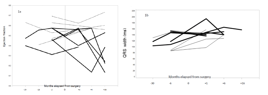
Figure 1a. Evolution of ejection fraction (a) and QRS complex width (b) during the pre- (negative numbers) and post-surgical period in 11 patients with perioperative left bundle branch block. Numbers on the “X” axis represent the echo- (a) and electro-cardiography
Figure 1b. average elapsed time (in months) in reference to time of surgery “0”. Solid lines represent patients with deteriorating ejection
In the LBBB group the average LV mass as calculated prior to surgery was 397 ± 53 and 240 ± 40 grams for the ‘EF deterioration’ and ‘stable EF' subgroups respectively (p=0.002). LV mass > 300 grams was more common in ‘EF deterioration’ group (100% versus 0%, p=0.001), suggesting that a higher myocardial mass might contribute to EF deterioration in patients with LBBB [3]. This result is in agreement with studies that have demonstrated an EF decline in patients with LV hypertrophy [3,4].
When considering the 11 LBBB patients combined the QRS width increased from 119 ± 26 ms to 144 ± 53 ms (p=0.01), in agreement with results of prior studies (Figure 1b) [5]. The QRS surpassed 120 ms pre-operatively in 2 patients in the “stable EF” and 4 in “EF deterioration” subgroup respectively (p=0.6), while in the remaining 5 patients this value was reached post-operatively. The QRS width was unchanged before and after surgery (94 ± 14 ms versus 95 ± 13 ms) in the 20 control patients.
We demonstrate a significant increase in QRS width post aortic valve replacement surgery in patients with peri-operative LBBB when compared with a control group, with a sharp decrease in EF in more than half of the LBBB patients versus no EF change in a control group without LBBB. Myocardial mass greater than 300 grams might contribute to EF deterioration in patients with LBBB.
The authors report no relationships that could be construed as a conflict of interest
- Devereux RB, Alonso DR, Lutas EM (1986) Echocardiographic assessment of left ventricular hypertrophy: comparison to necropsy findings. Am J Cardiol 57: 450-458. [Crossref]
- Nazif TM, Williams MR, Hahn RT (2014) Clinical implications of new onset left bundle branch block after transcatheter aortic valve replacement: analysis of the PARTNER experience. Eur Heart J 35: 1599-1607. [Crossref]
- Drazner MH, Rame JE, Marino EK (2004) Increased left ventricular mass is a risk factor for the development of a depressed left ventricular ejection fraction within five years: the Cardiovascular Health Study. J Am Coll Cardiol 43: 2207-2215. [Crossref]
- Angheloiu GO, Saul M, Edelman K (2013) Predictors of left ventricular function deterioration in patients with left bundle branch block and ejection fraction >50%. Congest Heart Fail 19: 1-4. [Crossref]
- Follath F, Ginks WR (1972) Changes in the QRS complex after aortic valve replacement. Br Heart J 1972 34: 553-560. [Crossref]