Abstract
Epithelioid hemangioma (EH) is a rare benign vascular lesion that presents as a nodular mass in the skin, usually in the head and neck region. It is usually asymptomatic, and its exact etiology is still unknown. We describe ultrasound, CT and CT-PET features of an uncommon disease presentation in the upper arm area. A radical excision was performed, and tissue was submitted for histopathological diagnosis: an immunohistochemical examination showed the expression of CD31 marker and confirmed the diagnosis of EH.
Keywords
Epithelioid hemangioma, CT, PET
Introduction
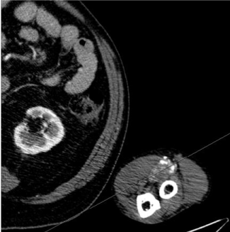
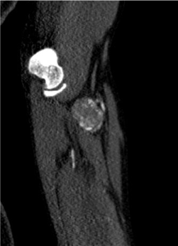
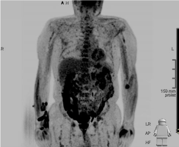
Epithelioid hemangioma (EH), once known as angiolymphoid hyperplasia with eosinophilia, is an uncommon vascular proliferative lesion that manifests with an expanding smooth-surfaced nodule. It is usually located in the head and neck area, particularly in the region around the ear (approximately 85% of cases), but less common sites include the long bones of arm and legs. The lesion lies mainly in the sub-cutis and it usually ranges from 0.5 to 2 cm in diameter, while larger nodules may extend deeply into the muscle or the fascia [1]. It is most commonly associated with pain or pruritus, but it may manifest with pulsation and spontaneous bleeding [2,3]. About half of the patients develop multifocal lesions. Significant lymph node enlargement and eosinophilia are occasionally present [4]. The peak incidence occurs between the second and the fourth decades, and the mean age of presentation is 37. Women are more affected than men [5]. In this paper, we present a case of EH occurring in the deep soft tissues of the left arm of a 76-year-old man: the age of the patient, the clinical presentation and the location of the tumor were uncommon. EH wasn’t initially considered in the differential diagnosis: malignant sarcoma was the preferred diagnosis, and angiosarcomas were considered. Correct identification of EH is pivotal as its treatments largely differs from those of other vascular tumors. Case report at the head 76-year-old man presented with an asymptomatic mass in correspondence of left elbow crease, steady-growing for approximately 2 months. There was no history of trauma and the patient denied any pain or numbness. His past medical history included a permanent pacemaker implantation and coronary artery angioplasty with stenting 3 months prior to assessment. Clinical examination revealed a non-tender swelling in the distal volar part of the arm due to a firm, oval, deep mass with a maximum diameter of 3 cm. The overlying skin color was normal and the axillary lymph nodes were not palpable. Passive and active motion of the left elbow and hand were unaffected; neurological examination was normal. Routine blood examination, including complete blood count, liver and renal function tests, did not reveal any abnormality, with a normal absolute eosinophil count of 400 cells/µL (5.8% of total white cell count). There was no evidence of proteinuria. A Doppler ultrasound examination revealed a well-defined hypoechogenic mass of approximately 3 x 3 x 2 cm in diameter with marked hypervascularity in the anterior compartment of the arm. Computed tomography (CT) of the left arm was performed, with and without intravenous administration of contrast material (iopromide 370 mg/ml, volume 100 ml, injection rate 4,5 ml/s) (Figure 1,2). The pre-contrast images revealed a soft-tissue mass of relative isoattenuation to muscle with heterogeneous enhancement following IV contrast medium injection. The lesion had no clear cleavage plane from the brachialis muscle and was non-dissociable from the proximal ulnar artery. A delayed phase scan showed further irregular fill in. The patient underwent PET-CT, showing intense FDG uptake at the arm mass (Figure 3). Given the patient’s age, the clinical presentation and the imaging characteristics of the lesion, sarcoma was the favored diagnosis; malignant vascular tumors were included in the differential for the marked hypervascularization and vascular invasion. The patient was taken to the operating room where a radical excision of the lesion and of the involved distal ulnar artery was performed. Histological examination revealed a central core of micro-macro vascular proliferation with epithelioid endothelial cells containing eosinophilic cytoplasm and vesicular nuclei. Diffuse and dense infiltration of lymphocytes and eosinophils, with multiple lymphoid follicles was also present. Peripherally marked vascular proliferation consisting of blood vessels of varying sizes was detected. Fine irregular fibrosis was also noted. There was no evidence of malignancy. Immunohistochemical stains demonstrated that the tumor cells were positive for the endothelial marker CD34, CD31 and FL1. The findings were considered consistent with EH. A diagnosis of EH was made and the patient was dismissed with no further treatment.

Figure 1. Axial CT arterial phase acquisition.

Figure 2. Coronal multiplanar reconstruction of CT arterial phase acquisition.

Figure 3. Positron Emission Tomography
Discussion
The definition of vascular tumors comprises a very heterogeneous group, such as hemangioma, angiosarcoma and their epithelioid variants. Their clinical behaviors, treatments and prognosis differ significantly, and it is of fundamental importance to distinguish them effectively. The World Health Organization (WHO) recognizes 3 different classes of soft tissues epithelioid vascular tumors according to their level of malignancy: EH, a benign tumor with a good prognosis, hemangioendothelioma (EHE), with a metastatic rate of 31% and angiosarcoma, a high-grade malignancy with a metastatic rate greater than 50% [6]. Differential diagnosis between them can be challenging: clinical and radiological features overlap, and pathologic characteristics of EH might also be seen in other vascular tumors. The radiological findings in EH, in fact, include locally aggressiveness, multifocality and other features suggestive of malignancy [7]. Because of its epithelioid nature, EH might also be misdiagnosed as metastatic carcinoma [6]. Distinguishing EH from EHE is critical, because the latter exhibits a more aggressive clinical course, with higher rates of multifocality and distant spread. Ultrasonography (US) coupled with color Doppler US is considered the imaging modality of choice for initial assessment and characterization of soft-tissue lesions of presumed vascular origin and it provides information about the degree of vascularity of a lesion. However, US has the disadvantages of a limited field of view, restricted penetration, and operator dependency [4,8]. MRI is the most valuable imaging modality for classification of vascular neoplasms: it allows an exact definition of the extension and the anatomic relationship to adjacent structures of the lesion, it provides optimal soft-tissue contrast and allows multiplanar image acquisition [9]. As our patient could not undergo MRI due to a pacemaker, a CT scan was performed, providing valuable information for therapy planning. CT scans, however, are of limited value in the characterization of these lesions, with imaging sufficiently characteristic to suggest the correct diagnosis in only a minority of cases [10,11]. To our knowledge, there are no works describing PET/CT characteristic and usefulness in patients affected by EH: it may be a valuable tool to detect multifocality or reactive adenopathy. Many authors consider EH to be a reactive lesion: 10% of EH occur following trauma and it usually is associated with prominent inflammatory response. Some author suggests that reactive condition caused by trauma leads to arteriovenous malformation and thus trigger cellular proliferation and growth. EH shows a tendency towards multifocality and local recurrence, which seems to prove a neoplastic origin. EH probably encompasses many different entities linked by epithelioid change of the endothelium, a feature encountered in both reactive and malignant vascular lesions [10]. Despite several modalities being tried, the best treatment of EH is largely debated: surgery is usually considered as first line treatment, but the lesions tend to recur. Medical therapies may be a viable alternative in some cases, especially in cosmetically sensitive sites, but few reports of success exist, mainly with systemic retinoids, topical/intra-lesional/systemic steroids, pentoxiphylline and indomethacin [12]. Minimally invasive approaches like cryotherapy, electrosurgery, laser therapy, radiotherapy and intra-lesional chemotherapy are under evaluation. Recurrence is common (about one third of the cases), but no metastases have been reported to date.
In conclusion, we presented a patient with an hypervascular lesion of the arm, thought initially to be malignant, but which proved to be an EH, an uncommon benign lesion: its presentation may be ambiguous, and its correct identification may preserve the patient from unnecessary aggressive treatments.
Conflict of interest
Authors declare no conflicts of interest
References
- Wells GC, Whimster IW (1969) Subcutaneous angiolymphoid hyperplasia with eosinophilia. Br J Dermatol 81: 1-14. [Crossref]
- Olsen TG, Helwig EB (1985) Angiolymphoid hyperplasia with eosinophilia. A clinicopathologic study of 116 patients. J Am Acad Dermatol 12: 781-796. [Crossref]
- Cheney ML, Googe P, Bhatt S, Hibberd PL (1993) Angiolymphoid hyperplasia with eosinophilia (histiocytoid hemangioma): evaluation of treatment options. Ann Otol Rhinol Laryngol 102: 303-308. [Crossref]
- Weiss SW, Enzinger FM (1982) Epithelioid hemangioendothelioma: a vascular tumor often mistaken for a carcinoma. Cancer 50: 970-981. [Crossref]
- Urabe A, Tsuneyoshi M, Enjoji M (1987) Epithelioid hemangioma versus Kimura's disease. A comparative clinicopathologic study. Am J Surg Pathol 11: 758-766. [Crossref]
- Errani C, Zhang L, Panicek DM, Healey JH, Antonescu CR (2012) Epithelioid hemangioma of bone and soft tissue: a reappraisal of a controversial entity. Clin Orthop Relat Res 470: 1498-1506. [Crossref]
- Antonescu C1 (2014) Malignant vascular tumors--an update. Mod Pathol 27 Suppl 1: S30-38. [Crossref]
- Paltiel HJ, Burrows PE, Kozakewich HP, Zurakowski D, Mulliken JB (2000) Soft-tissue vascular anomalies: utility of US for diagnosis. Radiology 214: 747-754. [Crossref]
- Flors L, Leiva-Salinas C, Maged IM, Norton PT, Matsumoto AH, et l. (2011) MR imaging of soft-tissue vascular malformations: diagnosis, classification, and therapy follow-up. Radiographics 31: 1321-1340.
- Kim JS, Chandler A, Borzykowski R, Thornhill B, Taragin BH (2012) Maximizing time-resolved MRA for differentiation of hemangiomas, vascular malformations and vascularized tumors. Pediatr Radiol 42: 775-784. [Crossref]
- Vilanova JC, Barceló J, Villalón M (2004) MR and MR angiography characterization of soft tissue vascular malformations. Curr Probl Diagn Radiol 33: 161-170. [Crossref]
- Singh S, Dayal M, Walia R, Arava S, Sharma R, et al. (2014) Intralesional radiofrequency ablation for nodular angiolymphoid hyperplasia on forehead: a minimally invasive approach. Indian J Dermatol Venereol Leprol 80: 419-421. [Crossref]


