Abstract
Background: Cardiovascular magnetic resonance imaging (CMR) represents the gold standard for routine examination of cardiac function and structure evaluation at rest and during exercise as well. Conventional stress CMR includes drug administration to increase heart rate (HR). The recently developed MR-conditional cardio stepper (CS) could be an alternative for real time stress CMR. So far, no data on cardiorespiratory responses exist for this device. Thus, this study evaluated cardiorespiratory responses to exhaustive testing on the CS compared to cycle and treadmill ergometry.
Methods: Forty-six healthy participants (24 men, 22 women, age: 31.4 ± 8.9 yrs, height: 174.8 ± 7.2 cm, weight: 70.3 ± 9.4 kg) performed three incremental exercise tests to volitional failure in a randomized crossover trial.
Results: Maximal values using the CS were significantly lower when compared to cycle and treadmill ergometry (HRmax: 170 ± 13 vs. 186 ± 9 vs. 190 ± 11 bpm, respectively; p<0.05, maximal oxygen uptake: 2971 ± 772 vs. 3531 ± 1023 vs. 3849 ± 1013 ml/min, respectively; p<0.05 and exercise duration: 539 ± 130 vs. 745 ± 157 vs. 710 ± 128 sec, respectively; p<0.05).
Conclusions: Although cardiorespiratory responses are not maximal when using the CS, the target heart rate, i.e. >85% of HRmax, for a stress CMR can be achieved.
Key words
stress cardiovascular magnetic resonance imaging, cardio stepper, cardiopulmonary exercise testing, MR conditional
Background
Cardiovascular magnetic resonance imaging (CMR) is used as a routine examination of cardiac function and structure evaluation [1], especially for coronary heart disease (CHD) diagnostics [2]. At rest, several perfusion defects of the heart, like coronary stenosis, remain undetected, especially in patients with unstable angina [3]. Therefore, stress tests during a CMR with a target heart rate of 85% of the theoretical maximal heart rate (220-age ± 10 bpm) are considered to be necessary [3-5]. To achieve this target heart rate, stress CMR is often carried out with drugs like Dobutamine and Adenosine [2,6]. However, this invasive procedure is associated with a potential risk of side effects like angina pectoris, hypertension, and even myocardial infarction etc. [6]. Considering these risks, alternative methods should be preferred. So far, several devices like treadmills, cycle ergometers and steppers for physical stress CMR were developed. Stress CMR on treadmills and upright cycle ergometers carried out outside the magnetic resonance tomograph (MRT) have the disadvantage of their inability to acquire cardiac images within 20 to 90 seconds of cessation of physical exercise, since this time is needed to get the post-stress image acquisition [7-9]. Consequently, post stress image acquisition at peak heart rate is not possible because of the exponentially decreasing heart rate after strenuous exercise [10]. To reduce time from finishing the stress test to post-stress imaging Lode developed a supine ergometer pedal (MRI Ergometer Pedal, Lode) for use during stress CMR. However, experiences in clinical settings show that performing supine cycle ergometer tests on the MRI Ergometer Pedal, with the goal of inducing high cardiac outputs, are not practicable due to the limited space in the MRT.
To overcome this limitation a short crank length of 60 mm was used to realize a real- time stress CMR [1]. However, according to Too and Landwer [11] and Martin and Spirduso [12] the optimal cycle crank length should be between 145 mm and 170 mm. Short cycle crank lengths result in a lower maximal power output, which might be insufficient to generate the needed heart rate increase for a stress CMR. Another commercially available device of Lode (MRI Ergometer Push/Pull, Lode) shows the same limitation with a maximum workload of 100 W. Using these devices, it is unlikely to reach the target heart rate of 85% of the theoretical maximal heart rate, which is necessary for a meaningful stress CMR [3-5]. This becomes particularly relevant for younger and trained subjects.
The cardio stepper (CS) developed by “Ergospect” (www.ergospect.com) generates the resistance via a hydraulic systems and allows a greater amplitude in the MRT due to step movements. This is of great advantage when compared to the above mentioned devices, because a clearly higher maximal power output and related maximal HR can be expected. However, until today no study exists that collected and compared cardiopulmonary standard values of maximal exercise testing on the CS with those of a standard treadmill or cycle ergometer.
Exercise tests using a cycle ergometer or a treadmill are among the most common tools for assessing the potential for and severity of CHD [13,14]. Furthermore, cardiorespiratory fitness testing is generally performed on these devices [15]. However, maximal cardiorespiratory responses attained on the cycle ergometer are somewhat lower compared with maximal treadmill values [16-18]. Smaller muscle involvement and greater regional muscle fatigue are thought to be responsible for this difference [19,20]. Such effects might also be expected for exercise testing with the CS. Additionally, it could be argued that the lack of familiarity with the CS exercise might lead to even lower values when compared to the cycle ergometer [21].
Therefore, the main goals of the current study were to evaluate if the target heart rate of 85% of the theoretical maximal heart rate can be achieved on the CS and to compare the maximal cardiorespiratory responses of the CS with those obtained by bicycle and treadmill testing. On the one hand we hypothesized that the target heart rate of 85% of the theoretical maximal heart rate will be reached using the CS but on the other hand we expected lower cardiopulmonary response achieved on the CS when compared to cycle and treadmill ergometry.
Methods
Subjects
A group of 46 healthy volunteers (24 men, 22 women, age: 31.4 ± 8.9 yrs, height: 174.8 ± 7.2 cm, weight: 70.3 ± 9.4 kg) were informed about the study aims and procedures and gave written informed consent to participate in the study. Volunteers completed a health history questionnaire to estimate potential risks associated with maximal exercise testing. Baseline characteristics of the subjects are shown in table 1. None of the subjects were professional athletes, but mostly well trained (6.5 ± 3.1 hours sport/week). During the study period one participant dropped out because of an injury not obtained during the exercise testing. The study was carried out in conformity with the ethical standards laid down in the 1975 declaration of Helsinki and has been approved by the Institutional Review Board of the Department of Sport Science, University of Innsbruck.
Table 1: Anthropometric data
| |
male
|
female
|
all
|
|
n=24
|
n=22
|
n=46
|
|
age [a]
|
31.8 ± 8.3
|
30.91 ± 9.7
|
31.4 ± 8.9
|
|
body weight [kg]
|
76.3 ± 7.3
|
63.7 ± 6.8
|
70.3 ± 9.4
|
|
body height [cm]
|
178.5 ± 6.8
|
170.8 ± 5.5
|
174.8 ± 7.2
|
|
BMI
|
23.9 ± 1.6
|
21.8 ± 1.7
|
22.9 ± 2.0
|
|
sport per week [h]
|
7.5 ± 3.2
|
5.5 ± 2.7
|
6.5 ± 3.1
|
The participants performed three incremental exercise tests to exhaustion at the same time of day, separated by 7 days within a 3-week period. To avoid learning effects the order of the different exercise tests was randomized. Test protocols were designed to yield similar test durations ranging between 7-17 minutes [22,23]. For the treadmill (quasar med, h/p/cosmos ) test a 3-min warm up period at a speed of 4 km/h and a slope of 12% followed by an adapted Bruce ramp protocol with 1 minute stages (American College of Sports Medicine 2006) was adopted. To perform the cycle ergometer test an electronically braked cycle ergometer (ergoline, ergoselect) was used. The saddle and handle bar positions of the ergometer were adjusted to resemble the subject’s own bike setup. Pedal rate had to be held over 70 rpm throughout testing. The test started after a warm up performed at 50 W for 3 min. Afterwards the workload (P) was increased by 25 W every minute until subjective exhaustion of the participant or the pedal rate dropped below 70 rpm. The stepper ergometer test was performed on the CS (Diagnostic pedal, Ergospect) with the same protocol that was used by the cycle ergometer test. All subjects were asked to hold a given rhythm throughout testing, which complied with 70 rpm pedal rate on the cycle ergometer. When the participants could not hold the rhythm anymore, the test was terminated. During all tests subjects were verbally encouraged to continue until exhaustion.
Physiological measurements
During the exercise tests, oxygen uptake (VO2), carbon dioxide output (VCO2) and minute ventilation (VE) were recorded continuously using a gas analysing system (Jaeger Oxycon pro, Carefusio). Heart rate (HR) was monitored by a Polar heart rate sensor (Polar heart rate sensor H2, Polar). Blood lactate concentration (La) was determined by spectrophotometry (C_line, EKF-Diagnostik Biosen). Arterialized blood samples were taken from an ear lobe prior to each testing session and directly after the end of each exercise. A subjective rating of perceived exertion (RPE), subdivided into systemic and muscular exertion, was obtained using the Borg scale [24-26]. The criteria used to confirm that VO2 had reached maximum values were: maximal respiratory exchange ratio (RERmax)>1.1, Lamax>8.0 mmol/l, HRmax>220 bpm minus age and a levelling off of VO2 despite increasing workload [5,27]. 30 sec means of VO2, VE and RER were taken as maximal values (HRmax, VO2max, VEmax, RERmax). According to [28] Pmax was charged in aliquot parts for each second for the cycle ergometer and cardio stepper.
CS description
The CS has been especially designed to stress test the cardiovascular system in a whole body MRT (figure 1). It is completely built of synthetic and fiber reinforced plastic materials to avoid interferences of the magnetic field and artefacts of the imaging. Therefore the device can be used in all scanners with a field strength of 1.5-7 Tesla and even higher. The design is similar to a “stairwalker” exercise machine with two feet. The two pedals are linked with a hydraulic system, the load on the pedals can be adjusted with a pneumatic driven, remote controlled hydraulic restrictor. Each pedal is equipped with a force sensor. The physical parameters power, work and frequency are calculated in combination with a pedal position sensor. In the “ergometer mode” the module is able to control the adjusted workload automatically (frequency-independent). The technical data are: Pedal displacement: 160 mm, Workload range: 25-450 watt, Frequency: 40-120 Steps per minute
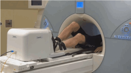
Figure 1. MR-conditional cardio stepper
Statistical analysis
Normal distribution of all data was checked with Kolmogorov-Smirnov-tests. A one-way analysis of variance (ANOVA) with repeated measures (cycle, treadmill, CS) and Friedman-tests were used to evaluate significant differences between dependent variables. Significant results were followed by post-hoc comparison using pairwise t-tests and Wilcoxon-tests for intra-group comparison (cycle versus stepper and treadmill versus stepper) and t-tests for independent variables and Mann-Whitney-U-tests for inter-group comparison (men versus women). Data are presented as mean and standard deviation (SD) and a probability level of p<0.05 was considered significant.
Results
The cardiorespiratory and metabolic responses during each trial (cycling vs. running vs. stepping) are shown in Table 2. HRmax, as well as HR at an RER of 1 (HRRER1) achieved on the cycle (p<0.001; p<0.001) or the treadmill (p<0.001; p<0.001) were lower on the CS. The HRmax was about 9.4% and 11.8% lower compared to the cycle ergometer or treadmill, respectively (figure 2). However, the target heart rate (85% of the expected maximal heart rate) could be reached with the CS. The average age of the sample was 31.8 years resulting in an expected maximal heart rate of 188.2 bpm. The HRmax of 170 ± 13 bpm achieved with the CS amounts to approximately 90%. 18.8% lower VO2max-rel values were observed on the CS when compared to the cycle ergometer (p<0.001) and 29.5% when compared to treadmill (p<0.001) (figure 2). Lower exercise durations (tmax) were detected between the CS and the cycle ergometer (-37.7%; p<0.001) as well as the treadmill (-24.1%; p<0.001). RPEmax-muscular values on CS were higher compared to the cycle (p=0.075) and significantly higher compared to the treadmill (p<0.001). RPEmax-system showed the lowest values compared to cycle (p<0.001) and treadmill (p<0.001). Pmax achieved with the CS (199 ± 54 W) was significantly lower (p<0.001) when compared to the values of the bicycle test (285 ± 65 watts).
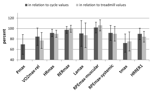
Figure 1. MR-conditional cardio stepper
Pmax, maximum power output; VO2max-rel, maximum relative oxygen uptake; HRmax, maximum heart rate; RERmax, maximum respiratory exchange ratio; Lamax, maximum lactate; RPEmax-muscular, maximum muscular rating of perceived exertion; RPEmax-systemic, maximum systemic rating of perceived exertion; tmax, time maximum; HRRER1, heart rate at RER=1; * significant difference from stepper (paired t-test).
Table 2. Maximal physiologic responses during the three different exercise tests
|
all (n=46)
|
ANOVA
|
|
| |
|
Cycle Ergometer
|
Treadmill
|
Cardio Stepper
|
F-value
|
p-value
|
|
|
Pmax [Watt]
|
285 ± 65*
|
-
|
199 ± 54
|
214.76
|
<0.001
|
|
|
VO2max-abs [ml/min]
|
3531 ± 1023*
|
3849 ± 1013*
|
2971 ± 772
|
-
|
<0.001
|
|
|
VO2max-rel [ml/min/kg]
|
49.9 ± 11.2*
|
54.4 ± 10.6*
|
42.0 ± 8.3
|
167.65
|
<0.001
|
|
|
HRmax [bpm]
|
186 ± 9*
|
190 ± 11*
|
170 ± 13
|
138.31
|
<0.001
|
|
|
VEmax [l/min]
|
138 ± 37*
|
142 ± 32*
|
112 ± 30
|
93.93
|
<0.001
|
|
|
RERmax
|
1.21 ± 0.10*
|
1.19 ± 0.08
|
1.18 ± 0.08
|
4.03
|
0.025
|
|
|
Lamax [mmol/l]
|
10.9 ± 2.9*
|
11.4 ± 3.1*
|
9.8 ± 2.7
|
10.96
|
<0.001
|
|
|
RPEmax-muscular
|
17.5 ± 1.8
|
16.9 ± 2.0*
|
17.9 ± 1.8
|
-
|
<0.001
|
|
|
RPEmax-systemic
|
16.7 ± 2.0*
|
17.0 ± 2.1*
|
14.9 ± 2.4
|
-
|
<0.001
|
|
|
tmax [sec]
|
745 ± 157*
|
710 ± 128*
|
539 ± 130
|
110.7
|
<0.001
|
|
|
VO2RER1-abs [ml/min]
|
2544 ± 861*
|
3120 ± 938*
|
2075 ± 633
|
75.18
|
<0.001
|
|
|
HRRER1 [bpm]
|
156 ± 20*
|
166 ± 16*
|
141 ± 17
|
91.91
|
<0.001
|
|
Pmax: maximum power output; VO2max-abs: maximum absolute oxygen uptake; VO2max-rel: maximum relative oxygen uptake; HRmax: maximum heart rate, VEmax: maximum minute ventilation; RERmax: maximum respiratory exchange ratio; Lamax: maximum lactate; RPEmax-muscular: maximum muscular rating of perceived exertion; RPEmax-systemic: maximum systemic rating of perceived exertion; tmax: time maximum; VO2RER1-abs: absolute oxygen uptake at RER=1; HRRER1: heart rate at RER=1; *significantly different from stepper (paired t-test)
Table 3 illustrates sex-related differences. There were found no sex-related differences on none of the three devices in HRmax. In addition, no sex-related differences for RERmax achieved on the CS were found (p=0.216). By contrast, Lamax and tmax differed significantly regarding sex (p=0.025; p<0.001). Females achieved 16.8% lower Lamax values and 28.5% lower tmax values than males.
Table 3: Gender-related maximal physiologic responses during the 3 different exercise tests
| |
male (n=24)
|
female (n=22)
|
|
Cycle
|
Treadmill
|
Cardio Stepper
|
Cycle Ergometer
|
Treadmill
|
Cardio Stepper
|
|
Pmax [Watt]
|
330 ± 51*
|
-
|
235 ± 41
|
237 ± 39*+
|
-
|
161 ± 37+
|
|
VO2max-abs [ml/min]
|
4265 ± 799*
|
4605 ± 764*
|
3556 ± 498
|
2732 ± 511*+
|
3024 ± 438*+
|
2334 ± 434+
|
|
VO2max-rel [ml/min/kg]
|
56.1 ± 10.5*
|
60.6 ± 10.0*
|
46.8 ± 6.7
|
43.1 ± 7.4*+
|
47.7 ± 6.3*+
|
36.8 ± 6.4+
|
|
HRmax [bpm]
|
185 ± 8*
|
190 ± 11*
|
170 ± 13
|
187 ± 10*
|
189 ± 11*
|
171 ± 13
|
|
VEmax [l/min]
|
165 ± 24*
|
166 ± 17*
|
136 ± 18
|
108 ± 23*+
|
114 ± 19*+
|
86 ± 15+
|
|
RERmax
|
1.20 ± 0.12
|
1.18 ± 0.09
|
1.17 ± 0.08
|
1.23 ± 0.09
|
1.20 ± 0.06
|
1.20 ± 0.09
|
|
Lamax [mmol/l]
|
12.1 ± 2.6*
|
12.7 ± 2.9*
|
10.7 ± 2.5
|
9.6 ± 2.7+
|
9.9 ± 2.6*+
|
8.9 ± 2.7+
|
|
RPEmax-muscular
|
17.5 ± 2.1*
|
17.4 ± 2.1*
|
18.4 ± 1.5
|
17.6 ± 1.5
|
16.3 ± 1.8*
|
17.4 ± 1.9
|
|
RPEmax-systemic
|
17.0 ± 1.8*
|
17.0 ± 1.9*
|
15.2 ± 1.7
|
16.4 ± 2.3
|
17.0 ± 2.4
|
15.4 ± 3.1
|
|
tmax [sec]
|
852 ± 123*
|
790 ± 107*
|
624 ± 99
|
628 ± 94*+
|
624 ± 85*+
|
446 ± 89+
|
|
VO2RER1-abs [ml/min]
|
2971 ± 922*
|
3676 ± 889*
|
2397 ± 652
|
2078 ± 473+
|
2513 ± 531*+
|
1724 ± 383+
|
|
HRRER1 [bpm]
|
152 ± 22*
|
163 ± 18*
|
136 ± 19
|
161 ± 16*
|
170 ± 13*
|
145 ± 13
|
Pmax: maximum power output; VO2max-abs: maximum absolute oxygen uptake; VO2max-rel: maximum relative oxygen uptake; HRmax: maximum heart rate, VEmax: maximum minute ventilation; RERmax: maximum respiratory exchange ratio; Lamax: maximum lactate; RPEmax-muscular: maximum muscular rating of perceived exertion; RPEmax-systemic: maximum systemic rating of perceived exertion; tmax: time maximum; VO2RER1-abs: absolute oxygen uptake at RER=1; HRRER1: heart rate at RER=1; *significantly different from stepper (paired t-test); +significantly different from males (unpaired t-test)
Discussion
The main outcome of the present investigation is that the target heart rate for a stress CMR can be achieved with the recently developed CS [3-5]. However, as expected the HR values using the CS were 10.5% and 8.6% lower when compared to the cycle ergometer and the treadmill, respectively. The wide workload range (25-450 watt) of the CS enables a stress CMR for nearly all participants, including young and well- trained persons to increase the heart rate to the required extent. By comparison, in other studies stress CMR was performed at heart rates between 110 and 120 bpm [1,29,30], which does not correspond to the required target heart rate of young people.
In the present study nearly all maximal values, except RPEmax-muscular during the cycling test and RERmax during the treadmill test, were lower on the CS when compared to the cycling and treadmill exercise, respectively. The main reasons responsible for the lower maximal values might be the supine position and the resulting less overall muscle mass involvement and the more pronounced local muscle fatigue on the CS [13,19,20,31]. This is supported by the high muscle metabolism values (lactate and RER) reached on the CS, which fulfilled the criteria for the attainment of maximal exertion. These high lactate and RER values could additionally be based on an isometric workload. Due to the restricted movement amplitude on the CS the contraction becomes rather comparable to an isometric muscle contraction, and tends to result in a faster muscle fatigue [32]. This is directly expressed in lower time values (CS tmax: 539 ± 130 sec), which could be responsible for the lower maximal values (i.e. VO2max) reached on the CS. The faster muscle fatigue on the CS is also supported by the high RPEmax-muscular and the lower RPEmax-system values.
There were no sex-related differences in HRmax during the exercise on the CS which allows to conclude that cardiopulmonary exercise testing on the CS results in similar responses between men and women.
Some limitations have to be mentioned. For the CS a workload increase of 25 W every minute may be too high, mainly for women as the exercise test duration (446 ± 89 sec) was of borderline [22,23]. Consequently, a lower increase of the workload is recommended for further studies. Furthermore, the fitness level of our young, healthy and well-trained participants was clearly higher compared to the general population and even more when compared to patients. Typically, patients with an indication for a stress CMR are older and less fit. The transfer of our data to the patients might therefore be limited. However, due to the very low minimum workload of 25 W and the minimum load increases of 1 W the device should also be applicable to this population. Additionally, it has to be mentioned that, especially older patients, will need some time to get used to this new technique.
Conclusion
In conclusion, although the maximal cardiorespiratory responses on the CS are somewhat lower compared to maximal treadmill or cycle exercise, the target heart rate for a stress CMR of 85% of the theoretical maximal heart rate can be achieved with the MR-conditional CS even in young and healthy subjects.
Competing interests
Authors SB, FM, HG and MB have no conflicts of interest or financial ties to disclose. KM worked as research assistant in the project in which the CS was developed and tested.
Authors contributions
SB, FM, HG, MB, KM: Conceived and designed the experiments. SB, FM: Performed the experiments. SB, FM, HG: Analyzed the data. SB, FM, HG, MB, KM: Wrote the paper.
References
- Gusso S, Salvador C, Hofman P, Cutfield W, Baldi JC, et al. (2012) Design and testing of an MRI-compatible cycle ergometer for non-invasive cardiac assessments during exercise. Biomed Eng Online 18: 11-13.
- Kelle S, Nagel E (2010) Bildgebende Verfahren in der Ergometrie (MRT). Springer Medizin Verlag Heidelberg 109-120.
- Arai AE (2007) Myocardial stress perfusion imaging using CMR. Magnetom Flash 2: 34-41.
- Kramer CM, Barkhausen J, Flamm SD, Kim RJ, Nagel E (2013) Standardized cardiovascular magnetic resonance (CMR) protocols 2013 update. J Cardiovasc Magn Reson 8: 15-91. [Crossref]
- Such U, Meyer T (2010) Die maximale Herzfrequenz. Deutsche Zeit Sportmed 61: 310-311.
- Jochims M, Bruder O, Jensen C, Sabin G (2007) High-dose dobutamine stress cardiac MR imaging for detection of myocardial ischemia. Magnetom Flash 2: 42-49.
- Thavendiranathan P, Dickerson JA, Scandling D, Balasubramanian V, Pennell ML, et al. (2014) Comparison of treadmill exercise stress cardiac MRI to stress echocardiography in healthy volunteers for adequacy of left ventricular endocardial wall visualization: A pilot study. J Magn Reson Imaging 39: 1146-1152. [Crossref]
- Foster EL, Arnold JW, Jekic M, Bender JA, Balasubramanian V, et al. (2012) MR-compatible treadmill for exercise stress cardiac magnetic resonance imaging. Magn Reson Med 67: 880-889. [Crossref]
- Rerkpattanapipat P, Gandhi SK, Darty SN, Williams RT, Davis AD, et al. (2003) Feasibility to detect severe coronary artery stenoses with upright treadmill exercise magnetic resonance imaging. Am J Cardiol 92: 603-606. [Crossref]
- [Crossref] Hottenrott K, Hoos O, Esperer HD (2006) Heart rate variability and physical exercise. Current status. Herz 31: 544-552.
- [Crossref] Martin JC, Spirduso WW (2001) Determinants of maximal cycling power: crank length, pedaling rate and pedal speed. Eur J Appl Physiol 84: 413-418.
- Too D, Landwer GE (2000) The effect of pedal crank arm length on joint angle and power production in upright cycle ergometry. J Sports Sci 18: 153-161. [Crossref]
- Saitoh M, Matsunaga A, Kamiya K, Ogura MN, Sakamoto J, et al. (2005) Comparison of cardiovascular responses between upright and recumbent cycle ergometers in healthy young volunteers performing low-intensity exercise: assessment of reliability of the oxygen uptake calculated by using the ACSM metabolic equation. Arch Phys Med Rehabil 86: 1024-1029. [Crossref]
- Gibbons RJ, Zinsmeister AR, Miller TD, Clements IP (1990) Supine exercise electrocardiography compared with exercise radionuclide angiography in noninvasive identification of severe coronary artery disease. Ann Intern Med 112: 743-749. [Crossref]
- McArdle WD, Katch FI, Katch VL (1996) Exercise physiology: energy, nutrition and human performance. Williams and Wilkins, Baltimore.
- Elsais WM, Mohammad WS (2011) Effect of different exercise modes on cardiovascular responses in male runners. World J Sport Sci 5: 191-196.
- [Crossref] Basset FA, Boulay MR (2000) Specificity of treadmill and cycle ergometer tests in triathletes, runners and cyclists. Eur J Appl Physiol 81: 214-221.
- Schuh A, Baranek G, Entleutner V, Senn E (1993) Unterschiede im Leistungsverhalten von Gesunden zwischen Fahrrad-und Laufbandergometrie. Phys Med Rehab Kuror 3: 33-37.
- May LJ, Punn R, Olson I, Kazmucha JA, Liu MY, et al. (2014) Supine cycling in pediatric exercise testing: disparity in performance measures. Pediatr Cardiol 35: 705-710. [Crossref]
- Walsh-Riddle M, Blumenthal JA (1989) Cardiovascular responses during upright and semi-recumbent cycle ergometry testing. Med Sci Sports Exerc 21: 581-585. [Crossref]
- [Crossref] Neary PJ, Wenger HA (1986) The effects of one- and two-legged exercise on the lactate and ventilatory threshold. Eur J Appl Physiol Occup Physiol 54: 591-595.
- Midgley AW, Bentley DJ, Luttikholt H, McNaughton LR, Millet GP (2008) Challenging a dogma of exercise physiology: does an incremental exercise test for valid VO2 max determination really need to last between 8 and 12 minutes? Sports Med 38: 441-447. [Crossref]
- Amann M, Subudhi A, Foster C (2004) Influence of testing protocol on ventilatory thresholds and cycling performance. Med Sci Sports Exerc 36: 613-622. [Crossref]
- Whaley MH, Brubaker PH, Otto RM, Armstrong LE (2006) ACSM's guidelines for exercise testing and prescription, Williams and Wilkins, Philadelphia, PA.
- [Crossref] Zamunér AR, Moreno MA, Camargo TM, Graetz JP, Rebelo AC, et al. (2011) Assessment of Subjective Perceived Exertion at the Anaerobic Threshold with the Borg CR-10 Scale. J Sports Sci Med 10: 130-136.
- [Crossref] O'Sullivan SB (1984) Perceived exertion. A review. Phys Ther 64: 343-346.
- Astorino TA, Robergs RA, Ghiasvand F, Marks D, Burns S (2000) Incidence of the oxygen plateau at VO2max during exercise testing to volitional fatigue. J Exerc Physiol Online 3: 4.
- Wonsich M, Berent R, Klicpera M, Laimer H, Marko C, et al. (2008) Praxisleitlinien Ergometrie. J Kardiol 15: 3-16.
- Roest AA, Lamb HJ, van der Wall EE, Vliegen HW, van den Aardweg JG, et al. (2004) Cardiovascular response to physical exercise in adult patients after atrial correction for transposition of the great arteries assessed with magnetic resonance imaging. Heart 90: 678-684. [Crossref]
- Roest AA, Kunz P, Lamb HJ, Helbing WA, van der Wall EE, et al. (2001) Biventricular response to supine physical exercise in young adults assessed with ultrafast magnetic resonance imaging. Am J Cardiol 87: 601-605. [Crossref]
- A°strand PO, Rodahl K, Dahl HA, Stromme SB (2003) Textbook of work physiology: physiological bases of exercise, Human Kinetics, Champaign, IL.
- Barmeyer J (2010) Formen der Arbeitsbelastung und ihr Wirken auf das Kreislaufsystem. Das kardiologische Gutachten. Barmeyer J, Barmeyer A, Bojara W, von Dryander ST, Germing A, Grewe P, Jäger D, Lawo TH, Lemke B, Lindstaedt M, Machraoui A, Mehrhoff F, eds. Thieme, Stuttgart, pp.14-18.