New drug discovery and development efforts have traditionally relied on ethnopharmacological information and have focused on plants with medicinal properties. In the search for structurally novel and mechanistically unique lead compounds, these programs are increasingly turning to the bioactive molecules provided by the animal biodiversity. This not only involves bioactive constituents from marine and terrestrial invertebrates such as insects and arthropods, but also those from amphibians and other ‘higher’ vertebrates such as reptiles. The venoms of lizards and snakes are complex mixtures of dozens of pharmacologically active compounds. So far, these substances have brought us important drugs such as the angiotensin-converting enzyme inhibitors captopril and its derivates for treating hypertension and some types of congestive heart failure, and the glucagon-like peptide-1 receptor agonist exenatide for treating type 2 diabetes mellitus. These drugs have been developed from the venom of the Brazilian pit viper Bothrops jararaca (Viperidae) and that of the Gila monster Heloderma suspectum (Helodermatidae), respectively. Subsequently, dozens of potentially therapeutically applicable compounds from lizards’ and snakes’ venom have been identified, several of which are now under clinical evaluation. Additionally, components of the immune system from these animals, along with those from turtles and crocodilians, have been found to elicit encouraging activity against various diseases. Like the venoms of lizards and snakes, the immune system of the animals has been refined during millions of years of evolution in order to increase their evolutionary success. This paper addresses some of the bioactive compounds from reptiles, and elaborates on the therapeutic potential of some of them as anticoagulants and antiplatelet drugs, as well as wound healing-promoting, antileishmanial, antiviral, immunomodulating, antimicrobial, and anticancer compounds.
lizards, snakes, testudines, crocodilians, venoms, immune system components, bioactive compounds, novel therapeutics
New drug discovery and development programs have historically relied on the identification of novel lead compounds from plant origin. This is understandable when considering that plants have been the main, if not the only sources of therapeutics for managing human diseases for millennia [1]. Only in 1806, a pharmacologically active ingredient (morphine) from a plant (the opium poppy Papaver somniferum (Papaveraceae)) was for the first time isolated from a plant [2]. Currently, morphine is used for, among others, the palliation of severe chronic pain in, for instance, terminal cancer patients [2], and serves as a precursor for a large number of opioid medications such as the antitussive codeine and the antidiarreal agent loperamide [2]. The identification of morphine from P. somniferum was soon followed by many others such as, among others, the central nervous system stimulant caffeine from the beans of the coffee plant Coffea arabica (Rubiaceae) in 1819 [3], the antimalarial quinine from the bark of the cinchona tree Cinchona officinalis (Rubiaceae) in 1820 [4], and the analgesic salicin from the bark of the white willow Salix alba (Salicaceae) in 1828 [5].
Since then, many more breakthrough drugs have been developed from plants, including the antineoplastic agents vincristine and paclitaxel from the periwinkle plant Catharanthus roseus (Apocynaceae) [6] and the Pacific yew Taxus brevifolia (Taxaceae) [7], respectively; the phytoestrogen diosgenin from yam species in the genus Dioscorea (Dioscoreaceae) that serves as a precursor of, among others, oral contraceptives and cortisone [8]; and the oral antihyperglycemic biguanide metformin from the French lilac Galega officinalis (Fabaceae) [9]. Other important sources of novel drugs were micro-organisms. The fungus Penicillium rubens (Trichocomaceae) and the actinomycete bacterial species Saccharopolyspora erythraea (Pseudonocardiaceae) gave the antibacterial agents penicillin [10] and erythromycin [11], respectively. Another bacterial species, Streptomyces nodosus (Streptomycetaceae), produced the antifungal and antileishmanial agent amphotericin B [12]. The ascomycete fungus Tolypocladium inflatum (Ophiocordycipitaceae) led to the immunosuppressive agent tacrolimus [13], and the soil-borne fungus Aspergillus terreus (Trichocomaceae) produced specific LDL-lowering HMG-CoA reductase-inhibiting statins such as simvastatin that reduce the risk of arterial blockage, a heart attack, a stroke, and diabetes mellitus [14].
More recently, investigators in the field of new drug discovery and development have also turned to the animal kingdom. This has led to a number of important drugs such as ziconotide, a powerful analgesic that was structurally based on the extremely potent conotoxins produced by predatory cone snails in the genus Conus (Conidae) [15]; and trabectedin, an orphan drug for treating soft-tissue sarcomas and ovarian cancer that was first identified in the mangrove tunicate Ecteinascidia turbinata (Perophoridae) [16]. Various reptiles have also yielded important and life-saving drugs. The angiotensin-converting enzyme inhibitor captopril and its derivatives for treating hypertension and some types of congestive heart failure, have been developed from the venom of the Brazilian viper Bothrops jararaca (Viperidae) [17]. And exenatide for treating type 2 diabetes mellitus was originally isolated from the venom of the Gila monster Heloderma suspectum (Helodermatidae) [18] (Figure 1).
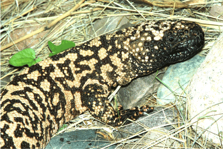
Figure 1. The Gila monster Heloderma suspectum (Helodermatidae) (from: https://images.app.goo.gl/dPs91WnmiHayS6S36)
In this paper, a number of bioactive compounds from several members of the reptilian suborders Lacertilla (lizards) and Serpentes (snakes) - both belonging to the order Squamata or scaled reptiles - as well as the orders Testudines (turtles and alike) and Crocodylia (crocodiles and alike) have extensively been addressed for their potential as clinically useful drugs. The fourth reptilian order, the Rhynchocephalia (tuataras), and the squamate suborder Amphisbaenia (worm lizards) have been left out of this paper because of the lack of literature data on their potential medical applicability. Each of these sections is preceded by background information about the (sub)order that is dealt with. The paper is concluded with some remarks about the previsions of reptilian bioactive compounds for new drug development programs.
Evolutionary development of reptiles: Reptiles are a class of tetrapod animals that possess scaly skin and lungs, are ectotherms, and lay shelled eggs on land. They comprise the extant lizards, snakes, and worm lizards; turtles, tortoises, and terrapins; crocodiles, alligators, caimans, and gharials; tuataras; as well as their extinct relatives [19]. These animals probably originated from advanced reptiliomorphs during the Carboniferous period, 360 to 300 million years ago [20]. Because these reptiliomorphan ancestors had four legs, reptiles are classified as tetrapods despite the existence of species with vestigial limbs and a limbless appearance such as snakes [21]. The skin of reptiles is covered with a continuous layer of scales containing keratin and waxy lipids which help reduce water loss from the body and represented an important adaptation that permitted them to live on land [22]. On the other hand, the occlusive skin prohibits cutaneous respiration like in amphibians, necessitating the development of efficient lungs [23].
The ability of reptiles to produce terrestrially-adapted eggs enclosed in an amnion and protected by a hard outer shell can be regarded as one of the monumental events in the Earth’s evolution, and certainly one of the main determining factors for the evolutionary success of these animals. This made them, unlike their immediate predecessors, the amphibians, independent of water for their reproduction. Notably, even aquatic reptiles return to land to lay eggs [24], whereas land-adapted amphibia search for water to lay their vulnerable shell-less eggs in order to ensure offspring [25]. The shell of the reptilian egg provides protection for the developing embryo and allows water retention while being permeable for gas exchange [24]. Inside, the embryo is surrounded by the amnion, a membrane that forms a fluid-filled sac in which the embryo is suspended in its own aquatic environment [24]. These advantages allowed the early reptiles to branch out to drier environments and enabled them to fully colonize many terrestrial niches [24].
Until the Late Carboniferous, around 310 million years ago, the early reptile-like amniotes represented a small, unremarkable group when compared to their much more numerous amphibian ancestors which then dominated terrestrial life on Earth [26]. This radically changed with the occurrence of the Carboniferous Rainforest Collapse 305 million years ago and the Permian-Triassic extinction event 54 million years later, the largest-ever extinction event on Earth [27]. The Carboniferous Rainforest Collapse was a relatively minor extinction event that was characterized by global warming and the destruction of large parts of the tropical rain forests that then covered the former supercontinent Euramerica [27]. The subsequent Permian-Triassic mass extinction event, colloquially referred to as the the Great Dying, wiped out 90 to 96% of all forms of life that had survived the Carboniferous Rainforest Collapse [27,28]. The all-out winners were the relatively few surviving reptiles which were able to produce hard-shelled amniotic eggs under those harsh and drier conditions [28]. As a result, they could thrive, diversify, occupy vacant ecological niches, and replace the amphibians as the dominant tetrapods [29]. This was the basis of the ‘rise of the reptiles’ which reached its pinnacle during the Mesozoicum (Triassic, Jurassic, and Cretaceous), 252 to 66 million years ago, the geological period referred to as the Age of Reptiles [29].
Characteristics of reptiles: As mentioned above, the modern reptiles comprise a class of ectothermic, tetrapod vertebrates that lay shelled eggs on land, possess scales on their skin which protect them from desiccation, breathe through lungs, make use of internal fertilization, and have amniotic development. Belonging to the superclass Tetrapoda, reptiles have in principle four legs that project sideways from the body [21]. Snakes as well as legless lizards in the family Anguidae (slowworms) have completely lost their limbs during evolution and move by using their ventral scales and ribs [30]. Hence the name ‘Reptilia’ of this animal class, which is derived from the Latin expressions ‘reptilis’ and ‘rēpō’, meaning ‘creeping’ and ‘to creep’, respectively, obviously referring to the fact that many crawl by moving on their belly or by means of small and short legs. Reptiles can be found everywhere on Earth, particularly in temperate and tropical regions. Most reptiles live on land but some species, such as those in the order Crocodylia, are amphibious, living both in water and on land.
Like other ectotherms, reptiles use relatively little of their metabolic energy from food to sustain their bodily functions and are able to survive on about 10% of the calories required by similarly-sized endotherms [31]. On the other hand, reptiles have to rely on the environment as their main source of body heat instead of generating heat by their own metabolism as endotherms do [32]. For this purpose, they have developed behavioral adaptations to help regulate their body temperature, such as basking in sunny places to warm up, and finding shady spots or going underground to cool down [32]. Under extreme environmental conditions, some reptiles may enter periods of dormancy, temporarily stopping their growth, development, and physical activity, thus minimizing metabolic activity and conserving energy. For instance, in response to very hot or dry conditions, North American desert tortoises and certain species of crocodiles may enter a period of estivation [33], while certain lizards, tortoises, turtles, frogs, and snakes brumate in very cold temperatures [33].
In lizards, snakes, and tuataras, the non-permeable, watertight skin is entirely covered with overlapping epidermal scales [22]. The shells of turtles and the plates of crocodiles are covered by scutes which are of dermal rather than epidermal origin and are fused to tough, protective armor [22]. Reptiles continuously shed their skin during their lifetime through ecdysis, involving the formation of a new layer of skin under the old one, and the separation of the old skin from the new one with the aid of proteolytic enzymes and lymphatic fluid [22]. The shells from turtles, tortoises, and terrapins do not undergo ecdysis since they are made up of about fifty bones of their skeleton including their spines and rib cage and grow with the animals [22]. However, the scutes on the surface of the shells shed or peel away to make way for newer, larger scutes [22]. The scales of some reptile species are colored for camouflage or aposetism. Well-known examples are the Madagascan satanic leaf-tailed gecko Uroplatus phantasticus (Gekkonidae) that can rapidly change its body color to better match its environment, avoiding being spotted by an approaching predator or a potential prey [34,35]; the highly venomous New World coral snakes in the family Elapidae (Figure 2) which advertise their poisonousness with vividly red, yellow/white, and black aposematic colored banding [36]; and the non-venomous Mexican milk snake Lampropeltis triangulum annulata (Colubridae) that mimics the warning coloration of coral snakes [37].
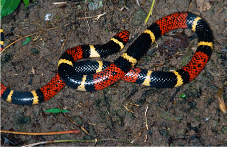
Figure 2. Aquatic coral snake Micrurus surinamensis (Elapidae) (from: https://images.app.goo.gl/ZPUuV2RzTzkUufaB7)
Most reptiles breathe through lungs which occupy a much smaller area of the body when compared to mammals but are larger than those of amphibians [23]. There are usually two lungs with the exception of snakes in which the left lung is rudimentary or has even entirely disappeared [23]. Most squamates and testudines have a three-chambered heart consisting of two atria and a ventricle, and two aortas leading to the systemic circulation [38]. Crocodilians have a four-chambered heart, similarly to birds, but also have two systemic aortas [38]. The blood is filtered by two relatively small kidneys and the urine is stored in a urinary bladder. In most species, uric acid is the main nitrogenous waste product [39]. The main exceptions are aquatic turtles which excrete most of their nitrogenous wastes as urea or ammonia [39]. In all reptiles, the urinogenital ducts empty into a cloaca along with the anus [39].
Most reptiles are carnivorous or insectivorous. As their meals are fairly simple to break down and digest, they have in general relatively simple and comparatively short digestive tracts [40]. However, being poikilotherms and because of their inability to masticate their food, digestion occurs slower than in mammals [40]. Turtles as well as some agamas and iguanas are the only groups of reptiles that are mostly herbivorous. They also ingest their meals whole but regularly swallow gastroliths to aid in digestion. Interestingly, salt water crocodiles are carnivorious but also use gastroliths, not to aid in the digestion of plant matter but as ballast to help stabilize them in the water or helping them to dive [41].
Like that of amphibians, the nervous system of reptilians basically consists of a central brain made up of a forebrain, a midbrain, and a hindbrain, as well as a spinal cord and nerves throughout the body including twelve pairs of cranial nerves [42]. As most reptiles are diurnal carnivorous hunters, they usually have excellent vision, allowing them to sharply detect colors (including ultraviolet wavelengths), shapes, motions, and depth at long distances [43]. Many snakes have a ‘third eye’ (the pineal gland) on the top of their head that cannot form images but is sensitive to changes in light and dark and can detect movement [43]. In addition, some snakes such as pit vipers, but also boas as well as pythons, have pits on both sides of the head behind the nostril and in front of the eye that are sensitive to infrared radiation, allowing them to sense the body heat of prey and to hunt in the dark (Figure 3) [44].
Figure 3. Pit viper with clearly visible pit located between the eye and the nostril (from: https://images.app.goo.gl/GDtm464p7zZ2oHBw6)
Many reptiles use chemically sensitive organs located in the nose and the roof of the mouth to find their prey [45]. Part of the epithelium of the nose consists of olfactory sensory cells for detecting airborne odors similarly to other vertebrates [45]. There is often a second chemoreceptor in the roof of the mouth - Jacobson’s organ - that serves as a short-range chemoreceptor of non-airborne odors [46]. Notably, the nerve connecting Jacobson’s organ to the brain is a branch of the olfactory nerve [46]. The use of Jacobson’s organ is most obvious in snakes, which rapidly flick their tongue in and out and transfer with each retraction odor particles adhering to the tongue to the roof of the mouth near the opening of the organ.
Reptiles reproduce sexually, although some species are capable of asexual reproduction [47]. The male specimens of snakes and lizards have paired copulatory organs known as hemipenes which are situated in a pouch at the base of the tail just caudal to the cloaca [48]. These organs engorge with sexual excitement and only one is used in each session to penetrate the cloaca of the female and deposit semen [48]. Male turtles and crocodilians have a single median phallus [48]. Tuatara lack copulatory organs and mating occurs by appositioning of the cloacae of the male and the female as the male discharges sperm [48]. All turtles, crocodiles, and tuataras as well as some species of lizard and snake are oviparous [47]. Sea snakes, garter snakes, boas, pit vipers, and spitting cobras, as well as skinks and night lizards have placenta-like structures capable of transferring nutrients to the foetus, give birth to live young, and are thus viviparous [47], and slowworms, the antenatal anaconda, and the adder are ovoviviparous, i.e., the embryos develop inside eggs that remain in the mother’s body until they are ready to hatch, but the foetus is not sustained through a placenta but from the egg [47]. Asexual reproduction by parthenogenesis has been described in the lizard families of racerunners, wall lizards, dragon lizards, and chameleons, as well as in the family of blind snakes [49].
Classification of reptilia: As of December 2020, the class Reptilia comprised 4 orders, 92 families, 1,216 genera, and 11,440 species [19]. The 4 orders are the Squamata, the Testudines, the Crocodylia, and the Rhynchocephalia (Scheme 1) [19]. The Squamata represents the largest reptilian order, encompassing over 11,000 known species or more than 95% of all known reptiles [19]. The Squamata include 3 suborders, the Lacertilia (lizards, 6,972 species), the Serpentes (snakes, 3,879 species), and the Amphisbaenia (worm lizards; 201 species) (Scheme 1), although some classifications place the Amphisbaena within the Lacertilia [19]. Based on archeological finds, the origin of the Squamata has been dated to the Early Triassic, 251.9 to 247.2 million years ago [50,51]. The name ‘Squamata’ is derived from the Latin word ‘squamatus’ for ‘scaly’ and ‘having scales’, referring to a distinguishing characteristic of this reptilian order, the horny scales or shields they bear on their skin [22]. Another typical feature of squamates is the presence of movable quadrate bones, enabling them to move the upper jaw relative to the neurocranium [52]. This is most evident in snakes, which are able to open their mouth very wide to accommodate comparatively large prey [52]. The Squamata are also characterized by the presence of two hemipenes in the male members [48] and the production of offspring by both viviparity and ovoviviparity in addition to the usual oviparity in most reptiles [47].
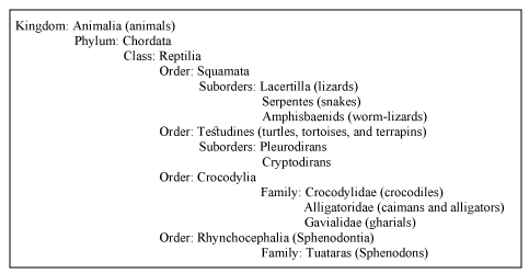
Scheme 1. Taxonomy of reptilia
The Testudines, also referred to as Chelonia, comprise 361 species of turtles, tortoises, and terrapins, and represent about 3% of all reptiles [19]. Like other reptiles, these animals are ectothermic, have scales covering their skin, breathe through lungs, and lay eggs on land. However, unique to the Testudines is the bony or cartilaginous shell developed from their ribs and other parts of their skeleton that encases their bodies, acting as a shield [22]. Testudines are among the oldest reptile groups, more ancient than snakes or crocodilians, their earliest known members dating from the Middle Jurassic, 174.1 to 163.5 million years ago [53]. An important aspect of their reproductive biology is the ability of the females to store viable sperm in their oviducts for long periods of time [54].
The Crocodylia comprise an order of large, predatory, semi‐aquatic reptiles which encompass 26 known species of crocodiles, alligators, caimans, and gharials [19]. They represent roughly 0.3% of all living reptile species [19]. Crocodilians first appeared 95 million years ago in the Late Cretaceous period (145 to 66 million years ago) and are the closest living relatives of birds [55]. Crocodilians have a unique body form that allows the eyes, ears, and nostrils to be above the water surface while the rest of the animal is hidden below [56]. The tail is long and robust, the skin is covered with bony scales, and the back is protected by thick, bony plates, enabling them to withstand harsh, dry conditions and to survive in both land and water [56]. The relatively long snout varies considerably in shape and proportion among families and genera [56].
The Rhynchocephalia or tuataras represent the smallest reptilian order, measuring up to 80 centimeters and weighing about 1 kilogram, and harboring only two living lizard-like (sub)species, S. punctatus punctatus and S. punctatus guntheri (Sphenodontidae) [19]. They arose during the Age of Reptiles in the Mesozoic era, 252 to 66 million years ago [50], and are currenty only found in small, relatively inaccesible, islands off the coast of New Zealand [57]. Although resembling lizards, several unique features of the skull and jaws clearly distinguish tuataras from squamates. Firstly, their dentition is unique among living species, consisting of two rows of teeth in the upper jaw that overlap one row on the lower jaw [58]. Secondly, they possess a third eye on the top of the head with a retina, lens, and nerve endings [58]. This so-called parietal eye is only visible in hatchlings and becomes covered in scales and pigments after four to six months, and is not used for vision but to help judge the time of day or season [59]. Tuataras are the sole surviving members of the Rhynchocephalia which was well represented by many species during the age of the dinosaurs in the Mesozoic, some 200 million years ago [58].
Lizards
Generalities about lizards: The squamate suborder Lacertilia or Sauria, commonly known as lizards, is a widespread group of scaled reptiles consisting of almost 7,000 extant species in 55 families and subfamiles [19]. Well-known lizard species that are extensively addressed further in this section are the Gila monsters in the family Helodermatidae and the monitor lizards in the family Varanidae [19]. The names ‘Lacertilia’ and ‘Sauria’ of this reptilian suborder are derived from the Latin words ‘lacerta’ for ‘lizard’ and ‘ilia’ for ‘similar to’, and the Ancient Greek word ‘saûros’ or ‘saúra’ for ‘lizard’ or ‘reptile’. Among the smallest species of lizard are the dwarf geckoes Sphaerodactylus ariasae and S. parthenopion from the Dominican Republic and the Virgin Islands, respectively, both in the family Sphaerodactylidae and measuring 1.6 to 2.0 centimeters from the snout to the base of the tail [60], as well as the 2- to 3-centimeters long Madagascan dwarf chameleon Brookesia micra (Chamaeleonidae) [61]. The largest lizard is the 3-meters long and approximately 70-kilograms heavy Komodo dragon Varanus komodoensis (Varanidae) found in Indonesia [62] (Figure 4).
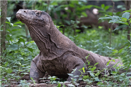
Figure 4. The Komodo dragon Varanus komodoensis (Varanidae) (from: https://images.app.goo.gl/eaLt1Hu9YMs4z2dN6)
A characteristic of lizards (that they share with snakes) is the movable quadrate bone, differentiating them from tuataras which have more rigid diapsid skulls [50]. Most lizards have rounded torsos, elevated heads on short necks, long tails, and four limbs, alternatingly using the right and left ones, resulting in a typical side-to-side locomotion. Some, such as the slowworm Anguis fragilis (Anguidae) are legless, and have long snake-like bodies [63]. Others such as the forest-dwelling flying lizards in the genus Draco (Agamidae) are able to glide over distances as far as sixty meters [64]. Still other species such as geckoes (Gekkota) and chameleons (Chamaeleonidae), can adhere to smooth vertical surfaces including glass and ceilings by using adhesive pads with millions of tiny hair-like structures with the aid of van der Waals forces [65]. And when fleeing from predators, basilisk lizards in the genus Basiliscus (Corytophanidae) can gather sufficient momentum to run across water for a brief distance while holding most of their body out of the water [66].
Lizards are found on all continents, particularly in tropical habitats where they mainly live on the ground, in rocks, on trees, and underground [19]. However, they are highly adaptable and can be encountered in rather extreme environments with the exception of Antarctica and most oceanic island chains [19]. For instance, the red tail toad-headed lizard Phrynocephalus erythrurus (Agamidae) lives on the Qiangtang Plateau in northern Tibet at a height of 4.5 to 5.3 kilometers above sea level [67]. And the marine iguana Amblyrhynchus cristatus (Iguanidae) from the Galápagos Islands is completely adapted to sea where it almost exclusively feeds on algae [68].
All lizards possess senses of sight, touch, olfaction, and hearing like other vertebrates, but depending on the habitat they live in, a particular sense is more prominently developed. For instance, fossorial skinks heavily rely on olfaction and touch [69], while geckoes largely depend on acute vision to hunt and to estimate the distance to their prey before striking [70]. Lizards make use of internal fertilization, and copulation involves the male inserting one of its hemipenes into the female’s cloaca [48]. The majority of species is oviparous, parental care is uncommon and the female usually abandons the eggs after laying them. Various species of racerunners in the family Teiidae reproduce by parthenogenesis, i.e., the production of young from unfertilized eggs [71].
Most lizards are carnivorous, preying on a large variety of animals ranging from insects to larger vertebrates [72]. In their turn, many lizards are preyed on by, among others, hawks, owls and eagles, as well as snakes, weasels, and even larger species of lizards [72]. For these reasons, they have developed a number of effective defensive mechanisms. These involve, among others, trying to outrun a predator or escape into a hole or a crack [73]; inflating their body to resemble an intimidating spiny balloon [74]; playing dead [75]; squirting a stream of foul-tasting blood from a pouch beneath their eyes on the opponent [76]; changing the colors and patterns on their skin to resemble their surroundings [77]; and autotomize their tail to distract the opponent and create an opportunity to flee [78].
In addition, some lizards in the family Helodermatidae (such as the Gila monster H. suspectum as well as the Mexican beaded lizard H. horridum and the Guatemalan beaded lizard H. charlesbogerti) and some monitor lizards in the genus Varanus (such as the Komodo dragon V. komodoensis, the spotted tree monitor V. scalaris, and the lace monitor V. varius) use venom to subdue their prey and defend themselves [79-81]. Unlike snakes which produce venom in their upper jaw [79-81], these lizards produce venom in modified salivary glands in their lower jaw [79-81]. Each gland has a separate duct leading to the base of the teeth, and the venom is delivered to the victim by chewing on the wound caused by biting [79-81]. Furthermore, monitor lizards feed on a broad spectrum of food items including decaying animals [82], and their saliva contains highly septic bacteria which are also delivered to bitten prey and predators [83,84]. However, these animals regularly engage in vicious territorial fights [85], sometimes inflicting serious wounds on each other which do not seem to cause serious harm [86]. This has led to the hypothesis that varanids have a robust innate immune system that protects them against potential sepsis due to bites from other monitor lizards [86].
Bioactive compounds from lizards: Lizards have been used for centuries in the ethnomedical practices of various societies throughout the world. Medieval manuscripts from Azerbaijan mention the use of the Caucasian agama Paralaudakia caucasia (Agamidae) and the common wall gecko Tarentola mauritanica (Phyllodactylidae) for treating, among others, leprosy and sexual impotence [87]. The oil extracted from the fat of spiny-tailed lizards in the genus Uromastyx (Agamidae) also has a long use as a topical cure for impotence and as an aphrodisiac in regions in northern Africa and India [88]. Other parts and products from these lizard species are promoted in Malaysia as a treatment for over twenty diseases including diabetes mellitus, heart disease, hypertension, gout, kidney problems, and sexual dysfunction [89]. In traditional chinese medicine, toad-head agamas in the genus Phrynocephalus (Agamidae) are dried and crushed and also used to treat, among others, erectile dysfunction [90]. In Mozambique, the tails from chameleons are processed into a medicinal preparation against asthma [91]. And in some, communities the meat and blood from various monitor lizards is consumed to promote strength, vitality, stamina, and sexual drive [92].
The venom of helodermatid and varanid lizards contains a large variety of proteins and peptides including helokinestatins, exendins, kallikreins, natriuretic peptides, serotonin, phospholipases A2 (PLA2s), three-finger toxin-like peptides (3FTXs), cysteine-rich secretory proteins (CRiSPS), and hyaluronidases [79-81]. These compounds, both alone and together, are responsible for the tissue breakdown, inflammation, edema, hypothermia, hypotension, peripheral smooth muscle paralysis, promotion of fibrinogen cleavage, inhibition of platelet aggregation, and continuous bleeding following a bite of one of these animals [79-81]. Thus, the venoms of these lizard species represent rich sources of pharmacologically active compounds.
Recognizing this, the venom of the Gila monster H. suspectum has thoroughly been investigated, which resulted in the identification of the 39-amino acid peptide exendin-4 that reduced fasting and postprandial blood glucose in patients with type 2 diabetes mellitus [18]. Exendin-4 appeared to improve β-cell sensitivity to glucose and to act as an agonist of glucagon-like peptide 1 (GLP-1), a hormone from the digestive tract that helps regulate insulin and glucagon secretion [18]. Subsequent efforts led, as mentioned before, to the development of the synthetic exendin-4 analogue exenatide (Byetta®), the first GLP-1 agonist for managing type 2 diabetes mellitus [18]. Similarly, the anticoagulant activities in monitor lizards’ venom and components of the resilient innate immune system of these animals may represent new lead compounds for treating thrombotic disorders and novel antiseptic wound healing-stimulating compounds, respectively.
Anticoagulants and antiplatelet drugs from monitor lizards: Hemostasis is a complex and intricately regulated process that provides the body the ability to rapidly stop bleeding after injury in order to prevent extensive blood loss and infection [93]. To this end, multiple components of the blood clotting system are activated in response to damage to blood vessels [93]. This occurs in a series of events that includes platelet aggregation and blood vessel constriction to stop the bleeding; the formation of a plug consisting of platelets, fibrin, and blood cells to block further blood loss; and the breakdown of the fibrin clot (fibrinolysis) once the wound has healed [93]. In a healthy individual, clotting and fibrinolysis are balanced, i.e., the blood develops clots and breaks them down exactly when needed. A too low clotting rate in the blood results in bleeding disorders such as hemophilias, clotting factor deficiencies, von Willebrand disease, hypercoagulable states, and deep venous thrombosis [94]. On the other hand, increased clotting of the blood can lead to the formation of dangerous clots in the blood vessels and the development of thrombotic disorders such as coronary artery disease, deep vein thrombosis, ischemic stroke, myocardial infarction, pulmonary embolism, and heart failure [95].
The latter conditions can be treated with anticoagulants or antiplatelet drugs [96]. Anticoagulants prolong the clotting time, thereby reducing fibrin formation, while antiplatelet drugs prevent the aggregation of platelets [96]. However, in the end, both classes of drugs prevent clots from forming and increasing in size, reducing the risk of circulation blockage and a heart attack or a stroke [96]. Examples of anticoagulants are vitamin K antagonists such as warfarin, direct thrombin inhibitors such as dabigatran, direct factor Xa inhibitors such as apixaban, and low-molecular weight heparin anticoagulants such as dalteparin [96]. Some commonly used antiplatelet drugs are glycoprotein platelet inhibitors such as eptifibatide (that has been derived from a protein in the venom of the southeastern pygmy rattlesnake Sistrurus miliarius barbouri (Viperidae) [97]), as well as platelet aggregation inhibitors such as aspirin, and protease-activated receptor-1 antagonists like vorapaxar [96].
As mentioned above, varanid lizards’ venom has the capacity to prevent blood from clotting. Converging lines of evidence have indicated that this takes place through at least two mechanisms, namely by cleaving fibrinogen and by blocking platelet aggregation. Support for the former mechanism is provided by the capacity of the venom from various species of Varanus to cleave fibrinogen [98]. However, this process occurs differently from that normally done by thrombin to produce a clot to cover a wound [98]. Using thromboelastography, the Varanus venom was observed to cause the fibrin chains to crosslink erroneously, producing a non-functional clot in a process called destructive, non-clotting cleavage [81,99].
Evidence for antiplatelet activity of varanid lizards’ venom came from the potent blocking effects of the venoms from the lace monitor V. varius and the Komodo dragon V. komodoensis on platelet aggregation and blood clotting [80,100,101]. These activities have been attributed to (type III) PLA2s in the venoms of the animals [102] which have been found to promote bleeding and inhibit platelet aggregation [100,101,103]. PLA2s have been reported to remove, among others, the fatty acid from the second position of the glycerol backbone of phospholipids including arachidonic acid, inhibiting the biosynthesis of thromboxanes by platelets and in this way, the formation of thrombi [104].
Although preliminary, these observations suggest that the coagulotoxic compounds in the venom from varanid lizards may represent potential new lead compounds for designing and developing novel drugs for treating blood clotting disorders. Importantly, although helodermatid lizards’ venom may also have this capacity, varanid lizards’ venom is for various reasons believed to be preferable for this purpose. Firstly, as mentioned above, varanid lizards’ venom probably prevents blood clotting by cleaving fibrinogen ánd blocking platelet aggregation [98-103] while helodermatid lizerds’ venom only exerts antiplatelet activity [105]. Secondly, the venom from varanid lizard species is extremely diverse and complex [49] while that from helodermatid lizard species is probably highly conserved [106], presumably because of the much larger global variation in ecological niches and prey utilized by the former species when compared to the latter [80]. Thus, varanid lizards’ venoms may comprise a richer variety of toxins for stopping blood from clotting when compared to those from helodermatid lizard species, presenting the opportunity to develop a range of new anticoagulants and/or antiplatelet compounds with different potencies.
Wound healing-promoting compounds from monitor lizards: A wound can be described as the loss or the disruption of the cellular, anatomical, and functional continuity of living tissues as a result of trauma [107]. Wounds can be classified as open wounds such as incisions, lacerations, and abrasions, and closed wounds such as crush injuries and various types of hematomas [108]. Wounds can also be distinguished according to the level of microbial contamination, i.e., clean wounds, contaminated wounds, infected wounds, and colonized wounds [109]. Obviously, wounds that are abundantly infected with bacteria can seriously impede the healing process and can result in life-threatening complications [109].
The body deals with a lesion by setting into motion a cascade of events with the aim to repair the injured tissues. This involves a complex and dynamic, but highly regulated cascade of biochemical and cellular events that entails four overlapping phases: hemostasis; inflammation; proliferation; and maturation and remodelling [107]. Hemostasis involves the formation of a fibrin clot by the aggregation of thrombocytes [107]. During inflammation, bacteria and cell debris are removed by white blood cells [107]. In the proliferation phase, the wound begins to close as the wound area is rebuild with new granulation tissue (largely consisting of collagen and extracellular matrix) that is revascularized by infiltrating blood vessels and subsequently covered by epithelial cells [107]. And during maturation and remodelling, newly formed collagen increases tensile strength to the wound area while cellular and angiogenic activities cease [107].
The orderly and timely manifestation of these processes is imperative to restore the anatomic and functional integrity of the injured site [107]. Thus, failure at any stage in the wound healing process may result in the development of chronic wounds, i.e., wounds that do not heal spontaneously within three months [110]. Examples of such wounds are diabetic, vascular, and pressure ulcers, and they represent an increasing burden to patients, their families, health care professionals, and health care systems [111]. This is for an important part due to the rising global incidence of conditions that impede wound healing such as diabetes mellitus, obesity, and vascular disorders [111]. For this reason, chronic wounds are anticipated to become important public health concerns of the near future [111].
There are various options for managing chronic wounds, including several forms of debridement and the use of antiseptics and antibiotics; stimulation of the intrinsic process of wound healing using, among others, growth factors and cytokines; as well as wound support with proper dressings until the wound area has closed [112]. Several lines of evidence suggest that compounds in monitor lizards’ blood plasma may also be useful for managing wounds. This supposition is mainly based on the various above-mentioned studies reporting no obvious harm in monitor lizards which have received bacteria-laden bites from conspecifics [83,84]. This suggests that these animals possess a robust innate immune system, allowing them to effectively deal with the inflammatory phase of the wound healing cascade, preventing the development of chronic wounds.
Indeed, the serum of the Komodo dragon V. komodoensis potently and rapidly inhibited the growth of cultures of the potentially pathogenic bacterial strains Streptococcus epidermitis, Salmonella typhimurium, Providencia stuartii, and Shigella flexneri, and moderately to strongly those of Escherichia coli, Staphylococcus aureus, and Klebsiella oxytoca [86]. These activities could be ascribed, at least partly, to the powerful antimicrobial peptide VK25 in the blood plasma of V. komodoensis [113]. A synthetic analog of VK25 designated DRGN-1 effectively killed cultured drug-resistant strains of Pseudomonas aeruginosa and S. aureus by permeabilizing their plasma membrane [113]. DRGN-1 also substantially promoted tissue regeneration in both uninfected wounds and biofilm wounds in BALB/c mice composed of P. aeruginosa and S. aureus [113], and stimulated the migration of HEKa keratinocytes in a scratch-wound closure assay, possibly through activation of the EGFR/STAT1/3 pathway [113]. Interestingly, among forty-eight novel potential cationic antimicrobial peptides identified in V. komodoensis plasma, seven exhibited activity against P. aeruginosa and S. aureus [114]. Notably, the sequencing, assembly and analysis of the genome of V. komodoensis revealed the presence of a variety of genes with important roles in host-defense and innate immunity, many of which were present in clusters, supporting a robust innate immune system of these animals [115]. These insights and developments raise the hope to identify varanidian antimicrobial peptides that can be used as novel treatments for chronic wounds.
Snakes
Generalities about snakes: The squamate suborder Serpentes or snakes comprises about 3,900 species in 30 families of ectothermic, amniote reptilians with elongated, legless bodies that move creeping on the floor [19]. A few well-known snake families are the Pythonidae (pythons), Boidae (boas), Colubridae (colubrids), Elapidae (elapids), and Viperidae (vipers and pit vipers). The name ‘Serpentes’ of this reptilian suborder comes from the Latin word ‘serpĕre’ meaning ‘to creep’. However, some species retain a pelvic girdle with a pair of vestigial claws on either side of the cloaca [21]. Although belonging to the same order as lizards, snakes lack the eyelids and external ears present in lizards [116]. As mentioned before, snakes also have very flexible skulls because of the presence of several rotational joints and mandibles, enabling them to swallow prey much larger than their head [52]. To accommodate their narrow bodies, snakes’ paired organs (such as kidneys) are positioned in front of each other instead of side by side, and most species have only one functional lung [117].
Snakes are encountered on every continent in almost all temperate to tropical terrestrial and aquatic habitats including deserts, forests (on the ground as well as in trees), oceans, streams, and lakes, but also in unusual locations. For instance, the Himalayan keelback Herpetoreas platyceps (Colubridae), the Himalayan pit viper Gloydius himalayanus (Viperidae), and the Tibetan hot-spring snake Thermophis baileyi (Colubridae) can be found in the Himalaya Mountains in habitats over 3,000 meters elevation [118-120]. There are also completely pelagic snakes such as the venomous yellow-bellied sea snake Hydrophis platurus (Elapidae) [121] and the coral reef snakes in the subfamily Hydrophiinae (Elapidae) which are widespread throughout the Indian and Pacific Oceans [121].
Several lines of evidence suggest that snakes have probably descended from burrowing lizards during the Cretaceous Period, 145 to 66 million years ago [122]. These subterranean ancestors of snakes evolved bodies streamlined for burrowing, eventually became limbless, and developed a brille (the transparent scales covering the eyes) and lost their external ears to cope with dirt and damage to corneas and ears [122]. The smallest known species of snake is the tiny, 10.4 centimeters-long Barbados thread snake Tetracheilostoma carlae (Leptotyphlopidae) [123]. Among the largest species are the reticulated python Malayopython reticulatus (Pythonidae) from South Asia and Southeast Asia, with an average length of 6.5 meters and an average weight of 59 kilograms [124], the Burmese python Python bivittatus (Pythonidae) that can become 5 meters long and weigh 75 kilograms [125], and the South American green anaconda Eunectes murinus (Boidae) that can reach a length of up to 5.21 meters and a weight of 30 to 70 kilograms [126].
Snakes mainly locate prey and predators by smell, using their forked tongues to detect airborne particles and passing them to Jacobson’s organ [46]. Pit vipers, some boas, and pythons can detect warm-blooded prey and predators using their ‘pits’ [44]. Vision is of secondary importance in most snakes, only allowing the animals to track movements [127]. As a general rule, vision is best in arboreal snakes and weakest in burrowing snakes [127]. Furthermore, snakes lack external ears and eardrums but have fully formed inner ear structures which allows them to detect vibrations traveling through the ground [128].
Like lizards, snakes employ internal fertilization for their reproduction, copulation involving the male inserting one of its forked hemipenes into the female’s cloaca [48]. Most species of snake lay eggs which they abandon shortly after laying [129]. Ring-necked spitting cobras in the genus Hemachatus (Elapidae), garter snakes in the genus Thamnophis (Colubridae), rattlesnakes in the genera Crotalus and Sistrurus (Viperidae), and anacondas in the genus Eunectes (Boidae), are ovoviviparous and retain the eggs within their body until they are almost ready to hatch [129]. The common boa Boa constrictor (Boidae) and the green anaconda E. murinus are fully viviparous, nourishing their young through a placenta as well as a yolk sac [130]. A few snake species such as the eastern copperhead Agkistrodon contortrix and the cottonmouth Agkistrodon piscivorus - both North American snake species in the family Viperidae - can reproduce by facultative parthenogenesis [131].
All snakes are carnivorous, eating small animals including lizards, frogs, other snakes, small mammals, birds, eggs, fish, snails, worms, and/or insects [21]. On the other hand, many snakes are prey for, among others, centipedes, scorpions, weasels, ferrets, birds, and other reptiles including other snakes [132]. For these reasons, snakes have developed an elaborate arsenal of characteristic defenses including, among others, defensive stances, hissing, threat displays, strike poses, attacking, and biting (see, for instance, references [133,134]). Although some snakes may bite, only a few families are venomous and they primarily use their venom to kill and subdue prey and initiate digestion rather than for self-defense. Examples of venomous snake families are the Viperidae (among others, rattlesnakes, bushmasters, and pit vipers), Atractaspididae (such as stiletto snakes and snake-eaters), Elapidae (including, among others, Australian copperheads, sea snakes, coral snakes, cobras, kraits, and mambas), and some Colubridae (such as tree snakes, the boomslang Dispholidus typus, keelback snakes, and garter snakes) [19].
Snakes produce their venom in their salivary glands which are located in the back of their head. Venom is delivered by contracting muscles in the head, exercising pressure on the venom glands [135]. More ‘advanced’ venomous snakes such as viperids and elapids inject their venom through their hollow fangs in the upper jaw [79-81,135] while rear-fanged snakes such as the boomslang have a groove on their teeth (also in the upper jaw) to channel venom into the wound [79-81,135]. However, the Mozambique spitting cobra Naja mossambica (Elapidae) spits its venom on an attacker [136]. And keelback snakes as well as garter snakes do not produce their venom themselves: the former sequester toxins from the poisonous toads they eat and secrete them from nuchal glands [137], the latter use the neurotoxin tetrodotoxin from the rough-skinned newt Taricha granulosa (Salamandridae) they prey on [138].
Bioactive compounds from snakes: Snakes are symbols in many religions and myths. In Christianity and Judaism, a serpent appears in the first book of the Bible when it tempts the first couple, Adam and Eve, with the forbidden fruit from the tree of knowledge of good and evil [139]. Snakes also played prominent roles in ancient Egypt as a decoration of the crown of the pharaohs, in the form of the primordial snake god Nehebkau, and in the ritual suicide of Cleopatra [140]. In Greek mythology, Medusa was one of the three Gorgon sisters who had snakes for hair and whose gaze turned those who looked at her to stone [141]. In ancient Greece, the serpent was also seen as the symbol of medicine, presumably because of the association of the periodic renewal of its skin with healing [142]. For this reason, Asclepius, the Greek god of medicine, carried a serpent wound around his rod, a symbol still used today to denote medicine [142]. Two other medical symbols involving snakes are also still used today, namely the Caduceus that stands for medicine in general, and the Bowl of Hygieia that symbolizes pharmacy [142].
Snakes also have a long use in the traditional medical practices of many cultures. In Mexico, rattlesnake pills made of dried ground rattlesnake flesh are indicated for curing a wide variety of ailments, including impurities in the face, cancer, itching, rheumatism, varicose veins, stress, heart disease, diabetes mellitus, hemorrhoids, and sexual impotence [143]. However, ingestion of the pills has been associated with salmonellosis [144]. In South African Zulu culture, traditional healers are attired in python skin because pythons are associated with power [145]. In many other traditional African cultures, the head of a python is believed to reverse spells and bad luck, prevent potential accidents from happening, and help attract a marital partner [146]. In many African societies python fat is topically applied to relieve rheumatic pains [146], and in various Asian countries the blood from live cobras is mixed with an alcoholic beverage and drunk to increase sexual virility [147]. Well-known is the Japanese custom of leaving the habu snake Protobothrops flavoviridis (Viperidae) to steep in rice wine or grain alcohol, turning it into habushu or habu sake [148].
Like those of helodermatid and varanid lizards [79-81], snake venoms consist of complex mixtures of proteins and non-proteins with a broad range of biological and pharmacological activities [149,150]. These include, among others, enzymes such as secreted PLA2s, L-amino acid oxidases (LAAOs), phosphodiesterases, acetylcholinesterases, snake venom metalloproteinases (SVMP), and snake venom serine proteases (SVSP), as well as non-enzymatic compounds such as protease inhibitors, natriuretic peptides, 3FTXs, CRiSPs, Kunitz-type inhibitors, and disintegrins [149,150]. These compounds, either alone or at certain combinations, elicit three broad types of pharmacological effects, namely hemotoxicity, neurotoxicity, and cytotoxicity [150]. For instance, certain PLA2s and 3FTX act on pre- or postsynaptic junctions as antagonist of ion channels and nicotinic or muscarinic receptors, causing paralysis and respiratory failure (see, for instance, reference [151]), while other PLA2s and 3FTXs, along with SVMPs, cause local tissue damage resulting in swelling, blistering, bruising, and necrosis, and systemic effects such as hypovolemic shock (see, for instance, reference [152]).
A myriad of drug development studies have explored the pharmacological activities of the many bioactive compounds in snake venoms [150]. This has resulted in the development of the above-mentiond angiotensin-converting enzyme inhibitor captopril and its derivatives from the venom of the Brazilian viper B. jararaca [17], as well as various other important drugs. Eptifibatide inhibits platelet aggregation, reducing the risk of acute cardiac ischemic events, and has been derived from a disintegrin in the venom of the rattlesnake S. miliarius barbouri [97]. Tirofiban also prevents the blood from clotting and has been developed from the venom of the saw-scaled viper Echis carinatus (Viperidae) [153]. Batroxobin is a thrombin-like serine protease that cleaves fibrinogen, reducing fibrinogen levels, and has been derived from the venom of the Brazilian Viperidae members B. moojeni and B. atrox [154]. And the anti-ageing substance Syn-Ake® that reduces the formation of wrinkles by relaxing the facial muscles, is based on a synthetic tripeptide that has been developed on the basis of the structure of waglerin 1 in the venom of the temple viper Tropidolaemus wagleri (Viperidae) [155].
Antileishmanial compounds from snakes: Leishmaniasis is a group of parasitic diseases caused by kinetoplastid flagellates of the genus Leishmania (Trypanosomatidae). These parasites comprise two subgenera, Leishmania and Viannia [156] which are found in both the Old and the New World, and only in the New World, respectively [156]. The parasites are spread by female sand flies of the genera Phlebotomus in the Old World and Lutzomyia in the New World [157] when they draw a blood meal from an infected host (including a human being) [157]. The Leishmania parasites exist as non-flagellated amastigote forms inside the host’s macrophages and as flagellated promastigotes in the gut of the sandfly [157]. Twenty-one of the thirty known Leishmania species are pathogenic to humans [157], and dependent on the species they cause a broad spectrum of disease forms ranging from cutaneous leishmanisasis and mucocutaneous leishmanisasis to severe viscerotropic forms of leishmaniasis and post kala-azar dermal leishmaniasis [158]. Cutaneous leishmaniasis is in some cases self-limiting but may cause serious mutilation in other cases [158], while visceral leishmaniasis - characterized by invasion of the parasites into bone marrow, liver, and spleen - is invariably lethal if left untreated [158].
The various forms of leishmaniasis are endemic in nearly hundred countries throughout the world [159], particularly in poor populations, disaster zones, and war-torn regions in tropical and subtropical parts of the world [160], producing each year 0.7 to 1 million new cases [159]. Treatment is mainly with systemically given pentavalent antimonals such as sodium stibogluconate and meglumine antimoniate; amphotericin B and its analogues (particularly liposomal amphotericin B); oral miltefosine; and/or paromomycin [158]. However, these drugs cause serious side-effects, evoke resistance, are very costly and beyond the reach of many endemic areas, require prolonged treatment, and/or must be prepared and administered by complicated procedures [158]. These considerations indicate an urgent need to develop novel, more efficacious agents against the various forms of leishmaniasis.
Snake venoms and some of their ingredients displayed substantial antiparasitic activities including activity against leishmaniasis [161]. For instance, the whole crude venoms of the Brazilian viper B. jararaca and the Sonoran lyresnake Trimorphodon biscutatus lambda (Colubridae), as well as those of the cascabel rattlesnake Crotalus durissus terrificus (Viperidae) (Figure 5) and the Marajó and Cerrado lancehead vipers Bothrops marajoensis and B. lutzi inhibited the growth of promastigotes of L. (L.) major [162,163], and L. (L.) amazonensis [164-166], respectively. Both leishmanial species are known to cause cutaneous leishmaniasis [158]. The crude venoms of B. lutzi and B. marajoensis, along with those from the horned desert viper Cerastes cerastes (Viperidae) and the Egyptian cobra Naja haje (Elapidae), also inhibited the growth of promastigotes of members of the L. donovani complex [165-167], the causative agents of visceral leishmaniasis [158].
Figure 5. The cascabel rattlesnake Crotalus durissus terrificus (Viperidae) (from: https://images.app.goo.gl/dP4RjuPpKtpteCrG7)
Various lines of evidence have implicated PLA2s, LAAOs, and CriSPs in the antileishmanial activities of the venoms of these and other snake species. The involvement of snake venom PLA2s in these effects was supported by the substantial inhibitory actions of these compounds on the growth, development, and infective capacity of promastigotes and amastigotes from L. (L.) major [163], L. (L.) amazonensis, [168], L. (L.) infantum [169], and L. (L.) amazonensis [170]. Importantly, the antileishmanial effects have been associated with perturbation of mitochondria, nuclei, and plasma membranes of the parasites [162,168] as well as the release of cytotoxic cytokines from host neutrophils against the parasites [170]. The latter observations were consistent with the cytotoxic effects of a fraction of the venom from the Central Asian cobra Naja naja oxiana (Elapidae) towards L. (L.) infantum promastigotes and amastigotes following stimulation of the immune system [171]. This venom also promoted the production of reactive oxygen species and apoptotic-like mechanisms, and inhibited the activity of arginase, a critical enzyme in the metabolism of Leishmania parasites [171].
Support for antileishmanial activity of LAAOs came from the notable inhibitory effects of LAAO-containing fractions from the above-mentioned snake venoms on the growth of promastigotes of L. (L.) amazonensis, L. (L.) infantum, L. (V.) braziliensis, L. (L.) donovani, and L. (L.) major [165,172-176]. Further fractionation of the LAAO-containing fraction of the C. durissus terrifucus venom yielded the proteins gyroxin, crotamine, and convulxin which also elicited important activity against L. (L.) amazonenesis promastigates, were not cytotoxic towards cultured J774 mouse BALB/c monocyte macrophages, and displayed activity in a BALB/c mouse model of leishmaniasis [164]. The antileishmanial effects of the LAAOs might be attributed to their ability to provoke oxidative stress by forming oxygen radicals and hydrogen peroxide [177,178].
An antileishmanial activity of a snake venom CRiSPs has so far only been reported for the venom of the prairie rattlesnake Crotalus viridis viridis (Viperidae) called crovirin [179]. This compound displayed promising activity against L. (L.) amazonensis promastigotes and amastigotes, and was only cytotoxic to normal mammalian cells including peritoneal macrophages at considerably higher concentrations than those required for its antileishmanial effect [179]. CRiSPs are believed to mainly function as ion channel blockers [179,180], but whether the antileishmanial activity of the C. viridis viridis CRiSP is associated with such a mechanism is so far not known. Nevertherless, together, these data support the potential of snake venoms as sources for new classes of drugs against leishmaniasis.
Antiviral compounds from snakes: Pathogenic viruses have plagued mankind since its existence and first caused epidemics when human beings settled in more densely populated agricultural communities from the Neolithic period on, roughly 12,000 years ago [181]. Under these conditions, viruses were able to rapidly spread and become endemic [181]. Viruses having livestock (or domesticated plants) as hosts also increased in frequency, and some evolved to mutated versions that were able to infect humans [182]. Among the best known examples of old and devastating viruses from zoonotic origin are the smallpox viruses which have caused many massive pandemics throughout human history [183], killing an estimated 300 million people around the world in the twentieth century alone [184]. These viruses probably first emerged in agricultural communities in India about 11,000 years ago and probably descended from the poxviruses of rodents [185].
Other viruses that have caused epidemics following the establishment of large human communities were measles, mumps, rubella, and polio, and they are believed to have emerged at the same time as the smallpox viruses [182]. However, influenza A viruses are responsible for the most shattering epidemics in human history, claiming 40 million to 50 million fatalities in the early twentieth century as the ‘Spanish flu’ [186]. The flu pandemic returned in 1957 as ‘Asian flu’ and then in 1968 as ‘Hong Kong flu’ that killed about 3 million people [186]. Some of the influenza viruses are believed to have crossed the species barrier from ducks and waterfowl to pigs and from there to humans [182].
Relatively recently emerging zoonotic viruses thought to have emerged through one or more animal hosts are the human immunodeficiency virus (HIV), the Ebola viruses, and the severe acute respiratory syndrome coronaviruses (SARS-CoVs) [187]. HIVs are the causative agents of the acquired immunodeficiency syndrome (AIDS), Ebola viruses cause severe and often fatal hemorrhagic fever, and SARS-CoVs cause serious respiratory problems [187]. Despite its virulence and rapid spread, SARS-CoV-1 did not cause the pandemic that was feared [188]. However, SARS-Cov-2 that first emerged in Wuhan, China in November 2019, has rapidly spread around the world and is responsible for the current pandemic with an enormous case-fatality rate in particularly older people and those with pre-existing comorbidities [188].
Today, many previously fatal viral diseases such as measles, mumps, yellow fever, and poliomyelitis can be treated with vaccines [189], while various others such as those causing herpes, AIDS, and hepatitis can be kept in check with antiviral drugs directed at specific stages in the viral life cycle [190]. However, there are still no vaccines or effective therapeutics available for, among others, the dengue virus and the Ebola virus, although these viruses pose massive public health threats to tens of millions of people worldwide (see, for instance, references [191,192]). Moreover, as illustrated by the 2009 influenza pandemic [193] and the present SARS-CoV-2 pandemic [188], more virulent drug-resistant strains of viruses can rapidly develop and spread around the world, necessitating continuous efforts to prevent and contain outbreaks. These considerations indicate an urgent need of more efficacious antiviral treatments.
Various lines of evidence suggest that ingredients in snake venoms may represent lead compounds of novel antiviral drugs [194,195]. Firstly, the use of the venom of the black-necked spitting cobra Naja nigricollis (Elapidae) (Figure 6) from sub-Saharan Africa as well as isolated cytotoxins from the venom led to lysis of human erythrocytes infected with the Sendai virus while leaving normal cells largely unaffected [196]. Furthermore, the crude venom of the cascabel rattlesnake C. durissus terrificus, as well as crotoxin, the main ingredient of the venom, inhibited measles virus, dengue virus, and yellow fever virus infection of cultured Vero African green monkey kidney cells [197,198] while not affecting the viability of normal Vero cultures [197,198]. Crotoxin has been found to consist of two components, crotapin and PLA2 [199], both of which also displayed antiviral effects which added to each other’s effect [197,198]. This was confirmed in subsequent studies using cultured Huh 7.5 human hepatocellular carcinoma cells infected with the JFH-1 hepatitis C virus strain [200]: crotoxin was found to interfere with the entry into as well as the release of virus particles from the host cells, while crotapotin prevented the release of virus particles and PLA2 perturbed the entry and replication of the virus [200].
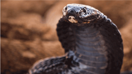
Figure 6. The black-necked spitting cobra Naja nigricollis (Elapidae) (from: https://images.app.goo.gl/vkum2vsDp1MMoXMh7)
The antiviral activities seemed to be associated with interference with the initial steps of the viral life cycle, i.e., the adsorption of virus particles to the host cell’s plasma membrane and their penetration into the cell [197,198]. Further studies showed that the activity of PLA2 in the C. durissus terrificus venom against the dengue virus and other enveloped viruses may involve, in addition, the cleavage of the glycerophospholipids in the viral lipid bilayer envelope, resulting in partial exposure of the viral genomic RNA [201]. Support for the importance of PLA2 in the antiviral effects of C. durissus terrificus venom has been provided by the phospholipase activity and virucidal effects of recombinant PLA2 against the chikungunya virus, the dengue virus, the yellow fever virus, and the Zika virus [202].
Snake venoms and some of their ingredients also elicited anti-HIV activity. Several snake venom PLA2s protected human primary blood leukocytes from infection by primary HIV-1 isolates, presumably by interfering with the process of uncoating of virus particles, thus halting the infection [203]. The PLA2 crotoxin in the venom of C. durissus terrificus showed in vitro activity against HIV that was probably associated with the destabilization of cell-surface heparans which are involved in the attachment of HIV to the plasma membrane of host cells and their entry into the cells [204]. Furthermore, a LAAO isolated from the venom of Stejneger's pit viper Trimeresurus stejnegeri (Viperidae) inhibited HIV-1 infection of C8166 human T cell leukemia in culture and replication of the virus in the cells at concentrations that had little effect on the viability of the host cells [205]. Immunokine®, an oxidized derivative of the α-toxin extracted from the venom of the Indochinese spitting cobra Naja siamensis (Elapidae), inhibited the infection of lymphocytes by HIV and feline immunodeficiency virus (FIV) by blocking the chemokine receptors CCR5 and CXCR4 [206]. Importantly, administration of a preparation with the trade name Samayz® containing various components derived from snake venom in conjunction with antiretroviral therapy to a patient with multidrug-resistant HIV has reportedly led to a decreased viral load and an elevated CD4+ cell count [207], providing additional support for the anti-HIV activity of snake venom ingredients.
Testudines
Generalities about testudines: The order Testudines or Chelonii consists of turtles, tortoises, and terrapins. Like other reptiles, these animals are ectothermic, have scales covering their skin, breathe through lungs, and lay eggs on land. However, unique to the Testudines is the bony shell that encases their body [22, 208]. This characteristic is captured in the names of this reptilian order which are based on the classical Latin word ‘testudo’ for ‘tortoise’ and the ancient Greek word ‘khelone’ for ‘interlocking shields or armor’. Unlike the claws, nails, horns, and beaks of other animals, the shells of testudines contain nerve endings which respond to mechanical stimuli such as pressure and vibration but also to those causing pain [208]. The testudines’ bony shell is not an exoskeleton but a modification of the ribcage with elements of the vertebral column, vertebrae, clavicles, and interclavicles, is not shedded, and grows with the animals [22,208]. The dorsal part of the shell is the carapace that can be tall and rounded, flat, or some shape in between, the ventral part is the plastron that covers most or part of the bottom of the animals [208]. The carapace and the plastron are connected to each other by a bony bridge or a flexible ligament [208]. In many species, the shell is covered with scutes which are regularly replaced by ecdysis [22,208], but in softshell turtles it is covered with leathery or rubbery skin [22,208].
The distinction between turtles, tortoises, and terrapins is not based on taxonomic criteria but on somewhat arbitrary and geographical considerations. The term ‘turtle’ can be used to refer to all species of the order Testudines including tortoises, but not all turtles are tortoises. This is, because tortoises belong to the Testudinidae family that is a group within the larger Testudines order. Furthermore, in the USA, ‘turtle’ refers to testudinians that are aquatic or semi-aquatic and ‘tortoise’ to terrestrial species, while in the United Kingdom freshwater species are called ‘terrapins’ and salt water species are called ‘turtles’. And in Australia, all turtles not residing in the ocean are named ‘tortoises’. In this paper, ‘turtles’ refers to sea turtles that rarely leave the ocean (such as the leatherback sea turtle Dermochelys-coriacea (Dermochelyidae; Figure 7), ‘tortoises’ to turtles that spend most of their time on land (such as the giant tortoises in the genus Chelonoidis (Testudinidae) from the Galápagos Islands), and ‘terrapins’ to turtles that spend time both on land and in brackish, swampy water (such as the North American common snapping turtle Chelydra serpentina (Chelydridae).
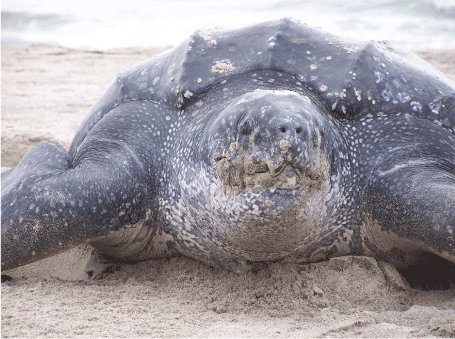
Figure 7. The leatherback sea turtle Dermochelys coriacea (Dermochelyidae) (from: https://images.app.goo.gl/FwwZ4R88LnRnvSXPA)
The Testudines are among the most ancient reptiles alive, with only the tuataras considered more primitive [209]. The largest extant testudian is the leatherback sea turtle D. coriacea that can attain a total length of up to 3 meters and a weight of more than 900 kilograms [210]. It is the fourth-heaviest modern reptile behind three species of crocodilians [210]. Its carapace is covered by leathery skin and oily flesh and it is found in all tropical and subtropical oceans and even within the Arctic Circle [210]. Some of the smallest testudines - an average carapace length of 10 centimeters and a weight of slightly more than 100 grams - are the speckled Cape tortoise Chersobius signatus (Testudinidae) from the western part of South Africa [211], the flattened musk turtle Sternotherus depressus (Kinosternidae) that is endemic to southern USA [212], and the bog turtle Glyptemys muhlenbergii (Emydidae) from eastern USA [213].
The approximately 361 extant species of turtles, tortoises, and terrapins are placed in 14 families and 97 genera [19]. Based on the way they retract their head and neck within their shells, testudines are grouped in two suborders, the Pleurodira and the Cryptodira [214]. The pleurodirans are also called side-necked turtles and fold their long neck sideways to insert their head within the shell. The pleurodirans comprise 3 extant families including the Podocnemididae family of South American river turtles [215]. The cryptodirans or hidden-necked turtles lower their neck and pull their head straight back to conceal it within their shell [216]. They comprise the remaining 11 families and include, among others, the highly migratory sea turtles in the families Dermochelyidae and Cheloniidae [217].
Testudines heavily rely on scent to acquire food, obtain a mate, find appropriate nesting areas, recognize and localize predators, and many of their other daily activities [218]. Some species can see color in the range between near ultraviolet to red, and many probably have exceptional night vision [218]. Most species can hear sound but are less sensitive to high frequencies [218]. Testudines do not have teeth but instead hard, flat surfaces on their jaws that allow them to grip and tear off bits of plants or animals for feeding [218]. Testudines display a wide variety of mating behaviors but do not form pair-bonds or social groups [219]. Most species bury their eggs in soil, sand, or rotting vegetation, but some lay them on the ground in the open. Neither parent incubates the eggs or attend them in any way; instead, the eggs are incubated by environmental heat [219]. The young break free of the egg using an egg tooth or caruncle after 45 to 90 days of development and fend for themselves after hatching [219].
Testudines do not have fangs or venoms to defend themselves. When threatened or under attack, many simply withdraw into their shells. Species with reduced shells that do not offer sufficient protection make use of additional defensive tactics when confronted with danger. For instance, musk turtles such as Sternotherus odoratus and mud turtles such as Kinosternon subrubrum - both in the family Kinosternidae - release a foul musky odor from two scent glands underneath their carapace to deter predators [220]; Bell’s hinge-back tortoise Kinixys belliana (Testudinidae) exudes a strong-smelling viscid material from its cloaca when handled; and the radiated tortoise Astrochelys radiata (Testudinidae) regurgitates its stomach contents when under predator attack [221]. Although most turtles can bite, only a few use biting as a defense. A few notable examples are the common snapping turtle C. serpentine as well as the alligator snapping turtle Macrochelys temminckii (Chelydridae) and the North American softshell turtles in the genus Apalone (Trionychidae) [222].
Bioactive compounds from Testudines: The meat of turtles, tortoises, and terrapins has long been considered a delicacy and is included in exclusive soups and stews in, among others, Chinese, Japanese, African, and Anglo-American cuisines, often along with the skin and the intestines. The species most commonly consumed are the Chinese softshell turtle Pelodiscus sinensis (Trionychidae), the Chinese three-striped box turtle Cuora trifasciata (Geoemydidae), the Chinese big-headed turtle Platysternon megacephalum (Platysternidae), the gopher tortoise G. polyphemus, and the common snapping turtle C. serpentina. Dishes and beverages prepared with parts of testudians are also consumed for their presumed health benefits. In Chinese culture, for instance, turtles are symbols of long life, personal wealth, fertility, strength, and a happy household [223], and turtle ingredients are believed to help maintain youthful beauty in females and improve sexual performance in males [223,224]. Furthermore, in Chinese traditional medicine, the shells and other parts of turtles and tortoises are processed into tablets, powders, and ointments for treating a myriad of conditions ranging from coughs and headache to a difficult childbirth and cancer [225,226].
In western Africa, parts from sea turtles including the leatherback D. coriacea are also believed to have healing properties and are used to treat a variety of disorders including malaria, seizures, fever, anemia, hepatitis, sexual underperformance, rickets in children, as well as conditions caused by evil spirits [227,228]. And in several Latin American and Caribbean countries, parts from turtles and tortoises would treat a myriad of conditons ranging from respiratory complaints and earache to rheumatism, diabetes mellitus, and cancer [229]. Some of these products are even available in pre-packaged form or included into cosmeceuticals. A few examples are the fat from the Amazon river turtle Podocnemis expansa (Podocnemididae) and the green sea turtle C. mydas that are marketed as crema de tortuga (turtle cream) [230] or included into manufactured bathing soaps, lotions, skin care products, anti-wrinkle formulations, and nail creams [231].
These applications are supported by the results from pharmacological studies indicating, among others, the high vitamin E content of the fat from the green sea turtle C. mydas [230] and the presence of abundant amounts of phenolic compounds and fatty acids with meaningful antibacterial and antioxidant activities in the fat from the Amazon river turtle P. expansa [232]. Notably, earlier studies [233] had provided indications for the antiinflammatory, antigargalesthetic (anti-itching and anti-urticarian), and analgesic activities of turtle and tortoise oil, and hinted on their potential usefulness in the prophylaxis and/or treatment of cardiovascular diseases and psoriasis. So far, studies on the pharmacological properties of parts of testudines and their potential medicinal applicability are scant. Hereunder, the potential usefulness of some of these preparations as immunomodulating compounds has been addressed.
Cancer-immunomodulating compounds from testudines: Immunomodulation involves therapeutic interventions aimed at modifying the immune response in such a way that the ratio of the different groups of immune cells are brought back into balance so that the immune system can function correctly [234]. Immunomodulation can involve either immunosuppression or immunostimulation. Immunosuppression is useful to diminish the immune response against transplanted organs in order to prevent rejection [235], and to treat autoimmune diseases such as pemphigus, lupus, or allergies [236, 237]. Immunosuppression can be accomplished by interfering with antigen presentation, T cell activation, or T cell proliferation [234] using, among others, anti-CD 154 monoclonal antibodies, cyclosporine A, rapamycin, or corticosteroids [238].
Immunostimulation is applied to augment the immune response and restore healthy immune function in cases of, among others, infectious diseases, primary or secondary immunodeficiency, and neoplastic disease [234]. Infectious diseases can be prevented by vaccination, the most effective immunomodulatory technique [239]. Primary immune defects are often manageable by allogeneic stem cell transplantation [240], while secondary immunodeficiencies caused by, for instance, malnutrition or HIV, are most effectively managed by treating the cause [241]. And immunomodulating therapies of cancer attempt to improve the immune system’s ability to attack and eliminate cancer cells using, among others, immune checkpoint inhibitors that remove the inhibitory effects of immune checkpoint pathways, thereby (re)activating cancer-targeting cytotoxic T cells; cytokines that regulate immune cell maturation, growth, and responsiveness; agonists that activate pathways for promoting T cell-mediated adaptive immune responses; and adjuvants that help attack cancer cells by stimulating general immune responses [242,243].
Preparations from testudines may also possess immunostimulatory properties. For instance, administration of the blood plasma, a fraction of the spleen extract, or an extract from the blood cells from the Russian tortoise Agrionemys horsfieldii (Testudinidae) - considered one of the most radioresistant animals [244] - to irradiated mice led to restoration of the mice’s bone marrow cell populations and an increased survival rate of the animals [245-248]. Numbers of peripheral blood leukocytes, spleen colonies, and bone marrow mitoses, as well as RNA-synthetic activity of irradiated bone marrow cells were also increased in animals treated with the tortoise preparations [246,247]. Moreover, an aqueous extract of the dried shell from the red-eared slider turtle Trachemys scripta elegans (Emydidae) led to an increase in serum levels of IgG as well as those of various lymphocyte populations in both normal BALB/c mice and mice treated with the immunosuppressive agent cyclophosphamide [249].
The immunomodulating substance in the spleen of the Russian tortoise A. horsfieldii was identified as an oligopeptide with a molecular weight up to 2,000 D that had been designated FTGN [244]. This compound accelerated repopulation of irradiated bone marrow at very low concentrations both in vivo and ex vivo, and a fluorescence-labeled derivative of FTGN selectively bound to CD34+ stem cells [244]. Thus, FTGN may play an important role in the proliferation of CD34+ cells and the repopulation of hematopoietic lineages after irradiation [244]. Interestingly, FTGN also protected the laboratory animals from alopecia [244]. These findings suggest that parts from testudines may contain an immunostimulating activity that may be useful in cancer patients with bone marrow damage as a result of radiotherapy or chemotherapy and in individuals undergoing autologous or allogenic bone marrow transplantation.
Incidentally, orally administered powder from the meat of the Chinese softshell turtle P. sinensis attenuated the increase in diameter and weight of experimental solid tumors in a C3H/He mouse model and prolonged the survival of the animals [250]. The powder did not affect ascites tumors in the mice, suggesting that it did not directly kill tumor cells and may act by activating the immune system [250]. Other studies on the antitumor activity of testudines’ parts reported cytotoxic effects of the extract from the shell of the Greek tortoise Testudo graeca (Testudinidae) and of peptides isolated from enzymatic hydrolysates of proteins from the Chinese three-striped box turtle C. trifasciata to various animal and human carcinoma cell lines [251,252]. Furthermore, peptides from the Chinese softshell turtle P. sinensis altered microRNA expression in the AGS human gastric adenocarcinoma cell line involving, among others, upregulation of several tumor suppressor miRNAs [253].
Together, these observations suggest that testudines-derived preparations may suppress cancerous growth through immunomodulating as well as direct anticancer effects. This supposition needs to be verified but if true, it supports the use of parts from various soft-shelled turtles in traditional Chinese medicine to improve the prognosis of cancer patients undergoing radiotherapy and chemotherapy [254].
Crocodilians
Generalities about crocodilians: The order Crocodylia comprises a total of 26 recognized species [19] which are placed in three families, namely the Crocodylidae (including the true crocodiles in the genus Crocodylus), the Alligatoridae (including the genera Alligator and Caiman), and the Gavialidae (that only consists of the gharial and the false gharial) [19]. The three crocodilian families can be distinguished from one another on the basis of the shape of the snout [56]. Gharials have a long narrow snout with adult males developing a bulbous growth at the tip called a ghara that is used to enhance vocal communication [56]. Alligators and caimans have a broad snout in which the fourth tooth of the lower jaw cannot be seen when the mouth is closed [56]. And Crocodylidae family members have various snout shapes but the fourth tooth of the lower jaw is always visible when the mouth is closed [56].
In addition to their long flattened snouts, crocodilians are characterized by their large, solidly built, lizard-like appearance. Hence the name of this group of reptiles that is derived from the Ancient Greek and Latin words ‘krokódeilos’ and ‘crocodilus’, respectively, both meaning ‘small lizard’. They have, furthermore, thick skin that is covered with non-overlapping scales; laterally compressed tails; eyes, ears, and nostrils at the top of the head, allowing them to stalk their prey with most of their body underwater; conical, peg-like teeth for efficiently seizing and holding prey rather than mastication since they swallow food unchewed; and massive, powerful jaw-closing muscles, enabling the animals to exert an enormous bite force [56]. As in birds, crocodilians have a four-chambered heart and a unidirectional system of air flow around the lungs [56]. All crocodilians are excellent swimmers and can also move well on land, although their limbs are reduced in size [56]. Species living in locations without a shoreline even climb trees to catch sunlight [255].
Crocodilians are mainly found in freshwater, saltwater, and brackish habitats with relatively high temperatures where they spend most of their time in water. The main exceptions are the American alligator Alligator mississippiensis (Alligatoridae) (Figure 8) [256] that inhabits the more temperate south-eastern parts of the, USA and the Chinese alligator Alligator sinensis (Alligatoridae) [257] that is encountered in the relatively moderate areas around the Yangtze River in China. The smallest living crocodilian is Cuvier’s dwarf caiman Paleosuchus palpebrosus (Alligatoridae) that mainly inhabits flooded forests near the major rivers and lakes in the central Amazon basin [258]. An adult typically weighs 6 to 7 kg with males growing to an average length of 1.4 meters and females usually not exceeding 1.2 meters [258]. The largest species of crocodylian in the world is the saltwater crocodile Crocodylus porosus (Crocodylidae) from the saltwater habitats and brackish wetlands throughout the tropical regions of Asia and the Pacific [259]. Females rarely surpass 3 meters, but males grow to a length of 4.6 to 5.2 meters and can weigh as much as 1,200 kilograms [259].
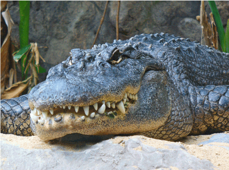
Figure 8. American alligator Alligator mississippiensis (Alligatoridae) (from: https://images.app.goo.gl/AAhg2HhNoVT7uEZZ9)
Figure 9. The Siamese crocodile Crocodylus siamensis (Crocodylidae) (from: https://images.app.goo.gl/55RgKVqBcwvtNA9CA)
Crocodilians are typically solitary, although dominant males usually monopolize reproductive females and patrol and defend territories harboring the females [260]. Copulation occurs in the water [260]. Females lay eggs in holes or in mounds and, unlike most other non-avian reptiles, care for their hatched young [260]. The number of eggs laid in a single clutch ranges from ten to fifty [260]. Crocodilian eggs are protected by hard shells made of calcium carbonate [260]. The incubation period is two to three months [260]. The gender of the hatchlings is determined by the temperature at which the eggs incubate: constant nest temperatures above 32 °C produce more males, while those below 31 °C produce more females [260].
Crocodilians are largely carnivorous, feeding on fish, crustaceans, molluscs, birds, and mammals, although several species have also been observed to consume fruit [261]. Some species like the Indian gharial Gavialis gangeticus (Gavialidae) and the Australian freshwater crocodile Crocodylus johnsoni (Crocodylidae) are specialized in feeding on fish, insects, and crustaceans, while the Chinese alligator A. sinensis and the South American broad-snouted caiman Caiman latirostris (Alligatoridae) specializes in eating hard-shelled molluscs [261]. On the other hand, the saltwater crocodile C. porosus and the American alligator A. mississippiensis have generalized diets and opportunistically feed on invertebrates, fish, amphibians, other reptiles, birds, and mammals [261]. Conversely, crocodilians have a few predators and natural enemies of their own [262]. Several birds of prey such as eagles and many carnivorous mammals such as wild pigs hunt young crocodilians [262]. Big cats such as lions, tigers, jaguars, and leopards sometimes attack, kill, and eat adult caimans, crocodiles, and alligators [262]. Large pythons may prey on crocodiles, territorial hippopotamuses regularly kill large crocodiles, and elephants will not hesitate to attack crocodiles on sight in order to protect their young [262]. Relatively small crocodile species such as the Australian freshwater crocodile C. johnsoni are even victims of larger and more dangerous species like the saltwater crocodile C. porosus [262]. However, the most important defensive mechanism of crocodilians that is believed to be at the basis of their evolutionary success is their robust innate immune system.
Bioactive compounds from crocodilians: Similarly to those from other reptiles [263], parts from crocodilians have a long use in various forms of traditional medicine on different continents. Crocodile oil extracted from the fatty tissues of the animals has been used for centuries in the traditional medical systems of Ancient Egypt, China, and India for treating a wide range of ailments including burns, alopecia, and wounds; chest ailments and respiratory conditions; gastrointestinal obstructions; rheumatism; high blood pressure; cancer, and as a vaginal contraceptive solution [264-266]. Based on centuries-old information from Amazonian Amerindian tribes [267], many Brazilians use the fresh fat from the broad-snouted caiman C. latirostris, the black caiman Melanosuchus niger (Alligatoridae), and Cuvier's dwarf caiman P. palpebrosus to alleviate rheumatism [268-270]. The teeth from these species are also worn as protection against snake bites [271]. Crocodile oil is incorporated into cosmetics and personal care products due to its anti-aging properties observed in human volunteers [272], antimicrobial and antiinflammatory activities in preclinical and clinical studies [264], and wound-healing actions in laboratory rats [265].
More recent insights have revealed a unique asset of crocodilians - although comparable to that mentioned above for monitor lizards [86] - that makes them even more interesting for pharmacological evaluations. This involves the apparent robust and resilient immune system of these animals that enables them to thrive in heavily infested aquatic environments [273], fight off life-threatening infections, and rapidly recover from severe injuries with almost no symptoms of microbial infections [274]. Indeed, even gaping wounds and missing limbs sustained in battles with predators, prey, or conspecifics do not seem to seriously incapacitate crocodilians [274]. Furthermore, being opportunistic feeders, many crocodilians scavenge dead and rotting flesh or other germ-infested foods, aggressively steal it from other predators, or let their food deliberately decompose in order to be better able to tear apart and digest the softened flesh without falling ill [275].
Crocodilians are also capable of withstanding chronic exposure to high concentrations of heavy metals such as arsenic, cadmium, cobalt, chromium, mercury, nickel, lead, and selenium as well as high levels of radiation, and still live for up to a 100 years without developing cancer or other debilitating ailments [276,277]. These observations and those mentioned in the preceding alinea support that the immune system of crocodilians is able to very efficiciently deal with parasitic and microbial infections as well as carcinogenic insults [278,279]. This quality may represent a formidable evolutionary adaptation of these animals that may explain why they were among the few survivors of the catastrophic Cretaceous-Tertiary extinction event [277]. It may also explain why they were able to thrive in the unsanitary, pest-infected habitats they have lived in since their origin [280].
A number of thorough investigations have supported the presence of antiparasitic, antimicrobial, antiviral, and anticancer entities in the serum of crocodilians [281]. These discoveries may pave the way for a new generation of drugs against infectious diseases and cancer, and are more comprehensively addressed hereunder. Of note, naturally-occurring antibiotic compounds (in particular antibiotic peptides) acting in host defense against infection are also present in other organisms including bacteria, archaea, protists, fungi, plants, and other animals [281,282] including monitor lizards [113-115]. These compounds elicited broad-spectrum activity against bacteria, yeasts, fungi, viruses, and cancer cells [281,283]. They display a net positive charge of +2 to +9 due to the presence of positively charged amino acids such as arginine, lysine, and histidine, and contain a high ratio of nonpolar amino acids [283,284]. As a result, they are able to readily bind to the negatively charged bacterial membranes [283,284], after which they insert into the cell membrane through their nonpolar part, and are taken up into the cell [285].
Antimicrobial compounds from crocodilians: As mentioned above, infectious diseases have been a major cause of morbidity and mortality throughout human history [181]. These diseases probably became more common with the establishment of large human settlements and the movement of people from rural to urban areas and the domestication of animals [181]. In addition to the earlier mentioned smallpox and flu pandemics caused by pathogenic viruses [183,186], these developments were accompanied by serious global and regional outbreaks caused by pathogenic bacteria and parasites such as the plague [286], cholera [287], tuberculosis [288], and syphilis [289], as well as malaria [290], leishmaniasis [159,160], and intestinal infections [291]. These ailments were for centuries major causes of death until they were replaced by non-communicable diseases from the second half of the twentieth century on [292].
Despite the progress accomplished in recent decades, morbidity and mortality due to infectious diseases is still very common. According to estimates of the World Health Organization (WHO), there were in 2010 globally 300 to 500 million cases of malaria, 333 million cases of sexually transmitted diseases, 33 million cases of HIV/AIDS, 14 million cases of tuberculosis, and 3 to 5 million cases of cholera [293]. Furthermore, every third death in the world was attributable to an infectious disease, most notably a lower respiratory infection (3.46 million), a diarrheal disease (2.46 million), HIV/AIDS (1.78 million), tuberculosis (1.34), malaria (1.1 million), or measles (900,000) [293]. Thus, despite the broad armamentarium of currently available antiparasitic, antimicrobial, and antiviral compounds [294], these developments indicate a need of novel forms of treatment of infectious diseases.
One way to go forward may involve the exploration of the antimicrobial peptides in the serum of crocodilians. Particularly the serum of the American alligator A. mississippiensis and the Siamese crocodile Crocodylus siamensis (Crocodylidae) have been investigated for the presence of these compounds. In both cases, crude fresh and freeze-dried serum as well as partially purified serum samples were found to be cytotoxic against a broad variety of bacterial strains as well as clinical isolates of several pathogenic bacteria [295-301]. The crude sera also inhibited the growth of various fungal species [296,298] but did not affect the viability of the murine macrophage-like cell line RAW 264.7 [300]. In addition, the A. mississippiensis serum elicited activity against HIV-1, West Nile virus, and Herpes simplex virus type 1 [296,302].
Partial purification of the crude sera led to the identification of several low-molecular-weight, hydrophobic, and cationic peptides with antibacterial activity - including activity against multidrug-resistant strains [301] - that were able to perturb and damage bacterial membranes and penetrate into the cytoplasmic space [295,296,299-301,303,304]. And using microparticles to harvest cationic peptides from biological samples followed by the de novo sequencing of the captured peptides, forty-five potential antimicrobial peptides were identified from the plasma of A. mississippiensis [305]. Eight of these peptides were chemically synthesized and were active against various bacterial strains [305]. Furthermore, both the peptides leucrocin I–IV isolated from C. siamensis white blood cells and the synthetic leucrocin I derivative NY15 inhibited the growth of various bacterial reference strains and clinical isolates, targeted bacterial plasma membranes, and were non-toxic to mammalian cells at bacteriolytic concentrations [306,307]. And a peptide obtained by hydrolysis of C. siamensis hemoglobin exerted bactericidal activity against E. coli, S. aureus, as well as K. pneumoniae and P. aeruginosa via a mechanism that might involve iron dysregulation and oxidative stress [308].
The antibacterial effects of crocodilian antimicrobial peptides may be mediated, at least partly, by antioxidant and antiinflammatory activities [309-312]. These activities have been associated with the hemoglobin fraction, hemoglobin hydrolysates, crude leukocyte extract, and plasma from the animals [309,311,312]. Thus, the immune system of crocodilians seems to contain protective molecules that may serve as lead compounds for a new generation of antibiotics. One of the first medications emerging from this concept was an anti-acne gel containing crude C. siamensis leukocyte extract [313] that displayed considerable in vitro antibacterial activity against Propionibacterium acnes and substantial inhibitory effects on proinflammatory markers and signs of inflammation in infected mouse ears [313].
Anticancer compounds from crocodilians: Cancer refers to a group of over 200 distinct disease forms that can start in almost any organ or tissue of the body, and is characterized by the uncontrolled growth of abnormal cells, their invasion into adjacent tissues, and their spread to distant organs via blood and lymph vessels [314]. The latter process, metastasis, is a major cause of death from cancer [314]. The biological events at the basis of cancer are aberrations in oncogenes, tumor suppressor genes, and/or repair genes [314] which lead to the transformation of normal cells to a precancerous lesion and the subsequent progression to a malignant tumor in a multistage process [314]. These changes are the result of biological or internal factors such as older age, being male or female, and/or inherited genetic defects; environmental and/or occupational exposures to, among others, carcinogenic chemicals, radioactive materials, radon, UV radiation, fine particulate matter, and/or asbestos; as well as lifestyle-related factors such as inappropriate dietary habits, lack of physical activity, and the use of tobacco and/or alcohol [315].
According to estimates of the International Agency for Research on Cancer, there were an estimated 19.3 million new cancer cases and almost 10.0 million cancer deaths in 2020 [316]. The most commonly diagnosed cancers were female breast cancer (11.7%), lung cancer (11.4%), colorectal cancer (10.0%), prostate cancer (7.3%), and stomach cancer (5.6%), and the leading causes of cancer death were lung cancer (18%), colorectal cancer (9.4%), liver cancer (8.3%), stomach cancer (7.7%), and female breast cancer (6.9%) [316]. Notably, cancer was in 2019 the first or second leading cause of death before the age of 70 years in 112 of 183 countries throughout the world and ranked third or fourth in a further twenty-three countries [317]. The therapeutic modalities for treating cancer include surgery, radiation therapy, chemotherapy, immunotherapy, targeted therapy using small-molecule drugs or monoclonal antibodies, and/or hormonal therapy [318]. Nevertheless, most cancers remain fatal, particularly when detected at an advanced stage [314]. This implies a need for more efficacious forms of treatment of neoplastic disease. Many efforts are being dedicated to this goal, including improved early diagnosis, better supportive care, the development of highly specific targeted therapies, and the identification of novel, more efficacious antineoplastic compounds.
As mentioned before, crocodilians can survive up to 100 years without developing cancer while being regularly exposed to well-known cancer-causing compounds [276,279]. Various lines of evidence support that their immune system is able to very efficiciently deal with carcinogenic insults in addition to microbial and parasitic infections. Firstly, extracts of C. siamensis leukocytes [319,320], a fraction of C. siamensis bile called ESC-3 [321], as well as serum and heart lysates, a gall bladder lysate, and the bile of the mugger crocodile C. palustris (Crocodylidae) [322] inhibited the proliferation of various tumor cell lines in culture. The C. siamensis leukocyte extract did not affect the viability of non-cancerous cells [320] but stimulated the overproduction of reactive oxygen species in cancer cells [320] and inhibited the formation of colonies as well as the migration and invasion of these cells [319,320]. The latter observation might be attributed to a decrease in the activities and levels of matrix metalloproteinase-2 and -9 [319] following the disruption of signaling via vascular-endothelial growth factor- and integrin-mediated pathways [319].
Notably, the C. siamensis leukocyte extract and the ESC-3 bile fraction stimulated the expression of pro-apoptotic proteins such as caspase-3, caspase-8, p53, and XIAP, and inhibited that of apoptosis-regulating proteins such as Bcl-2, causing the cancer cells to undergo apoptosis through the mitochondria-dependent pathway [319-321]. Further investigations revealed that the anticancer activities in crocodilian preparations were attributable to the same peptides involved in their antimicrobial activities. For instance, the KT2, RT2, and RP9 peptides derived from the crude C. siamensis leukocyte extract were cytotoxic against various tumor cell lines and killed these cells by apoptosis [323,324] while not affecting the viability of non-cancerous cell lines [324]. The cytotoxic effects might be attributed to interference with intracellular signaling pathways involved in the regulation of the cell cycle [324,325], and the resulting apoptosis might also be associated with a decreased expression of pro-apoptotic genes and an increased expression of tumor suppressor genes [324]. These changes in expression and levels of apoptosis-promoting and apoptosis-countering proteins were also observed in HCT116 human colon cancer xenografts in BALB/c nude mice treated with the RT2 or the KT2 peptide, in addition to a decrease in volume and weight of the xenografts [326, 327].
Parallel cell cuture studies showed that the RT2 and the KT2 peptides interacted with the cancer cell’s plasma membrane, were internalized by endocytosis, and caused nuclear condensation and clear signs of apoptosis, supporting that they exerted their cancer cell killing effects in a comparable manner as their antimicrobial effects [328]. It thus seems that the cancer-fighting activities of crocodilians are associated with the antimicrobial peptides that are part of their innate immune system [276,279,329]. These (positively charged) anticancer peptides presumably exert their cancer cell-killing actions by the same molecular principles as their antimicrobial effects, namely perturbation of the cancer cells’ plasma membranes through electrostatic interactions [329]. This is, because cancer cell membranes generally overexpress anionic molecules, exhibiting a net negative charge [330] that is similar to that of bacterial cells [331]. As a result, the anticancer peptides selectively perturb cancer cell membranes while leaving those of healthy mammalian cells unaffected [330], resulting in either apoptosis through the mitochondrial lytic system, or necrosis via cell membrane lysis [332]. Further research in these crocodilian compounds may open the door to novel modalities for treating cancer.
Similarly to those from many invertebrates [333-335] and amphibians [336], the exploration of bioactive compounds from reptiles may yield structurally novel and mechanistically unique lead compounds for developing breakthrough medicines. This paper has lifted a corner of the veil, reviewing the opportunities for the development of anticoagulants and antiplatelet drugs as well as wound healing-promoting, antileishmanial, antiviral, immunomodulating antimicrobial, and anticancer compounds from various species, genera, or families of reptiles. The ACE inhibitors captopril and its analogues, the GLP-1 agonist exenatide, and the platelet inhibitor eptifibatide are living proof of these opportunities [17,18,97].
So far, only a fraction of the complex venomous mixtures and the resilient immune system from a relative handful of reptiles has been investigated. This suggests that this animal class represents a considerable and so far largely untapped source of novel therapeutics. Indeed, the venoms of helodermatid lizards may yield second generation GLP-1 agonists against type 2 diabetes mellitus [337] and novel treatments for Alzheimer’s disease [338]. The hemotoxic, neurotoxic, and cytotoxic compounds in the venoms of snakes [150] may produce promising lead compounds for treating a variety of human ailments in addition to thrombotic disorders [150,195]. Further exploration of the biology of varanids, testudines, and crocodilians may produce surprising results that open new avenues for further exploration. For instance, the conditioned media of bacteria from the gut of the Amboina box turtle Cuora amboinensis (Geoemydidae) exerted potent and broad antibacterial activity including activity against methicillin-resistant Staphylococcus aureus while not affecting the viability of human cells [339], suggesting another clinical application of a reptile species.
An important condition for these previsions to become reality is meticulous conservation of this class of animals. However, habitat destruction, pollution, climate change (many turtles have environmental sex determination), and introduced invasive species result in enormous biodiversity loss including reptiles [340]. While the growing demand for reptiles throughout the world maintains the unsustainable overexploitation of many reptilian species for food, pets, leather, as well as traditional and ritual, purposes which further contributes to the decline of this class of animals [263,341-343]. In fact, at this moment, the International Union for Conservation of Nature and Natural Resources (IUCN) has classified 332 reptilian species as critically endangered, 588 as endangered, and 538 as vulnerable [344].
For these reasons, the impact on biodiversity loss due to human causes (habitat destruction, pollution, and climate change) must be controlled through proper national legislation, enforced controls, as well as cultural shifts in public attitudes towards the environment. Obviously, procedures against the release of invasive non-native species that can be harmful to reptile populations must also be prohibited. Furthermore, international agreements must be made to restrict the trade in vulnerable and endangered reptile species, and threatened species must be saved from imminent extinction by expanding populations in ex situ captive breeding programs and reintroducing them in the wild following proper habitat protection, changes in cultural attitudes, and strengthened international trade regulations. Hopefully, these steps can avert the alarming decline in reptile populations and the loss of a potentially valuable source of lead compounds for innovative therapeutics.
- Mans DRA (2013) From forest to pharmacy: plant-based traditional medicines as sources for novel therapeutics. Academia J Med Plants 1: 101-110.
- Trescot AM, Datta S, Lee M, Hansen H (2008) Opioid pharmacology. Pain Physician 11: S133-S153. [Crossref]
- Fredholm B, Bättig K, Holmén J, Nehlig A, Zvartau E, et al. (1999) Actions of caffeine in the brain with special reference to factors that contribute to its widespread use. Pharmacol Rev 51: 83-133. [Crossref]
- Reyburn H, Mtove G, Hendriksen I, Von Seidlein L (2009) Oral quinine for the treatment of uncomplicated malaria. BMJ 339: b2066. [Crossref]
- Desborough MJR, Keeling DM (2017) The aspirin story - from willow to wonder drug. Br J Haematol 177: 674-683.
- Van Der Heijden R, Jacobs DI, Snoeijer W, Hallard D, Verpoorte R, et al. (2004) The Catharanthus alkaloids: pharmacognosy and biotechnology. Curr Med Chem 11: 607-628.
- Kingston DG, Newman DJ (2007) Taxoids: cancer-fighting compounds from nature. Curr Opin Drug Discov Devel 10: 130-144.
- Tong WY, Dong X (2009) Microbial biotransformation: recent developments on steroid drugs. Recent Pat Biotechnol 3: 141-153.
- Hadden DR (2005) Goat’s rue - French lilac - Italian fitch - Spanish sainfoin: Gallega officinalis and metformin: the Edinburgh connection. J R Coll Physicians Edinb 35: 258-260.
- Miller EL (2002) The penicillins: a review and update. J Midwifery Womens Health 47: 426-434.
- Shafia S, Chandluri P, Ganpisetti R, Lakshmi BVS, Swami PA, et al. (2016) Erythromycin use as broad spectrum antibiotic. World J Pharm Med Res 2: 23-26.
- Cuddihy G, Wasan EK, Di Y, Wasan KM (2019) The development of oral amphotericin B to treat systemic fungal and parasitic infections: has the myth been finally realized? Pharmaceutics 11: 99. [Crossref]
- Pritchard D (2005) Sourcing a chemical succession for cyclosporin from parasites and human pathogens. Drug Discov Today 10: 688-691.
- Stossel TP (2008) The discovery of statins. Cell 134: 903-905.
- Mir R, Karim S, Kamal MA, Wilson CM, Mirza Z, et al. (2016) Conotoxins: structure, therapeutic potential and pharmacological applications. Curr Pharm Des 22: 582-589.
- Rinehart KL (2000) Antitumor compounds from tunicates. Med Res Rev 20: 1-27.
- Vane JR (1999) The history of inhibitors of angiotensin-converting enzyme. J Physiol Pharmacol 50: 489-498.
- Furman B (2012) The development of Byetta (exenatide) from the venom of the Gila monster as an anti-diabetic agent. Toxicon 59: 464-471.
- Uetz P, Freed P, Hošek J (2020) The reptile database. Downloaded from http://www.reptile-database.org. Accessed February 15, 2021.
- Laurin M, Reisz RR (1995) A reevaluation of early amniote phylogeny. Zool J Linn Soc 113: 165-223.
- Greene H, Cundall D (2000) Limbless tetrapods and snakes with legs. Science 287: 1939-1941.
- Rutland C, Cigler P, Kubale V (2019) Reptilian skin and its special histological structures. In: Rutland CS, Kubale V (eds). Veterinary anatomy and physiology (pp. 135-153). IntechOpen, London, UK.
- Carvalho O, Gonçalves C (2011) Comparative physiology of the respiratory system in the animal kingdom. Open Biol J 4: 35-46.
- Vickaryous MK, McLean KE (2011) Reptile embryology. Methods Mol Biol 770: 439-455.
- Hanken J (1989) Development and evolution in amphibians. Amer Sci 77: 336-343.
- Shine R (2005) Life-history evolution in reptiles. Annu Rev Ecol Evol Syst 36: 23-46.
- Sahney S, Benton MJ, Falcon-Lang HJ (2010) Rainforest collapse triggered Pennsylvanian tetrapod diversification in Euramerica. Geology 38: 1079-1082.
- Erwin D (1994) The Permo-Triassic extinction. Nature 367: 231-235.
- Ezcurra MD, Butler RJ (2018) The rise of the ruling reptiles and ecosystem recovery from the Permo-Triassic mass extinction. Proc R Soc B 285: 20180361.
- Gasc J-P, Gans C (1990) Tests on locomotion of the elongate and limbless lizard Anguis fragilis (Squamata: Anguidae). Copeia 4: 1055-1067.
- Nagy KA, Girard IA, Brown TK (1999) Energetics of free-ranging mammals, reptiles, and birds. Annu Rev Nutr 19: 247-1277.
- McCluskey ES (1982) Temperature regulation in tetrapod vertebrates: ectotherms vs. endotherms. Origins 9: 98-100.
- Wilsterman K, Ballinger MA, Williams CM (2020) A unifying, eco‐physiological framework for animal dormancy. Funct Ecol 00: 1-21.
- Couper P, Hoskin C, Cogger H, Amey A (2018) Saltuarius cornutus. The IUCN Red List of threatened species: e.T178216A83324062.
- Ratsoavina F, Glaw F, Ramanamanjato J-B, Rabibisoa N, Rakotondrazafy NA (2020) Uroplatus phantasticus (amended version of 2011 assessment). The IUCN Red List of Threatened Species: e.T172906A177285160.
- Brodie ED, Janzen FJ (1995) Experimental studies of coral snake mimicry: generalized avoidance of ringed snake patterns by free-ranging avian predators. Funct Ecol 9: 186-190.
- Ruxton G, Allen W, Sherratt T, Speed Ml (2018) Avoiding attack: the evolutionary ecology of crypsis, aposematism, and mimicry, 2nd ed. Oxford University Press. Oxford, UK.
- White FN (1978) Circulation: a comparison of reptiles, mammals, and birds. In: Piiper J (ed). Respiratory function in birds, adult and embryonic. Proceedings in Life Sciences (pp 51-60). Springer, Berlin, Heidelberg, Germany.
- Dantzler WH, Bradshaw SD (2009) Osmotic and ionic regulation in reptiles. In: Evans D (ed). Osmotic and ionic regulation: cells and animals (pp 443-503). CRC Press, Boca Raton (FL), USA.
- Stevens CE (2001) Digestive system of amphibians, reptiles and birds. Wiley, Hoboken (NJ), USA.
- Henderson DM (2003) Effects of stomach stones on the buoyancy and equilibrium of a floating crocodilian: a computational analysis. Can J Zool 81: 1346-1357.
- Wyneken J (2007) Reptilian neurology: anatomy and function. Vet Clin North Am Exot Anim Pract 10: 837-853.
- Brames H (2007) Aspects of light and reptile immunity. Iguana: conservation, natural history, and husbandry of reptiles. Intern Rept Conserv Found 14: 19-23.
- Goris R (2011) Infrared organs of snakes: an integral part of vision. J Herpetol 45: 2-14.
- Schwenk K (1995) Of tongues and noses: chemoreception in lizards and snakes. Trends Ecol Evol 10: 7-12.
- Døving KB, Trotier D (1998) Structure and function of the vomeronasal organ. J Exp Biol 201: 2913-2925.
- Blackburn DG (2018). Reproduction in reptiles. 10.1016/B978-0-12-809633-8.20651-1.]. In: Skinner MK (ed). Encyclopedia of reproduction, 2nd ed (pp 573-578). Academic Press, Cambridge (MA), USA.
- Wyneken J, Mader D (2002) The reproductive system of reptiles - anatomy, physiology and clinical perspectives. Proc Assoc Rept Amphib Vet 2002: 187-189.
- Cole CJ (1975) Evolution of parthenogenetic species of reptiles. pp 340-355. In: Reinboth R (ed). Intersexuality in the animal kingdom (pp 340-355). Springer, Berlin, Heidelberg. Germany.
- Jones, ME, Anderson CL, Hipsley CA, Müller J, Evans SE, et al. (2013) Integration of molecules and new fossils supports a Triassic origin for Lepidosauria (lizards, snakes, and tuatara). BMC Evol Biol 13: 208.
- Simȭes TR, Caldwell MW, Talanda M, Bernardi M, Palci A, et al. (2018) The origin of squamates revealed by a Middle Triassic lizard from the Italian Alps. Nature 557: 706-709.
- Palci A, Caldwell M, Hutchinson M, Konishi T, Lee M (2019) The morphological diversity of the quadrate bone in squamate reptiles as revealed by high‐resolution computed tomography and geometric morphometrics. J Anat 236: 210-227.
- Joyce WG (2015) The origin of turtles: a paleontological perspective. J Exp Zool B Mol Dev Evol 324: 181-193.
- Gist DH, Congdon JD (1998) Oviductal sperm storage as a reproductive tactic of turtles. J Exp Zool 282: 526-534.
- Nesbitt SJ (2011) The early evolution of archosaurs: relationships and the origin of major clades. Bull Am Mus Nat Hist 352: 1-292.
- Grigg G, Gans C (1993) Morphology and physiology of the Crocodylia. In: Fauna of Australia, vol 2A, Amphibia and Reptilia (pp. 326-336). Australian Government Publishing Service, Canberra, Australia.
- Hay JM, Sarre SD, Lambert DM, Allendorf FW, Daugherty CH (2010) Genetic diversity and taxonomy: a reassessment of species designation in tuatara (Sphenodon: Reptilia). Conserv Genet 11: 1063-1081.
- Jones MEH, Cree A (2012) Tuatara. Curr Biol 22: 986-987.
- Tosini G (1997) The pineal complex of reptiles: physiological and behavioral roles. Ethol Ecol Evol 9: 313-333.
- Hedges SB, Thomas R (2001) At the lower size limit in amniote vertebrates: a new diminutive lizard from the West Indies. Caribb J Sci 37: 168-173.
- Glaw F, Köhler J, Townsend TM, Vences M (2012) Rivaling the world’s smallest reptiles: discovery of miniaturized and microendemic new species of leaf chameleons (Brookesia) from northern Madagascar. PLoS One 7: e31314. [Crossref]
- Hocknull SA, Piper PJ, Van den Bergh GD, Due RA, Morwood MJ, et al. (2009) Dragon’s paradise lost: palaeobiogeography, evolution and extinction of the largest-ever terrestrial lizards (Varanidae). PLoS One 4: e7241.
- Gvoždík V, Jandzik D, Lymberakis P, Jablonski D, Moravec J, et al. (2010) Slow worm, Anguis fragilis (Reptilia: Anguidae) as a species complex: genetic structure reveals deep divergences. Mol Phylogenet Evol 55: 460-472.
- Mcguire J, Dudley R (2011) The biology of gliding in flying lizards (genus Draco) and their fossil and extant analogs. Integr Comp Biol 51: 983-990.
- Spinner M, Westhoff G, Gorb SN (2014) Subdigital setae of chameleon feet: friction-enhancing microstructures for a wide range of substrate roughness. Sci Rep 4: 5481.
- Hsieh ST, Lauder GV (2004) Running on water: three-dimensional force generation by basilisk lizards. Proc Nat Acad Sci 101: 16784-16788.
- Jin Y-T, Liu N-F (2010) Phylogeography of Phrynocephalus erythrurus from the Qiangtang Plateau of the Tibetan Plateau. Mol Phylogenet Evol 54: 933-940.
- Laurie A (1990) Population biology of marine iguanas (Amblyrhynchus cristatus). I. Changes in fecundity relates to a population crash. J Anim Ecol 59: 515-528.
- Ashton KG (2005) Life history of a fossorial lizard, Neoseps reynoldsi. J Herpetol 39: 389-395.
- Meiri S (2020) What geckos are - An ecological-biogeographic perspective. Isr J Ecol Evol 66: 1-11.
- Manriquez-Morán N, Cruz M, Méndez-de-la-Cruz F (2005) Reproductive biology of the parthenogenetic lizard, Aspidoscelis cozumela. Herpetologica 61: 435-439.
- Rocha C, Davor V (1998) Reptiles as predators and as preys in a restinga habitat of Southeastern Brazil. Ciência e cultura 50: 364-368.
- Cooper WE Jr., Sherbrooke WC (2010) Initiation of escape behavior by the Texas horned lizard (Phrynosoma cornutum). Herpetologica 66: 23-30.
- Sherbrooke WC (1991) Behavioral (predator-prey) interactions of captive grasshopper mice (Onychomys torridus) and horned lizards (Phrynosoma cornutum and P. modestum). Am Midl Nat 126: 187-195.
- Sherbrooke W, May CJ (2008) Body-flip and immobility behavior in regal horned lizards: a gape-limiting defense selectively displayed toward one of two snake predators. Herpetol Rev 39: 156-162.
- Sherbrooke WC, Middendorf III GA (2001) Blood-squirting variability in horned lizards (Phrynosoma). Copeia 4: 1114-1122.
- Berg P, Berg J, Berg R (2020) Predator-prey interaction between a boomslang, Dispholidus typus, and a flap-necked chameleon, Chamaeleo dilepis. Afr J Ecol 00: 1-6.
- Bateman PW, Fleming PA (2009) To cut a long tail short: a review of lizard caudal autotomy studies carried out over the last 20 years. J Zool 277: 1-14.
- Kwok HF, Ivanyi C, Morris A, Shaw C (2010) Proteomic and genomic studies on lizard venoms in the last decade. Proteom Insights 3: 25-31.
- Fry BG, Wroe S, Teeuwisse W, van Osch, Moreno K, et al. (2009) A central role for venom in predation by Varanus komodoensis (Komodo dragon) and the extinct giant Varanus (Megalania) priscus. Proc Nat Acad Sci USA 106: 8969-8974.
- Koludarov I, Jackson TNW, Op den Brouw B, Dobson J, Dashevsky D, et al. (2017) Enter the dragon: the dynamic and multifunctional evolution of anguimorpha lizard venoms. Toxins 9: 242.
- Shine R, Somaweera R (2019) Last lizard standing: the enigmatic persistence of the Komodo dragon. Glob Ecol Conserv 18: e00624.
- Bull JJ, Jessop TS, Whiteley M (2010) Deathly drool: evolutionary and ecological basis of septic bacteria in Komodo dragon mouths. PLoS One 5: e11097. [Crossref]
- Goldstein EJC, Tyrrell KL, Citron DM, Cox CR, Recchio IM, et al. (2013) Anaerobic and aerobic bacteriology of the saliva and gingiva from 16 captive Komodo dragons (Varanus komodoensis): new implications for the "bacteria as venom" model". J Zoo Wildl Med 44: 262-272.
- Auffenberg W (1981) The behavioral ecology of the Komodo monitor. University Presses of Florida, Gainesville (FL), USA.
- Merchant M, Henry D, Falconi R, Muscher B, Bryja J, et al. (2013) Antibacterial activities of serum from the Komodo dragon (Varanus komodoensis). Microbiol Res 4: e4.
- Alakbarli F, Hajiyeva E (2010) Medieval medical manuscripts from Baku in the memory of the World Programme of Unesco. Biomed Int 1. 37-39.
- Vyas R (1990) Notes on the capture of the spiny-tailed lizard (Uromastyx hardwickii) in Gujarat. Hamadryad 15: 28.
- Ching S, Or OC (2016) The use of spiny-tailed lizards Uromastyx spp. for medicinal purposes in Peninsular Malaysia. TRAFFIC Bull 28: 35-40.
- Lim PHC (2017) Asian herbals and aphrodisiacs used for managing ED. Transl Androl Urol 6: 167-175.
- Williams VL, Moshoeu TJ, Alexander GJ (2016) Reptiles sold as traditional medicine in Xipamanine and Xiquelene Markets (Maputo, Mozambique). S Afr J Sci 112: 7/8.
- Nijman V (2016) Perceptions of Sundanese men towards the consumption of water monitor lizard meat in West Java, Indonesia. Biawak 10: 22-25.
- Palta S, Saroa R, Palta A (2014) Overview of the coagulation system. Indian J Anaesth 58: 515-523.
- Peyvandi F, Garagiola I, Biguzzi E (2016) Advances in the treatment of bleeding disorders. J Thromb Haemost 14: 2095-2106.
- Roth M, Manwani D (2011) Thrombotic disorders. Pediatr Rev 32: 41-43.
- Bala MM, Paszek E, Lesniak W, Wloch-Kopec D, Jasinska K, et al. (2018) Antiplatelet and anticoagulant agents for primary prevention of thrombosis in individuals with antiphospholipid antibodies. Cochrane Database Syst Rev 7: CD012534.
- Lazarovici P, Marcinkiewicz C, Lelkes PI (2019) From snake venom’s disintegrins and C-type lectins to anti-platelet drugs. Toxins 11: 303.
- Wolberg AS (2007) Thrombin generation and fibrin clot structure. Blood Rev 21: 131-142.
- Dobson JS, Zdenek CN, Hay C, Violette A, Fourmy R, et al. (2019) Varanid lizard venoms disrupt the clotting ability of human fibrinogen through destructive cleavage. Toxins 11: 255.
- Fry BG, Vidal N, Norman JA, Vonk FJ, Scheib H, et al. (2006) Early evolution of the venom system in lizards and snakes. Nature 439: 584-588. [Crossref]
- Fry BG, Winter K, Norman JA, Roelants K, Nabuurs RJ, et al. (2010) Functional and structural diversification of the Anguimorpha lizard venom system. Mol Cell Proteomics 9: 2369-2390.
- Wüster W, Ramjan S, Jackson T, Martelli P, Kini R, et al. (2003) LC/MSLC/MS (liquid chromatography, mass spectrometry) analysis of Colubroidea snake venoms: evolutionary and toxinological implications. Rapid implications. Rapid Commun Mass Spectrom 17: 2047-2062.
- Fry BG, Vidal N, Norman JA, Vonk FJ, Scheib H, et al. (2005) Early evolution of the venom system in lizards and snakes. Nature 439: 584-588.
- Burke JE, Dennis EA (2009) Phospholipase A2 structure/function, mechanism, and signaling. J Lipid Res 50: S237-S242.
- Huang TF, Chiang HS (1994) Effect on human platelet aggregation of phospholipase A2 purified from Heloderma horridum (beaded lizard) venom. Biochim Biophys Acta 1211: 61-68.
- Koludarov I, Jackson TNW, Sunagar K, Nouwens A, Hendrikx I, et al. (2014) Fossilized venom: the unusually conserved venom profiles of Heloderma species (beaded lizards and gila monsters). Toxins 6: 3582-3595.
- Velnar T, Bailey T, Smrkolj V (2009) The wound healing process: an overview of the cellular and molecular mechanisms. J Int Med Res 37: 1528-1542.
- Percival NJ (2002) Classification of wounds and their management. Gen Surg 20: 114-117.
- Wolcott R, Cutting K, Dowd S, Percival S (2010) Types of wounds and infections. In: Percival S, Cutting K (eds). Microbiology of wounds (pp 219-232). CRC Press, Boca Raton (FL), USA.
- Hurd T (2013) Understanding the financial benefits of optimising wellbeing in patients living with a wound. Wounds Int 4: 13-17.
- Sen CK, Gayle M, Gordillo GM, Roy S, Kirsner R, et al. (2009) Human skin wounds: a major and snowballing threat to public health and the economy. Wound Repair Regen 17: 763-771.
- Pai DR, Madan SS (2013) Techniques in chronic wound management: review of the literature and recent concepts. J Nov Physiother 3: 134.
- Chung EMC, Dean SN, Propst CN, Bishop BM, Van Hoek ML, et al. (2017) Komodo dragon-inspired synthetic peptide DRGN-1 promotes wound-healing of a mixed-biofilm infected wound. NPJ Biofilms Microbiomes 3: 9.
- Bishop BM, Juba ML, Russo PS, Devine M, Barksdale SM, et al. (2017) Discovery of novel antimicrobial peptides from Varanus komodoensis (Komodo dragon) by large scale analyses and de novo-assisted sequencing using electron transfer dissociation mass spectrometry. J Proteome Res 16: 1470-1482.
- Van Hoek ML, Prickett MD, Settlage RE, Kang L, Michalak P, et al. (2019) The Komodo dragon (Varanus komodoensis) genome and identification of innate immunity genes and clusters. BMC Genomics 20: 684. [Crossref]
- Wiens JJ, Slingluff JL (2001) How lizards turn into snakes: a phylogenetic analysis of body-form evolution in anguid lizards. Evolution 55: 2303-2318.
- Perez D, Sheehy III CM, Lillywhite HB (2019) Variation of organ position in snakes. J Morphol 280: 1798-1807.
- Tillack F (2003) On the distribution and biology of the Himalayan mountain keelback, Amphiesma platyceps (Blyth, 1854) and a case of Amphigonia retardata (Serpentes: Colubridae: Natricinae). Sauria 25: 21-27.
- Hofmann S (2012) Population genetic structure and geographic differentiation in the hot spring snake Thermophis baileyi (Serpentes, Colubridae): indications for glacial refuges in southern-central Tibet. Mol Phylogenet Evol 63: 396-406.
- Chowdhury S, Purkayastha J, Chaudhuri A (2018) Gloydius himalayanus (Himalayan pitviper): geographic distribution notes. Herpetol Rev 49: 505.
- Rasmussen AR, Murphy JC, Ompi M, Gibbons JW, Uetz P, et al. (2011) Marine reptiles. PLoS One 6: e27373.
- Woltering JM (2012) From lizard to snake; behind the evolution of an extreme body plan. Curr Genomics 13: 289-299.
- Hedges SB (2008) At the lower size limit in snakes: two new species of threadsnakes (Squamata: Leptotyphlopidae: Leptotyphlops) from the Lesser Antilles. Zootaxa 1841: 1-30.
- Barker DG, Barker TM, David MA, Schuett GW (2015) A review of the systematics and taxonomy of Pythonidae: an ancient serpent lineage. Zool J Linn Soc 175: 1-19.
- Barker DG, Barten SL, Ehrsam JP, Daddono L (2012) The corrected lengths of two well-known giant pythons and the establishment of a new maximum length record for Burmese pythons, Python bivittatus. Bull Chicago Herp Soc 47: 1-6.
- Rivas JA, Ascanio RE, Muñoz MDC (2008) What is the length of a snake? Contemp Herpetol 2008: 1-3.
- Simões BF, Sampaio FL, Douglas RH, Kodandaramaiah U, Casewell NR, et al. (2016) Visual pigments, ocular filters and the evolution of snake vision. Mol Biol Evol 33: 2483-2495.
- Knight K (2012) Snakes hear through skull vibration. J Exp Biol 215: ii.
- Shine R (2003) Reproductive strategies in snakes. Proc Biol Sci 270: 995-1004.
- Blackburn D, Stewart J (2011) Viviparity and placentation in snakes. In: Sever DM, Aldridge RD (eds). Reproductive biology and phylogeny of snakes (pp. 119-181). CRC Press, Boca Raton (FL), USA.
- Booth W, Smith CF, Eskridge PH, Hoss SK, Mendelson JR, Schuett GW (2012) Facultative parthenogenesis discovered in wild vertebrates. Biol Lett 8: 983-985.
- Martins M (1996) Defensive tactics in lizards and snakes: the potential contribution of the Neotropical fauna. In: Del Claro K. (ed). Anais do XIV Encontro Anual de Etologia, Sociedade Brasileira de Etologia, pp 185-199. Universidade Federal de Uberlandia, Minas Gerais, Brazil.
- Kardong KV, Bels VL (1998) Rattlesnake strike behavior: kinematics. J Experiment Biol 201: 837-850.
- Averill-Murray R (2006) Natural history of the western hog-nosed snake (Heterodon nasicus) with notes on envenomation. Sonoran Herpetol 19: 98-101.
- Jackson K (2003) The evolution of venom‐delivery systems in snakes. Zool J Linn Soc 137: 337-354.
- Vermaak SS, Visser A, le Roux TLB (2010) A deadly bed partner: m'Fesi (Mozambique spitting cobra). SA Orthop J 9: 58-62.
- Hutchinson DA, Savitzky AH, Mori A, Burghardt GM, Meinwald J, et al. (2012) Chemical investigations of defensive steroid sequestration by the Asian snake Rhabdophis tigrinus. Chemoecology 22: 199-206.
- Williams BL, Brodie ED Jr, Brodie ED 3rd (2004) A resistant predator and its toxic prey: persistence of newt toxin leads to poisonous (not venomous) snakes. J Chem Ecol 30: 1901-1919.
- Downey ME (2011) The original choice: the prohibition of the tree of the knowledge of good and evil. Word in the World 4: 15-25.
- Retief FP, Cilliers L (2006) Snake and staff symbolism in healing. Acta Theol 26: 189-199.
- Dexter MR (2010) The ferocious and the erotic. “Beautiful” Medusa and the neolithic bird and snake. J Fem Stud Relig 26: 25-41.
- Wilcox RA, Whitham EM (2003) The symbol of modern medicine: why one snake is more than two. Ann Intern Med 138: 673-677.
- Rubio M (1998) Rattlesnake. Portrait of a predator. Smithsonian Inst Press, Washington, DC, USA.
- Waterman S, Juarez G, Carr S, Kilman L (1990) Salmonella arizona infection in Latinos associated with a rattlesnake folk medicine. Am J Public Health 80: 286-289. [Crossref]
- Simelane T, Kerley G (1997) Recognition of reptiles by Xhosa and Zulu communities in South Africa, with notes on traditional beliefs and uses. Afr J Herpetol 46: 49-53.
- D'Cruze N, Assou D, Coulthard E, Norrey JD, Megson D, et al. (2020) Snake oil and pangolin scales: insights into wild animal use at “Marché des Fétiches” traditional medicine market, Togo. Nat Conserv 39: 45-71.
- Chye PLM (2006) Traditional Asian folklore medicines in sexual health. Indian J Urol 22: 241-245.
- Somaweera R, Somaweera N (2010) Serpents in jars: the snake wine industry in Vietnam. J Threat Taxa 2: 1251-1260.
- Munawar A, Ali SA, Akrem A, Betzel C (2018) Snake venom peptides: tools of biodiscovery. Toxins 10: 474.
- Ferraz CR, Arrahman A, Xie C, Casewell NR, Lewis RJ, et al. (2019) Multifunctional toxins in snake venoms and therapeutic implications: from pain to hemorrhage and necrosis. Front Eco. Evol 7: 218.
- Casewell NR, Wuster W, Vonk FJ, Harrison RA, Fry BG, et al. (2013) Complex cocktails: the evolutionary novelty of venoms. Trends Ecol Evol 28: 219-229.
- Gutierrez JM, Rucavado A, Escalante T, Diaz C (2005) Hemorrhage induced by snake venom metalloproteinases: biochemical and biophysical mechanisms involved in microvessel damage. Toxicon 45: 997-1011.
- Zhao L, Jian Y, Li T, Wang H, Lei Z, et al. (2020) The safety and efficiency of tirofiban in acute ischemic stroke patients treated with mechanical thrombectomy: a multicenter retrospective cohort study. Biochem Res Int 2020: 5656173.
- Vu TT, Stafford AR, Leslie BA, Kim PY, Fredenburgh JC, et al. (2013) Batroxobin binds fibrin with higher affinity and promotes clot expansion to a greater extent than thrombin. J Biol Chem 288: 16862-16871.
- Thakre AD (2018) Snake venom peptides use in anti-ageing products. Int J Sci Res 7: 53-58.
- Lainson R, Shaw JJ (1987) Evolution, classification and geographical distribution. In: eters W, Killick-Kendrick R (eds). The leishmaniasis in biology and medicine (vol 1), pp 1-120. Academic Press, London, UK.
- Bañuls AL, Hide M, Prugnolle F (2007) Leishmania and the leishmaniases: a parasite genetic update and advances in taxonomy, epidemiology and pathogenicity in humans. Adv Parasitol 64: 1-109.
- Mans DRA, Kent AD, Hu RVPF, Schallig HDFH (2017) Epidemiological, biological, and clinical aspects of leishmaniasis with special emphasis on busi yasi in Suriname. J Clin Exp Dermatol Res 8: 2.
- Burza S, Croft SL, Boelaert M (2018) Leishmaniasis. Lancet 392: 951-970.
- Alvar J, Yactayo S, Bern C (2006) Leishmaniasis and poverty. Trends Parasitol 22: 552-557.
- Adade CM, Souto-Padrón T (2015) Venoms as sources of novel anti-parasitic agents. In: Gopalakrishnakone P (ed). Toxins and drug discovery. Toxinology, pp 1-31. Springer, Dordrecht, The Netherlands.
- Gonçalves AR, Soares MJ, de Souza W, DaMatta RA, Alves EW (2002) Ultrastructural alterations and growth inhibition of Trypanosoma cruzi and Leishmania major induced by Bothrops jararaca venom. Parasitol Res 88: 598-602.
- Peichoto ME, Tavares FL, Dekrey G, Mackessy SP (2011) A comparative study of the effects of venoms from five rear-fanged snake species on the growth of Leishmania major: identification of a protein with inhibitory activity against the parasite. Toxicon 58: 28-34.
- Passero LFD, Tomokane TY, Corbett CEP, Laurenti MD, Toyama MH, et al. (2007) Comparative studies of the antileishmanial activity of three Crotalus durissus ssp. venoms. Parasitol Res 101: 1365-1171.
- Costa Torres AF, Dantas RT, Toyama MH, Diz Filho E, Zara FJ, et al. (2010) Antibacterial and antiparasitic effects of Bothrops marajoensis venom and its fractions: phospholipase A2 and L-amino acid oxidase. Toxicon 55: 795-804.
- De Menezes RR, Torres AF, da Silva TS, de Sousa DF, Lima DB, et al. (2012) Antibacterial and antiparasitic effects of Bothropoides lutzi venom. Nat Prod Commun 7: 71-74. [Crossref]
- Fernandez-Gomez R, Zerrouk H, Sebti F, Loyens M, Benslimane A, et al. (1994) Growth inhibition of Trypanosoma cruzi and Leishmania donovani infantum by different snake venoms: preliminary identification of proteins from Cerastes cerastes venom which interacts with the parasites. Toxicon 32: 875-882.
- Nunes DC, Figueira MM, Lopes DS, De Souza DL, Izidoro LF, et al. (2013) BnSP-7 toxin, a basic phospholipase A2 from Bothrops pauloensis snake venom, interferes with proliferation, ultrastructure and infectivity of Leishmania (Leishmania) amazonensis. Parasitology 140: 844-854.
- Grabner AN, Alfonso J, Kayano AM, Moreira-Dill LS, Dos Santos APA, et al. (2017) BmajPLA2-II, a basic Lys49-phospholipase A2 homologue from Bothrops marajoensis snake venom with parasiticidal potential. Int J Biol Macromol 102: 571-581.
- De Moura AA, Kayano AM, Oliveira GA, Setúbal SS, Ribeiro JG, et al. (2014) Purification and biochemical characterization of three myotoxins from Bothrops mattogrossensis snake venom with toxicity against Leishmania and tumor cells. Biomed Res Int 2014: 195356.
- Nikpour S, Tabatabaie F, Sharifi I, Mostafavi M, Oliaee RT, et al. (2020) The fraction of the snake venom, its leishmanicidal effect, and the stimulation of an anti-leishmania response in infected macrophages. Endocr Metab Immune Disord Drug Targets 20: 1.
- Toyama MH, Toyama DO, Passero LF, Laurenti MD, Corbett CE, et al. (2006) Isolation of a new L-amino acid oxidase from Crotalus durissus cascavella venom. Toxicon 47: 47-57.
- Ciscotto P, Machado de Avila RA, Coelho EA, Oliveira J, Diniz CG, et al. (2009) Antigenic, microbicidal and antiparasitic properties of an L-amino acid oxidase isolated from Bothrops jararaca snake venom. Toxicon 53: 330-341.
- Paiva R d MA, de Freitas Figueiredo R, Antonucci GA, Paiva HH, de Lourdes Pires Bianchi M, et al. (2011) Cell cycle arrest evidence, parasiticidal and bactericidal properties induced by L-amino acid oxidase from Bothrops atrox snake venom. Biochimie 93: 941-947.
- Bregge-Silva C, Nonato MC, de Albuquerque S, Ho PL, Junqueira de Azevedo IL, et al. (2012) Isolation and biochemical, functional and structural characterization of a novel L-amino acid oxidase from Lachesis muta snake venom. Toxicon 60: 1263-1276.
- Carone SEI, Costa TR, Burin SM, Cintra ACO, Zoccal KF, et al. (2017) A new L-amino acid oxidase from Bothrops jararacussu snake venom: isolation, partial characterization, and assessment of pro-apoptotic and antiprotozoal activities. Int J Biol Macromol 103: 25-35.
- Zuliani JP, Kayano AM, Zaqueo KD, Neto AC, Sampaio SV, et al. (2009) Snake venom L-amino acid oxidases: some consideration about their functional characterization. Protein Pept Lett 16: 908-912.
- Samel M, Tõnismägi K, Rönnholm G, Vija H, Siigur J, et al. (2008) L-amino acid oxidase from Naja naja oxiana venom. Comp Biochem Physiol B Biochem Mol Biol 149: 572-580.
- Adade CM, Carvalho AL, Tomaz MA, Costa TF, Godinho JL, et al. (2014) Crovirin, a snake venom cysteine-rich secretory protein (CRISP) with promising activity against Trypanosomes and Leishmania. PLoS Negl Trop Dis 8: e3252.
- Tadokoro T, Modahl CM, Maenaka K, Aoki-Shioi N (2020) Cysteine-rich secretory proteins (CRISPs) from venomous snakes: an overview of the functional diversity in a large and underappreciated superfamily. Toxins (Basel) 12: 175.
- Krause RM (1992) The origin of plagues: old and new. Science 257: 1073-1078. [Crossref]
- Venkatesan G, Balamurugan V, Gandhale PN, Singh RK, Bhanuprakash V (2010) Viral zoonosis: a comprehensive review. Asian J Anim Vet Adv 5: 77-92.
- Simmons BJ, Falto-Aizpurua LA, Griffith RD, Nouri K (2015) Smallpox: 12 000 years from plagues to eradication: a dermatologic ailment shaping the face of society. JAMA Dermatol 151: 482. [Crossref]
- Ilic M, Ilic I (2017) The last major outbreak of smallpox (Yugoslavia, 1972): the importance of historical reminders. Travel Med Infect Dis 17: 69-70.
- Hughes AL, Irausquin S, Friedman R (2010) The evolutionary biology of poxviruses. Infect Genet Evol 10: 50-59.
- Wever PC, van Bergen L (2014) Death from 1918 pandemic influenza during the First World War: a perspective from personal and anecdotal evidence. Influenza Other Respir Viruses 8: 538-546.
- Parvez M, K, Parveen S (2017) Evolution and emergence of pathogenic viruses: past, present, and future. Intervirology 60: 1-7.
- Yang Y, Peng F, Wang R, Yange M, Guan K, et al. (2020) The deadly coronaviruses: the 2003 SARS pandemic and the 2020 novel coronavirus epidemic in China. J Autoimmun 109: 102434.
- Taylor MW (2014) Vaccines against viral infections. In: Viruses and man: a history of interactions, pp 355-377. Springer, Cham, Switzerland.
- Clercq E, Li G (2016) Approved antiviral drugs over the past 50 years. Clin Microbiol Rev 29: 695-747.
- Noble CG, Chen YL, Dong H, Gu F, Lim SP, et al. (2010) Strategies for development of Dengue virus inhibitors. Antiviral Res 85: 450-462.
- De La Guardia C, Lleonart R (2014) Progress in the identification of dengue virus entry/fusion inhibitors. Biomed Res Int 2014: 825039.
- Broor S, Chahar HS, Kaushik S (2009) Diagnosis of influenza viruses with special reference to novel H1N1 2009 influenza virus. Indian J Microbiol 49: 301-307.
- Da Mata ÉC, Mourão CB, Rangel M, Schwartz EF (2017) Antiviral activity of animal venom peptides and related compounds. J Venom Anim Toxins Incl Trop Dis 23: 3.
- Mohamed Abd El-Aziz T, Garcia Soares A, Stockand JD (2019) Snake venoms in drug discovery: valuable therapeutic tools for life Saving. Toxins (Basel) 11: 564.
- Borkow G, Ovadia M (1999) Selective lysis of virus-infected cells by cobra snake cytotoxins: a sendai virus, human erythrocytes, and cytotoxin model. Biochem Biophys Res Commun 264: 63-68.
- Petricevich VL, Mendonca RZ (2003) Inhibitory potential of Crotalus durissus terrificus venom on measles virus growth. Toxicon 42: 143-153.
- Muller VD, Russo RR, Cintra AC, Sartim MA, Alves-Paiva Rde M, et al. (2012) Crotoxin and phospholipases A2 from Crotalus durissus terrificus showed antiviral activity against dengue and yellow fever viruses. Toxicon 59: 507-515.
- Choumet V, Bouchier C, Délot E, Faure G, Saliou B, et al. (1996) Structure and function relationship of crotoxin, a heterodimeric neurotoxic phospholipase A2 from the venom of a South-American rattlesnake. Adv Exp Med Biol 391: 197-202.
- Shimizu JF, Pereira CM, Bittar C, Batista MN, Campos GRF, et al. (2017) Multiple effects of toxins isolated from Crotalus durissus terrificus on the hepatitis C virus life cycle. PLoS One 12: e0187857. [Crossref]
- Muller VD, Soares RO, dos Santos NN Jr, Trabuco AC, Cintra AC, et al. (2014) Phospholipase A2 isolated from the venom of Crotalus durissus terrificus inactivates dengue virus and other enveloped viruses by disrupting the viral envelope. PLoS One 9: e112351. [Crossref]
- Russo RR, Dos Santos Júnior NN, Cintra ACO, Figueiredo LTM, Sampaio SV, et al. (2019) Expression, purification and virucidal activity of two recombinant isoforms of phospholipase A2 from Crotalus durissus terrificus venom. Arch Virol 164: 1159-1171.
- Fenard D, Lambeau G, Valentin E, Lefebvre JC, Lazdunski M, et al. (1999) Secreted phospholipases A2, a new class of HIV inhibitors that block virus entry into host cells. J Clin Invest 104: 611-618.
- Villarrubia VG, Costa LA, Díez RA (2004) Fosfolipasas A2 segregadas (sPLA2): ¿amigas o enemigas? ¿Actores de la resistencia antibacteriana y antivirus de la inmunodeficiencia humana? Med Clin (Barc) 123: 749-757.
- Zhang YJ, Wang JH, Lee WH, Wang Q, Liu H, et al. (2003) Molecular characterization of Trimeresurus stejnegeri venom L-amino acid oxidase with potential anti-HIV activity. Biochem Biophys Res Commun 309: 598-604.
- Shivaji PG (2007) Therapeutic alternatives from venoms and toxins. Indian J Pharmacol 39: 260-264.
- Alrajhi AA, Almohaizeie A (2008) Snake venom preparation for drug-resistant human immunodeficiency virus. Ann Saudi Med 28: 292-293.
- Cordero GA (2017) The turtle’s shell. Curr Biol 27: R168-R169.
- Herrera-Flores JA, Stubbs TL, Benton MJ (2017) Macroevolutionary patterns in Rhynchocephalia: is the tuatara (Sphenodon punctatus) a living fossil? Palaeontology 60: 319-328.
- Stewart K, Johnson C, GodfreyMH (2007) The minimum size of leatherbacks at reproductive maturity, with a review of sizes for nesting females from the Indian, Atlantic and Pacific Ocean basins. Herpetol J 17: 123-128.
- Daniels SR, Hofmeyr MD, Henen BT, Baard EHW (2010) Systematics and phylogeography of a threatened tortoise, the speckled padloper. Anim Conserv 13: 237-246.
- Dodd CK Jr, Enge KM, Stuart JN (1988) Aspects of the biology of the flattened musk turtle, Sternotherus depressus, in northern Alabama. Bull Fla Mus Nat Hist 34: 1-64.
- Bailey KA, Guyer C (1998) Demography and population status of the flattened musk turtle, Sternotherus depressus, in the Black Warrior River basin of Alabama. Chelonian Conserv Biol 3: 77-83.
- Anquetin J, Tong H, Claude J (2017) A Jurassic stem pleurodire sheds light on the functional origin of neck retraction in turtles. Sci Rep 7: 42376. [Crossref]
- Ferreira G, Bronzati M, Langer M, Sterli J (2018) Phylogeny, biogeography and diversification patterns of side-necked turtles (Testudines: Pleurodira). R Soc Open Sci 5: 171773.
- Wise TB, Stayton CT (2017) Side-necked versus hidden-necked: a comparison of shell morphology between pleurodiran and cryptodiran turtles. Herpetologica 73: 18-29.
- Anthony EJ, Brunier G, Gardel A, Hiwat M (2019) Chenier morphodynamics on the Amazon-influenced coast of Suriname, South America: implications for beach ecosystem services. Front Earth Sci 7: 35.
- Lautenschlager S, Ferreira GS, Werneburg I (2018) Sensory evolution and ecology of early turtles revealed by digital endocranial reconstructions. Front Ecol Evol 6: 7.
- Pearse DE (2001) Turtle mating systems: behavior, sperm storage, and genetic paternity. J Hered 92: 206-211.
- Meshaka W, Gibbons J, Hughes D, Klemens M, Iverson J, et al. (2017) Kinosternon subrubrum (Bonnaterre 1789) - Eastern Mud Turtle. Chelonian Res Monogr 5: 1-16.
- Auffenberg W (1977) Display behavior in tortoises. Amer Zool 17: 241-250.
- Herrel A, O’Reilly JC, Richmond AM (2002) Evolution of bite performance in turtles. J Evol Biol 15: 1083-1094.
- Lee DS, Liao SK (2014) As effective as a mother’s kiss - Turtles in traditional Chinese medicine. Radiata 23: 4-29.
- Zhang H, Wu M-Y, Guo D-J, Wan C-W, Lau C-C, et al. (2013) Gui-ling-gao (turtle jelly), a traditional Chinese functional food, exerts anti-inflammatory effects by inhibiting iNOS and pro-inflammatory cytokine expressions in splenocytes isolated from BALB/c mice. J Funct Foods 5: 625-632.
- Chen T-H, Chang H-C, Lue K-Y (2009) Unregulated trade in turtle shells for Chinese traditional medicine in east and southeast Asia: the case of Taiwan. Chelonian Conserv Biol 8: 11-18.
- Li L, Cheung H-Y (2012) Turtle shell extract as a functional food and its component-based comparison among different species. Hong Kong Pharm J 19: 33-37.
- Fretey J, Segniagbeto G, Soumah M (2007) Presence of sea turtles in traditional pharmacopoeia and beliefs of West Africa. Marine Turtle Newsletter 116: 23-25.
- Martins S, Rocha F, Rodrigues E, Lopes SA, Abella E, et al. (2015). The use of sea turtles in traditional medicine in the Cape Verde Archipelago, West Africa. African Sea Turtle Newsletter 4: 12-15.
- Alves RRN, Alves HN (2011) The faunal drugstore: animal-based remedies used in traditional medicines in Latin America. J Ethnobiol Ethnomedicine 7: 9.
- Uppala L (2015) A review on active ingredients from marine sources used in cosmetics. SOJ Pharm Pharm Sci 2: 1-3.
- Alves RRN, Rosa IL (2010) Trade of animals used in Brazilian traditional medicine: trends and implications for conservation. Hum Ecol 38: 691-704.
- Brazil MV da S, Souza M de B, Ferraz VP, Martins BA, De Carvalho Neto MF, et al. (2020) Antibacterial, total phenols, antioxidant, and fatty acids of the lyophilized body fat of Podocnemis expansa (Schweigger, 1812) from farm in Acre State, Brazil. J Med Plants Res 14: 458-467.
- Tavares Martins S, Rocha F, Rodrigues E, Lopes S, Nuno L, et al. (2015) The use of sea turtles in traditional medicine in the Cape Verde Archipelago, West Africa. African Sea Turtle Newsletter 4: 12-15.
- Gea-Banacloche JC (2006) Immunomodulation. In: Runge MS, Patterson C (eds) Principles of molecular medicine, pp 893-904. Humana Press, Totowa (NJ), USA.
- Mohammad C, Abdeta D, Hirpa E, Melesie G (2015) Review on the influence of immune system on organ transplantation and its therapeutic strategy. Afr J Basic Appl Sci 7: 247-255.
- Yeh SW, Ahmed B, Sami N, Ahmed AR (2003) Blistering disorders: diagnosis and treatment. Dermatologic Therapy 16: 214-223.
- Maidhof W, Hilas O (2012) Lupus: an overview of the disease and management options. Pharm Therapeut 37: 240-249.
- Samuel S, Nguyen T, Choi HA (2017) Pharmacologic characteristics of corticosteroids. J Neurocrit Care 10: 53-59.
- Benn CS, Netea MG, Selin LK, Aaby P (2013) A small jab - a big effect: nonspecific immunomodulation by vaccines. Trends Immunol 34: 431-439.
- Castagnoli R, Delmonte OM, Calzoni E, Notarangelo LD (2019) Hematopoietic stem cell transplantation in primary immunodeficiency diseases: current status and future perspectives. Front Pediatr 7: 295. [Crossref]
- Bucciol G, Meyts I (2020) Recent advances in primary immunodeficiency: from molecular diagnosis to treatment. F1000Res 9: F1000 Faculty Rev-194.
- Carter S, Thurston DE (2018) Immuno-oncology agents for cancer therapy. Pharmaceut J 300: 7913.
- Zhang H, Chen J (2018) Current status and future directions of cancer immunotherapy. J Cancer 9: 1773-1781.
- Turdiev A, Filiutovich O, Mirkin F, Byk G (2019) A peptide from Testudo horsfieldii tortoise spleen as a potential helper for reducing acute radiation syndrome. J Pept Sci 25: e3202. [Crossref]
- Turdyev AA, Usmanov RB, Madzhidova DKh, Nigmatov Z (1985) Postradiation recovery of hematopoiesis in mice administered cell-free tissue extracts from the central Asiatic turtle. Radiobiologiia 25: 665-669.
- Turdyev AA, Usmanov RB, Iukel'son LIa, Nigmatov Z, Madzhidova DKh, et al. (1986) Therapeutic effect of purified components of a turtle spleen extract. Radiobiologiia 26: 685-688.
- Prus EK, Turdyev AA, Trifonov IuA, Voloshin SV, Usmanov RB (1994) The radiotherapeutic effect of the CM-5 fraction of turtle spleen extract. Radiat Biol Radioecol 34: 138-142.
- Turdyev AA, Aleksandrov VV, Usmanov RB, Batyrbekov AA, Ivanov VI, et al. (1998) Hemo- and immunostimulating effect of an extract from blood cells of the Central Asian tortoise. Radiat Biol Radioecol 38: 207-214.
- Xu JH, Li AX, Zhu XQ, Feng LQ, Xie MQ (2007) The effect of turtle (Trachemys scripta elegans) shell extract in normal and cyclophosphamide-treated mice. Food Agric Immunol 18: 83-93.
- Feng H, Yamazaki M, Matsuki N, Saito H (1996) Anti-tumor effects of orally administered soft-shelled turtle powder in mice. Biol Pharm Bull 19: 367-368.
- Bayazit V (2004) Cytotoxic effects of some animal and vegetable extracts and some chemicals on liver and colon carcinoma and myosarcoma. Saudi Med J 25:156-163.
- He S, Mao X, Zhang T, Guo X, Ge Y, et al. (2016) Separation and nanoencapsulation of antitumor peptides from Chinese three-striped box turtle (Cuora trifasciata). J Microencapsul 33: 344-354.
- Wu YC, Liu X, Wang JL, Chen XL, Lei L, et al. (2018) Soft-shelled turtle peptide modulates microRNA profile in human gastric cancer AGS cells. Oncol Lett 15: 3109-3120.
- Fu JJ, Tan SL, Li YG, Lv H, Zhu WF, et al. (2015) Adjuvant effects of snapping turtle co-peptide (STCP) on radiotherapy for cancer. J Jiangxi Uni Trad Chin Med 27: 68-71.
- Dinets V, Britton A, Shirley M (2013) Climbing behaviour in extant crocodilians. Herpetol Notes 7: 3-7.
- Elsey R, Woodward A, Balaguera-Reina SA (2018) Alligator mississippiensis. IUCN Red List of Threatened Species: e.T46583A3009637.
- Jiang H, Wu X (2018) Alligator sinensis. IUCN Red List of Threatened Species: e.T867A3146005.
- Campos Z, Sanaiotti T, Magnusson WE (2010) Maximum size of dwarf caiman, Paleosuchus palpebrosus (Cuvier, 1807), in the Amazon and habitats surrounding the Pantanal, Brazil. Amphibia-Reptilia 31: 439-442.
- Britton ARC, Whitaker R, Whitaker N (2012) Here be a dragon: exceptional size in saltwater crocodile (Crocodylus porosus) from the Philippines. Herpetol Rev 43: 541-546.
- Hale MD, Cloy-McCoy JA, Doheny BM, Parrott BB (2018) Reproductive biology of crocodilians. In: Skinner MK (ed.). Encyclopedia of reproduction (vol 6), pp. 646-653. Academic Press, Oxford, UK.
- Adame MF, Jardine TD, Fry B, Valdez D, Lindner G, et al. (2018) Estuarine crocodiles in a tropical coastal floodplain obtain nutrition from terrestrial prey. PLoS One 13: e0197159. [Crossref]
- Somaweera R, Brien M, Shine R (2013). The role of predation in shaping crocodilian natural history. Herpetol Monogr 27: 23-51.
- Alves RRN, Da Silva Vieira WL, Santana GG (2008) Reptiles used in traditional folk medicine: conservation implications. Biodivers Conserv 17: 2037-2049.
- Buthelezi S, Southway C, Govinden U, Bodenstein J, Du Toit K, et al. (2012) An investigation of the antimicrobial and anti-inflammatory activities of crocodile oil. J Ethnopharmacol 143: 325-330.
- Li HL, Deng YT, Zhang ZR, Fu QR, Zheng YH, et al. (2016) Evaluation of effectiveness in a novel wound healing ointment-crocodile oil burn ointment. Afr J Tradit Complement Altern Med 14: 62-72.
- Higgins V, Bryant M, Howell A, Battersby J (2017) Ordering adoption: materiality, knowledge and farmer engagement with precision agriculture technologies. J Rural Stud 55: 193-202.
- Costa-Neto EM (2005) Animal-based medicines: biological prospection and the sustainable use of zootherapeutic resources. An Acad Bras Cienc 77: 33-43.
- Alves RRN, Rosa IL (2006) From cnidarians to mammals: the use of animals as remedies in Wshing communities in NE Brazil. J Ethnopharmacol 107: 259-276.
- Alves RRN, Rosa IL (2007) Zootherapeutic practices among Wshing communities in north and northeast Brazil: a comparison. J Ethnopharmacol 111: 82-103.
- Alves RRN, Rosa IL (2007) Zootherapy goes to town: the use of animal-based remedies in urban areas of NE and N Brazil. J Ethnopharmacol 113: 541-555.
- Alves RRN, Pereira-Filho GA (2007) Commercialization and use of snakes on north and northeastern Brazil: implications for conservation and management. Biodivers Conserv 16: 969-985.
- Venter T (2012) Characterisation, toxicology and clinical effects of crocodile oil in skin products. PhD thesis. School of Pharmacy, North-West University (Potchefstroom Campus), Potchefstroom, South Africa.
- Wan QH, Pan SK, Hu L, Zhu Y, Xu PW, et al. (2013) Genome analysis and signature discovery for diving and sensory properties of the endangered Chinese alligator. Cell Res 23: 1091-1105.
- Preecharram S, Daduang S, Bunyatratchata W, Araki T, Thammasirirak S, et al. (2008) Antibacterial activity from Siamese crocodile (Crocodylus siamensis) serum. Afr J Biotechnol 7: 3121-3128.
- Gallagher A, Papastamatiou Y, Barnett A (2018). Apex predatory sharks and crocodiles simultaneously scavenge a whale carcass. J Ethol 36, 205-209.
- Siddiqui R, Mansur Ali S, Khan N (2016) Do crocodiles and alligators hold the key to treat cancer? BMJ 354: i3763. [Crossref]
- Khan NA, Soopramanien M, Siddiqui R (2019) Crocodiles and alligators: physicians' answer to cancer? Curr Oncol 26: 186.
- Lehner AF, Rumbeiha W, Shlosberg A, Stuart K, Johnson M, et al. (2013) Diagnostic analysis of veterinary dried blood spots for toxic heavy metals exposure. J Anal Toxicol 37: 406-422.
- Jeyamogan S, Khan NA, Siddiqui R (2017) Animals living in polluted environments are a potential source of anti-tumor molecule(s). Cancer Chemother Pharmacol 80: 919-924.
- Lee S, Siddiqui R, Khan NA (2012) Animals living in polluted environments are potential source of antimicrobials against infectious agents. Pathog Glob Health 106: 218-223.
- Van Hoek ML (2014) Antimicrobial peptides in reptiles. Pharmaceuticals 7: 723-753.
- Wang G, Li X, Wang Z (2016) APD3: the antimicrobial peptide database as a tool for research and education. Nucleic Acids Res 44: D1087-D1093.
- Zhang LJ, Gallo RL (2016) Antimicrobial peptides. Curr Biol 26: R14-R19.
- Ren SX, Shen J, Cheng AS, Lu L, Chan RL, et al. (2013) FK-16 derived from the anticancer peptide LL-37 induces caspase-independent apoptosis and autophagic cell death in colon cancer cells. PLoS One 8: e63641.
- Bahar AA, Ren D (2013) Antimicrobial peptides. Pharmaceuticals (Basel) 6: 1543-1575.
- Rollins SE, Rollins SM, Ryan ET (2003) Yersinia pestis and the plague. Am J Clin Pathol 119: S78-S85.
- Harris JB, LaRocque RC, Qadri F, Ryan ET, Calderwood SB (2012) Cholera. Lancet 379: 2466-2476.
- Pai M, Behr M, Dowdy D, Dheda K, Divangahi M, et al. (2016). Tuberculosis. Nat Rev Dis 2: 16076.
- Peeling RW, Mabey D, Kamb ML, Chen XS, Radolf JD, et al. (2017) Syphilis. Nat Rev Dis Primers 3: 17073.
- Kondrashin AV, Morozova LF, Stepanova EV, Turbabina NA, Maksimova MS, et al. (2018) On the epidemiology of Plasmodium vivax malaria: past and present with special reference to the former USSR. Malar J 17: 346.
- Alum A, Rubino JR, Ijaz MK (2010) The global war against intestinal parasites - should we use a holistic approach? Int J Infect Dis 14: e732-e738.
- Hunter DJ, Reddy KS (2013) Noncommunicable diseases. N Engl J Med 369: 1336-1343.
- World Health Organization (2010) Fact sheets on infectious diseases. Downloaded from: http://www.who.int/mediacentre/factsheets/en/. Accessed March 2, 2021.
- Gould K (2016) Antibiotics: from prehistory to the present day. J Antimicrob Chemother 71: 572-575.
- Merchant ME, Roche C, Elsey RM, Prudhomme J (2003) Antibacterial properties of serum from the American alligator (Alligator mississippiensis). Comp Biochem Physiol B Biochem Mol Biol 136: 505-513.
- Merchant ME, Leger N, Jerkins E, Mills K, Pallansch MB, et al. (2006) Broad spectrum antimicrobial activity of leukocyte extracts from the American alligator (Alligator mississippiensis). Vet Immunol Immunopathol 110: 221-228.
- Preecharram S, Daduang S, Bunyatratchata W, Araki T, Thammasirirak S (2008) Antibacterial activity from Siamese crocodile (Crocodylus siamensis) serum. Afr J Biotechnol 7: 3121-3128.
- Leelawongtawon R, Siruntawineti J, Chaeychomsri W, Sattaponpan C (2010) Antibacterial and antifungal activities from Siamese crocodile blood. J Med Assoc Thai 93: S58-S64.
- Preecharram S, Jearranaiprepame P, Daduang S, Temsiripong Y, Somdee T, et al. (2010) Isolation and characterisation of crocosin, an antibacterial compound from crocodile (Crocodylus siamensis) plasma. Anim Sci J 81: 393-401.
- Kommanee J, Preecharram S, Daduang S, Temsiripong Y, Dhiravisit A, et al. (2012) Antibacterial activity of plasma from crocodile (Crocodylus siamensis) against pathogenic bacteria. Ann Clin Microbiol Antimicrob 11: 22.
- Barksdale SM, Hrifko EJ, van Hoek ML (2017) Cathelicidin antimicrobial peptide from Alligator mississippiensis has antibacterial activity against multi-drug resistant Acinetobacter baumanii and Klebsiella pneumoniae. Dev Comp Immunol 70: 135-144.
- Merchant ME, Pallansch M, Paulman RL, Wells JB, Nalca A, et al. (2005) Antiviral activity of serum from the American alligator (Alligator mississippiensis). Antiviral Res 66: 35-38.
- Merchant ME, Roche CM, Thibodeaux D, Elsey RM (2005) Identification of alternative pathway serum complement activity in the blood of the American alligator (Alligator mississippiensis). Comp Biochem Physiol B Biochem Mol Biol 141: 281-288.
- Barksdale SM, Hrifko EJ, Chung EM, van Hoek ML (2016) Peptides from American alligator plasma are antimicrobial against multi-drug resistant bacterial pathogens including Acinetobacter baumannii. BMC Microbiol 16: 189.
- Bishop BM, Juba ML, Devine MC, Barksdale SM, Rodriguez CA, et al. (2015) Bioprospecting the American alligator (Alligator mississippiensis) host defense peptidome. PLoS One 10: e0117394. [Crossref]
- Pata S, Yaraksa N, Daduang S, Temsiripong Y, Svasti J, et al. (2011) Characterization of the novel antibacterial peptide Leucrocin from crocodile (Crocodylus siamensis) white blood cell extracts. Dev Comp Immunol 35: 545-553.
- Yaraksa N, Anunthawan T, Theansungnoen T, Daduang S, Araki T, et al. (2014) Design and synthesis of cationic antibacterial peptide based on Leucrocin I sequence, antibacterial peptide from crocodile (Crocodylus siamensis) white blood cell extracts. J Antibiot (Tokyo). 67: 205-212.
- Lueangsakulthai J, Jangpromma N, Temsiripong T, McKendrick JE, Khunkitti W, et al. (2017) A novel antibacterial peptide derived from Crocodylus siamensis haemoglobin hydrolysate induces membrane permeabilization causing iron dysregulation, oxidative stress and bacterial death. J Appl Microbiol 123: 819-831.
- Phosri S, Mahakunakorn P, Lueangsakulthai J, Jangpromma N, Swatsitang P, et al. (2014) An investigation of antioxidant and anti-inflammatory activities from blood components of crocodile (Crocodylus siamensis). Protein J 33: 484-492.
- Theansungnoen T, Yaraksa N, Daduang S, Dhiravisit A, Thammasirirak S, et al. (2014) Purification and characterization of antioxidant peptides from leukocyte extract of Crocodylus siamensis. Protein J 33: 24-31.
- Phosri S, Jangpromma N, Patramanon R, Kongyingyoes B, Mahakunakorn P, et al. (2017) Protective effect of crocodile hemoglobin and whole blood against hydrogen peroxide-induced oxidative damage in human lung fibroblasts (MRC-5) and inflammation in mice. Inflammation 40: 205-220.
- Lueangsakulthai J, Phosri S, Theansungnoen T, Jangpromma N, Temsiripong T, et al. (2018) Novel antioxidant and anti-inflammatory peptides from the Siamese crocodile (Crocodylus siamensis) hemoglobin hydrolysate. Biotechnol Appl Biochem 65: 455-466.
- Phupiewkham W, Lu Q, Payoungkiattikun W, Temsiripong T, Jangpromma N, et al. (2018) Development and characterization of an anti-acne gel containing Siamese Crocodile (Crocodylus siamensis) leukocyte extract. J Microbiol Biotechnol 28: 707-717.
- Idikio HA (2011) Human cancer classification: A systems biology-based model integrating morphology, cancer stem cells, proteomics, and genomics. J Cancer 2: 107-115. [Crossref]
- Yadav M, Chatterjee P, Tolani S, Kulkarni J, Mulye M, et al. (2018) A Nexus model of cellular transition in cancer. Biol Res 51: 23.
- Sung H, Ferlay J, Siegel RL, Laversanne M, Soerjomataram I, et al. (2021) Cancer statistics 2020: GLOBOCAN estimates of incidence and mortality worldwide for 36 cancers in 185 countries. CA Cancer J Clin 0: 1-41.
- World Health Organization (2020) Global health estimates 2020: deaths by cause, age, sex, by country and by region, 2000‐2019. WHO; 2020. Downloaded from: who.int/data/gho/data/themes/mortality‐and‐global‐health‐estimates/ghe‐leading‐causes‐of‐death. Accessed March 2, 2021.
- Zaigham Abbas and Sakina Rehman (2018). An overview of cancer treatment modalities. In: Shahzad HN (ed). Neoplasm, pp. 139-157. IntechOpen, London, UK.
- Patathananone S, Thammasirirak S, Daduang J, Chung J, Temsiripong Y, et al. (2016) Inhibition of HeLa cells metastasis by bioactive compounds in crocodile (Crocodylus siamensis) white blood cells extract. Environ Toxicol 31: 1329-1336.
- Phosri S, Jangpromma N, Chang LC, Tan GT, Wongwiwatthananukit S, et al. (2018) Siamese crocodile white blood cell extract inhibits cell proliferation and promotes autophagy in multiple cancer cell lines. J Microbiol Biotechnol 28: 1007-1021.
- Song W, Shen DY, Kang JH, Li SS, Zhan HW, et al. (2012) Apoptosis of human cholangiocarcinoma cells induced by ESC-3 from Crocodylus siamensis bile. World J Gastroenterol 18: 704-711.
- Siddiqui R, Jeyamogan S, Ali SM, Abbas F, Sagathevan KA, et al. (2017) Crocodiles and alligators: antiamoebic and antitumour compounds of crocodiles. Exp Parasitol 183: 194-200.
- Theansungnoen T, Maijaroen S, Jangpromma N, Yaraksa N, Daduang S, et al. (2016) Cationic antimicrobial peptides derived from Crocodylus siamensis leukocyte extract, revealing anticancer activity and apoptotic induction on human cervical cancer cells. Protein J 35: 202-211.
- Maijaroen S, Jangpromma N, Daduang J, Klaynongsruang S (2018) KT2 and RT2 modified antimicrobial peptides derived from Crocodylus siamensis Leucrocin I show activity against human colon cancer HCT-116 cells. Environ Toxicol Pharmacol 62: 164-176.
- Xu F, Na L, Li Y, Chen L (2020) Roles of the PI3K/AKT/mTOR signalling pathways in neurodegenerative diseases and tumours. Cell Biosci 10: 54.
- Maraming P, Maijaroen S, Klaynongsruang S, Boonsiri P, Daduang S, et al. (2018) Antitumor ability of KT2 peptide derived from leukocyte peptide of crocodile against human HCT116 colon cancer xenografts. In Vivo 32: 1137-1144.
- Maraming P, Klaynongsruang S, Boonsiri P, Maijaroen S, Daduang S, et al. (2018) Antitumor activity of RT2 peptide derived from crocodile leukocyte peptide on human colon cancer xenografts in nude mice. Environ Toxicol 33: 972-977.
- Maraming P, Klaynongsruang S, Boonsiri P, Peng SF, Daduang S, et al. (2019) The cationic cell-penetrating KT2 peptide promotes cell membrane defects and apoptosis with autophagy inhibition in human HCT 116 colon cancer cells. J Cell Physiol 234: 22116-22129.
- Gaspar D, Veiga AS, Castanho MA (2013) From antimicrobial to anticancer peptides. a review. Front. Microbiol 4: 294.
- Hoskin DW, Ramamoorthy A (2008) Studies on anticancer activities of antimicrobial peptides. Biochim Biophys Acta 1778: 357-375.
- Mader JS, Hoskin DW (2006) Cationic antimicrobial peptides as novel cytotoxic agents for cancer treatment. Expert Opin Investig Drugs 15: 933-946.
- Riedl S, Zweytick D, Lohner K (2011) Membrane-active host defense peptides - challenges and perspectives for the development of novel anticancer drugs. Chem Phys Lipids 164: 766-781.
- Mans DRA (2016) Exploring the global animal biodiversity in the search for new drugs - marine invertebrates. J Transl Sci 2: 170-179.
- Mans DRA, Shellice S, Ganga D, Kartopawiro J (2016) Exploring the global animal biodiversity in the search for new drugs - insects. J Transl Sci 3: 371-386.
- Mans DRA (2017) Exploring the global animal biodiversity in the search for new drugs - chelicerates, myriapods, and crustaceans. J Transl Sci 3: 1-18.
- Mans DRA, Pawirodihardjo J, Djotaroeno M, Friperson P (2020) Exploring the global animal biodiversity in the search for new drugs. Amphibians. J Transl Sci 7: 17-17.
- Good DJ (2012) Extending the reach of Exendin-4: new pathways in the control of body weight and glucose homeostasis. Endocrinology 153: 2051-2053.
- Hölscher C (2010) The role of GLP-1 in neuronal activity and neurodegeneration. Vitam Horm 84: 331-354.
- Akbar N, Khan NA, Sagathevan K, Iqbal M, Tawab A, et al. (2019) Gut bacteria of Cuora amboinensis (turtle) produce broad-spectrum antibacterial molecules. Sci Rep 9: 17012. [Crossref]
- Gibbons JW, Scott DE, Ryan TJ, Buhlmann KA, Tuberville TD, et al. (2000) The global decline of reptiles, deja’ vu amphibians. BioScience 50: 653-666.
- Alves R, Neto NAL, Santana GG, Vieira WLS, Almeida WO, et al. (2009) Reptiles used for medicinal and magic religious purposes in Brazil. Appl Herpetol 6: 257-274.
- Valencia-Aguilar A, Cortés-Gómez AM, Ruiz-Agudelo CA (2013) Ecosystem services provided by amphibians and reptiles in Neotropical ecosystems. Int J Biodivers Sci Ecosyst Serv Manag 9: 257-272.
- Auliya M, Altherr S, Ariano-Sánchez D, Baard E, Brown C, et al. (2016) Trade in live reptiles, its impact on wild populations, and the role of the European market. Biol Conserv 204. 103-119.
- IUCN 2021. The IUCN Red List of threatened species. Version 2021-1. Downloaded from https://www.iucnredlist.org. Accessed March 16, 2021.