Abstract
Pelvic organ prolapse (POP) is a gynecological condition that occurs when the pelvic viscera protrude into the vagina due to loss of normal support. The lifetime risk for at least one POP operation is 11%-19%. Caucasian women account for 67.6% of prolapse surgeries and have 4-5 times the risk of symptomatic prolapse in comparison to African American women. Differences in connective tissue, which vary by race, are a risk factor for developing POP. The extra-cellular matrix (ECM) proteins fibulin-3, fibulin-5 and HOXA11 have been implicated as being important for pelvic organ support in human and mouse models. This study is designed to determine the vaginal epithelium expression profiles for fibulin-3, fibulin-5, and HOXA11 from women of different ethnicities with POP and correlate these findings with their risk for developing POP.
Methods: Transcriptional analysis of vaginal epithelial tissue (VET) was performed using mRNA extracted from VET of African American, Caucasian, and Hispanic women. Quantitative real time (qRT-PCR) was employed to analyze vaginal epithelium expression of fibulin-3, fibulin-5, HOXA11 and matrix metalloproteinases-2, -9. Western blot analysis was performed on VET from African American and Caucasian women with and without POP. Immunofluorescent (IFA) staining was performed on VET specimens for filbulin-3, fibulin-5, and HOXA11 on one African American, Hispanic and Caucasian woman matched for age, parity and degree of prolapse.
Results: Vaginal epithelial explanted tissue (VET) was collected from 43 patients undergoing surgery for POP and 9 patients with benign non-POP disorders. Patient demographics in our study reflected high numbers of Caucasian women with POP that are younger at the time of surgery with greater disease severity. Caucasian women with POP (25) had the second highest average severity of POP disease (3.0) with (17/25) clinically presenting with grade 3/4 disease and the youngest average age at surgery of 49.0 years with the lowest average parity 2.2. African American and Hispanic women with POP had higher levels of expression and matrix organization of fibulin-5 in the vaginal epithelium compared to Caucasian women with POP. Western blot analysis revealed higher levels of fibulin-5 total protein expressed in vaginal epithelium of African American women when compared to Caucasian women with POP and without POP. Analysis of fibulin-3 total protein revealed lower levels of fibulin-3 in VET tissue from African American women without POP compared to controls and generally higher levels of fibulin-3 in Caucasian women with POP compared to controls. By immunofluorescent staining, Caucasian women with POP had the highest level of fibulin-3 expression however fibulin-3 matrix expression patterns were disorganized compared to controls without POP. Transcriptional expression levels of HOXA11observed in vaginal epithelium from the African woman and Hispanic woman were higher and showed a higher level of matrix organization than Caucasian women. We also observed transcriptional up regulation of MMP2 in African women with POP compared to African women without POP.
Keywords
extracellular matrix proteins, fibulins, immunohistochemistry, pelvic organ prolapse, racial disparity, tissue microarrays, vaginal epithelial cells
Conclusion
Investigation into the underlying causes of POP regarding changes in vaginal epithelium ECM proteins will allow us to better understand the mechanisms involved in this elusive medical condition that would provide treatment alternatives to surgery.
Introduction
Pelvic organ prolapsed (POP) is a condition where the pelvic viscera protrude into the vagina. POP is associated with a significantly poorer quality of life and symptoms such as difficulty defecating, lower abdominal pain, dyspareunia, and difficulty voiding the bladder [1,2]. POP affects 3.3 million American women resulting in $96.9 million for ambulatory care visits and over $1 billion in surgical care annually [3-6]. The lifetime risk of having one operation for POP is 11%-19% [7,8]. Overall, 2.9% of women report experiencing symptomatic POP [9]. The number of American women with POP will increase 46% from 3.3 to 4.9million between 2010 and 2050 and there will be an estimated 45% increase in pelvic floor disorders from 2008-2030 [5,10]. During 2008-2030 the population will only increase by 22% pointing to the effects of an aging population on the incidence of POP [10].
Prolapse has been associated with disorders of proteins of the extracellular matrix (ECM) including Cutis Laxa, Marfan syndrome and Ehlers-Danlos suggesting an important role of ECM molecules in the maintenance of pelvic support [11-13]. In addition, changes have been found in ECM proteins of women who have prolapse. The amount of collagen, immature and mature elastin, and also the elastin and collagen degrading enzyme, MMP-9, are increased in the vaginal epithelium of women with prolapse [14,15]. The amount of elastin increases with advanced prolapse and is proposed to be representative of a secondary response to prolapse and as prolapse progresses, the vagina remodels to accommodate the altered biomechanical stress of prolapse [14]. Despite the increase in elastin, the elastin fibers in POP patients are found to be disordered and globular compared with the controls [16].
Fibulin-3 and -5 are extracellular matrix proteins found highly expressed in tissues abundant with elastin [17]. Fibulin-3 and -5 are differentially distributed in elastogenic tissue suggesting tissue-specific roles in elastogenesis [17]. Fibulin-3 shows higher expression in the skin while Fibulin-5 shows higher expression in the aorta and lung [17]. Fibulin-5 is crucial to the maturation of elastic fibers where it acts as a scaffold protein that promotes higher organization and links developing elastic fibers to cells [17-19]. Fibulin-5-/- mice show generalized elastinopathy with fragmentation and reduction of the elastin fiber network [17].
Fibulin-3 knockout mice exhibit urogenital bulges containing pelvic organs as early as 2 months of age with 89-100% developing prolapse after 5 months of age [20,21]. Fibulin-5 knockout mice exhibit urogenital bulges as early as 2-3 months of age with 92% experiencing pelvic prolapse by 6 months [22,23]. In women with prolapse, fibulin-5 mRNA expression is significantly decreased in anterior vaginal wall tissue and uterosacral ligaments [16,24,25]. HOX are homeobox genes important for patterning and development of tissue and are not generally expressed at maturity. The persistent expression of HOX genes in the female reproductive system is thought to help maintain developmental plasticity [26]. HOXA11 is found expressed in the female genital tract and most highly in the uterine stroma [26]. HOXA11 is expressed in the female reproductive tract, and the expression decreases 75-fold in the uterosacral ligaments of women with POP compared to controls [27].
The incidence of POP varies with ethnicity and disproportionately effects Caucasian women. White women, who account for 67.6% of prolapse surgeries, have 4-5 times the risk of symptomatic prolapse and 40% higher risk of more severe prolapse in comparison to African American women, who account for only 3.8% of surgeries [28,29]. Symptomatic prolapse is half as prevalent among African American compared to white women, with an odds ratio of 0.4 [30]. Studies find Hispanic women to be at an increased or equal risk for POP and receive prolapse as a diagnosis at equal rates as compared to Caucasian women [31,32].
To date, little is known of the quantitative difference in fibulin-3, fibulin-5 and HOXA11 of vaginal epithelium of women of difference ethnicities with and without POP. In this study, we hypothesized that there would be decreased expression of protein and mRNA for fibulin-3, fibulin-5, and HOXA11 in women with prolapse compared to those with normal support. We also hypothesized that these markers would be decreased in Caucasian relative to African American women.
Materials and methods
Selection of vaginal epithelial explanted tissue
Vaginal epithelial explanted tissue was obtained from 52 women at Nashville General Hospital undergoing elective pelvic surgeries. Tissue used for the pelvic organ prolapse (POP) groups was obtained from women undergoing surgery for POP with or without urinary incontinence. POP was diagnosed and graded by the Baden-Walker System. Tissue for the control groups was obtained from women undergoing gynecological surgery for disorders other than prolapse such as menorrhagia, leiomyoma, endometrial hyperplasia, and adenomyosis. Tissue was not obtained from patients with gynecological cancer. Vaginal epithelial tissue was collected from patients self-identified as African American, Caucasian and Hispanic along with demographic data and severity of prolapse (Table 1). Specimens were procured according to the IRB policies of Meharry Medical College in conjunction with Nashville General Hospital.
| |
African American |
Caucasian |
Hispanic* |
Number of women |
19 |
25 |
7 |
Prolapse (n=) |
13 |
23 |
6 |
Age at surgery (y) |
54.6 |
49 |
59.2 |
Parity |
2.6 |
2.2 |
4.2 |
POP Severity |
|
|
|
Grade 2 |
3 |
5 |
0 |
Grade 3 |
9 |
12 |
2 |
Grade 4 |
1 |
5 |
3 |
Unknown |
0 |
1 |
1 |
Average severity |
2.8 |
3 |
3.6 |
No Prolapse (n=) |
6 |
3 |
1 |
Age at surgery (y) |
41.5 |
41.3 |
45 |
Parity |
1.8 |
1.3 |
2 |
Note: *non-white, non-black Hispanic |
Table 1. Demographic and clinical characteristics of women with and without pelvic organ prolapse (POP)
Vaginal tissue processing
All vaginal explanted tissues were processed approximately 15 minutes post acquisition from patients. Harvested explanted tissues were placed in sterile saline and transported to the bio safety level -3 (BSL-3) facility 5 minutes post-harvest. Tissues were washed 3x in phosphate buffered saline held at 37°C and sectioned with a scalpel. Dissected vaginal epithelial explanted sections from each patient were stored as fresh frozen tissue in liquid nitrogen for mRNA and total protein isolation as well as in buffered formalin for formalin fixed paraffin embedded tissue for immunohistochemistry.
RNA extraction and cDNA amplification
Fresh frozen vaginal explanted tissue was removed from liquid nitrogen, allowed to thaw on ice and homogenized in lysis buffer with tissue ruptor (Company and State) using a Qiagen Fibrous tissue RNA kit (Qiagen, Valencia, CA) according to manufacturers’ recommendations. Total RNA was extracted from explanted tissue lysates using a Qiagen RNesay Mini KIT (Qiagen, Valencia, CA) according to the manufacturer’s instructions. Extracted RNA was treated with 0.4 units/ml of RNAase free DNAase (Qiagen, Valencia, CA). RNA purity was first determined by spectrophotometric analysis, then resuspended in RNAase free water at a concentration of 250 ng/μl and stored at -80°C. RNA quality was assessed using an Agilent 2100 bioanalyzer. Messenger RNA in 1 μg of each sample was primed using oligo-dT and reverse transcribed with a High-Capacity cDNA reverse transcription kit (Applied Biosystems, Foster City, CA). Tissue specimens from patients are shown in Table 1.
Quantitative realtime RT-PCR
Real Time PCR was performed in 96 well optical plates (Sorenson Bioscience, Inc.) using cDNA and a MyiQ Single-Color Real-Time PCR Detection System (Bio-Rad Laboratories, Hercules, CA) in 25 μl reaction volumes. A master mix was made according to manufacturer’s instructions using SYBR Green Supermix (Bio-Rad Laboratories, Hercules, CA) and forward and reverse primers at a concentration of 250 nM per well, made in RNAase DNAase free H2O. Primer sequences for qPCR for fibulin-5, fibulin-3, HOXA11, MMP2 and MMP9 are shown in Table 2. The cDNAs from vaginal explanted tissue were diluted 1:3 using RNAase DNAase free H2O; 3μl of this dilution was added to each well. Control wells substitute water for cDNA. The cycling sequence included 95°C for 3 minutes, 95°C for 15 seconds, 60°C for 1 minute, 95°C for 1 minute, 55°C for 1 minute, and 55°C for 30 seconds for 81 cycles total. A GAPDH primer set shown in Table 2 was amplified and included for normalization. Data analysis was done with the Bio-Rad iQ5 Optical System Software Version 2. qRT-PCR primer used in the study for fibulin-3, fubulin-5 and HOXA11 are shown in Table 2.
Gene |
Primer |
Sequence (5’-3’) |
|
|
Fibulin-3 |
Forward |
5’ TGC CTG TGG TTG ACT CTT AGA A 3’ |
|
|
| |
Reverse |
5’ TGC CAT CAG ACA TCT TCC AG 3’ |
|
|
Fibulin-5 |
Forward |
5’ GCA TCA ATA CTG AAG GCG GG 3’ |
|
|
| |
Reverse |
5’ GCA GAT GAA AGA GCC GTA GG 3’ |
|
|
HOXA11 |
Forward |
5’ CTC AGT GTC TGG CTG CAG AG 3’ |
|
|
| |
Reverse |
5’ GCT TCC AAG CTC AGT TCA AGA 3’ |
|
|
GAPDH |
Forward |
5’ GAA GGT GAA GGT CGG AGT 3’ |
|
|
| |
Reverse |
5’ GAA GAT GGT GAT GGG ATT TC 3’ |
|
|
Table 2. Primer sequences used for quantitative RT-PCR analysis of fibulin-3, fibulin-5, HOXA11, and GAPDH
Immunofluorescent staining
Immunofluorescent staining was performed on the vaginal epithelial tissue of women with and without prolapse of different ethnic backgrounds. Optimal cutting temperature (OCT) compound cryopreserved tissue sections were brought to room temperature from -80°C. The OCT was removed from the tissue with acetone for 1 minute at room temperature and staining was performed. Tissue specimens were air dried for 15 min, hydrated in Tris saline (pH 7.4) for 5 min, and then incubated for 1 hour in a humidified chamber at 37°C with either a goat polyclonal antibody to Fibulin-5 (Santa Cruz, CA), a mouse monoclonal antibody to fibulin-3 (Santa Cruz, CA) or a rabbit polyclonal antibody to HOXA11(Abcam, MA). All antibodies were used at 1:50 dilution for immunofluorescent staining. Vaginal cells were washed 3 times with Tris saline and then incubated at 37°C for 30 min with a combination of either secondary donkey anti-goat, donkey anti-mouse or donkey anti-rabbit IgG antibodies conjugated with either rhodamine isothiocyanate (RITC) or with fluorescein isothiocyanate (FITC), (Jackson ImmunoResearch, West Grove, PA) at a 1:100 dilution in PBS. Cells were washed another 3 times in Tris saline and mounted with Vectashield mounting media (Burlingame, CA) containing 1.5 µg/ml of 4’,6-diamidino-2-phenylindole (DAPI). Fluorescence was photographed with a Nikon TE 2000S fluorescent microscope mounted with a charged coupled-device camera.
Western blot
Cell extracts were prepared using RIPA lysis buffer (50mM Tris pH 7.5, 150mM NaCl, 2mM EDTA pH 8.0, 1% NP40, 0.5% deoxycholate sodium, 0.1% SDS, and proteinase inhibitor). Lysates were placed on ice for 30 minutes, then clarified by centrifugation. Total protein was measured by BCA assay (Pierce;Thermo Fisher Scientific, Waltham, MA). Fifteen μg of protein lysates from paired mock and infected samples were fractionated in 4-20% SDS-PAGE gels, transferred to nitrocellulose membranes, blocked with 5% milk, 0.1% TBST (0.1% Tween 20, 20 mM Tris, 150 mM NaCl) and incubated at 4°C overnight with a monoclonal antibody to either major immediate early gene (MIE) [Mab 810 recognizes MIE IE1 and IE2, (Millipore, Temecula, CA)] or to pp65 (Vector Laboratories, Burlingame, CA), both at a 1:2000 dilution. Membranes were washed 5 times in 0.1% TBST and incubated for 1 h followed by incubation with a secondary antibody donkey anti-mouse peroxidase conjugate (Santa Cruz Biotech, Santa Cruz, CA) at a dilution of 1:10,000. Immunoreactive bands were detected with SuperSignal West Dura Extended Substrate (Pierce; Thermo Fisher Scientific) following exposure to x-ray film.
Results
Patient demographics reflect higher numbers of Caucasian women with POP that are younger at the time of surgery with greater disease severity.
The demographic characteristics of the patients who provided tissue are shown in Table 1. African American women (19) had the lowest average severity of POP disease (2.8) with only (1/19) clinically presenting with grade 4 disease and an average age at surgery of 54.6 years with an average parity 2.6 (Table 1). A summary of prolapse grade, age at surgery and parity for all patients were also determined (Table 3). The seven Hispanic women with POP in this study had the highest average severity of POP disease (3.6) with (3/7) clinically presenting with grade 4 disease and an average age at surgery of 59.2 years and the highest average parity of 4.2 Table 1). Caucasian women with POP (25) had the second highest average severity of POP disease (3.0) with only (17/25 clinically presenting with grade 3/4 disease and an average age at surgery of 49.0 years with the lowest average parity 2.2 (Table 1). The VET samples acquired from age matched controls (African American 6; Caucasian 3, Hispanics 1) were largely younger with lower parity (Table 1). Because of the lower number Hispanic control specimens acquired in this study, Hispanic women were not included in some of the analysis that required control tissue.
Grade of prolapse |
Number of samples |
Average age (y) |
Parity |
Grade 2 |
8 |
42.8 |
2 |
Grade 3 |
23 |
52.3 |
2.5 |
Grade 4 |
9 |
61.3 |
3.6 |
unknown |
2 |
|
|
Table 3. Clinical characteristics of women with pelvic organ prolapse according to prolapse grade
African American women express higher level of fibulin-5 in the vaginal epithelium among women with POP.
Fibulin-5 mRNA expression in vaginal epithelial explants from African American and Hispanic women are generally higher when compared Caucasian women (Figure 1A). Using age matched specimens from women with POP we examine fibulin-5 expression by qRT-PCR analysis using vaginal epithelial mRNA from 4 African American women, 5 Caucasian women and 4 Hispanic women. Higher levels of fibulin-5 mRNA expression was observed with African women (VET-2, -14,-23, and 27) and Hispanic women (VET-15, -16, 26, -33, and 49) compared to Caucasian women (Vet-8,-21, -30, and 34, Figure 1A). A comparative analysis of fibulin-5 mRNA expression by qRT-PCR in VET from aged-matched women with all grades of POP was performed (Figure 1B). Again, we observed that the level of fibulin-5 expression was highest among African American women followed by Hispanic women with the lowest for level of fibulin-5 expression observed in Caucasian women (Figure 1B). All gene specific primer sets used in this study are indicated (Table 2).
Fibulin-5 protein expression profiles show higher level of expression and organization in VET from African American and Hispanic women compared to Caucasian women
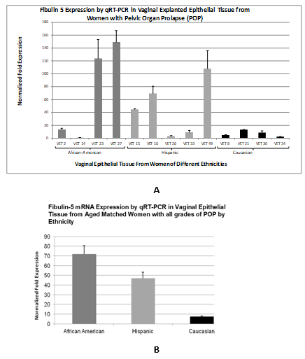
Figure 1. qRT-PCR results of fibulin-5 expression in vaginal epithelial tissue from women with pelvic organ prolapse. (A) mRNA extracted from vaginal tissue explants from African American, Hispanic and Caucasian women were utilized for qRT-PCR to determine fibulin-5 transcriptional expression. Expression values were normalized to GAPDH. (B) Shows fibulin-5 expression levels by qRT-PCR in vaginal epithelial explanted tissue from aged-matched women with all grades POP by ethnicity
We also selected one age and grade matched specimen from each ethnic group and determined fibulin-5 expression levels by qRT-PCR (Figure 2A). The highest level of fibulin-5 mRNA expression was observed with tissue from the African American woman followed by the Hispanic female with lowest level of fibulin-5 expression observed with tissue from the Caucasian female (Figure 2A). We then wanted to determine fibulin-5 protein expression levels in fresh frozen tissue specimens from these same 3 women with prolapse (Figure 2B).
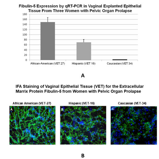
Figure 2. Transcriptional analysis of 3 women POP matched for age, parity and degree of prolapse. (A) Fibulin-5 expression by qRT-PCR in vaginal explanted epithelial tissue from an African American woman (VET-27), a Hispanic woman (VET-16) and a Caucasian woman (VET 34) all with POP closely matched for age, parity and degree of prolapse. (B) Shown are immunofluorescent staining patterns of OTC preserved fresh frozen vaginal epithelial tissue from the African American woman (VET-27), a Hispanic woman (VET-16) and a Caucasian woman (VET 34) using an antibody to fibulin-5. FITC Stained cells are green and DAPI was used to stain nuclei blue
Here we show that fibulin-5 expression levels were highest in the African American females and that the ECM staining patterns of fibulin-5 of the African American woman with POP appears to maintain a certain degree of organization when compared to fibulin-5 staining patterns in the control vaginal epithelium from the African American and the Caucasian women (Figure 2B). Fibulin-5 ECM staining patterns for the Hispanic female with POP appears somewhat less organized with less staining intensity when compared with fibulin-5 staining patterns of the African American female with prolapse (Figure 2B). The fibulin-5 ECM staining patterns in the vaginal epithelium of the Caucasian female with POP looked completely disorganized and poorly stained in comparison to both the African American and Hispanic females with POP and the Caucasian control (Figure 2B).
Higher levels of fibulin-5 total protein are expressed in vaginal epithelium of African American women when compared to Caucasian women with and without POP
We then examined total fibulin-5 protein expressed in vaginal epithelial tissue from aged matched and grade matched African American and Caucasian women by western blot analysis. Western blot analysis revealed higher levels of fibulin-5 total protein expressed in vaginal epithelium of African American women when compared to Caucasian women with POP and without POP. Analysis of fibulin-3 total protein revealed lower levels of fibulin-3 in VET tissue from African American women with POP compared to controls and we observed generally higher levels of fibulin-3 in Caucasian women with POP compared to controls (Figure 3).
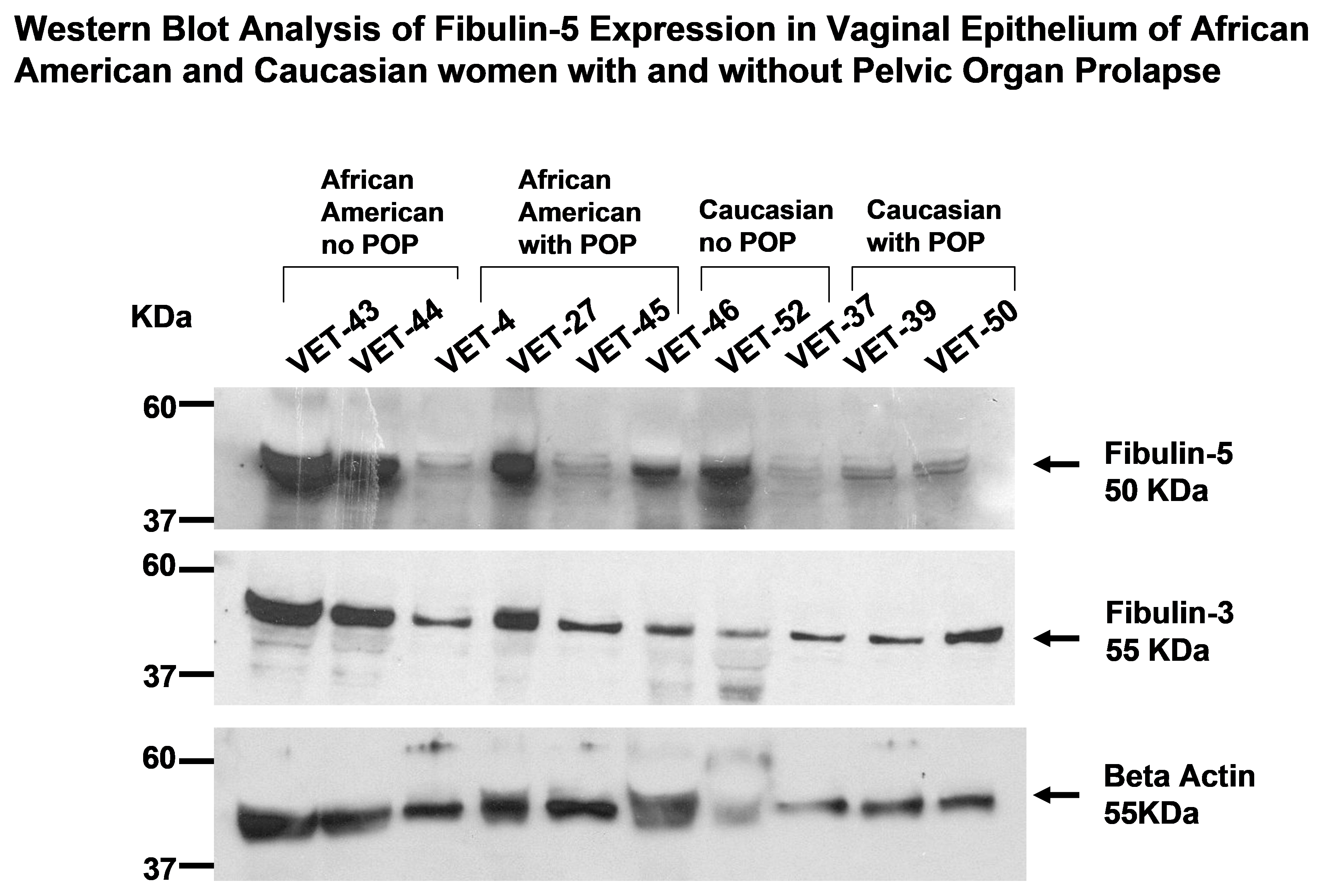
Figure 3. Western blot analysis of fibulin-5 in Vaginal epithelial tissue from women with and without POP. Ten micrograms of total protein extracted from fresh frozen VET tissue from African American and Caucasian women with and without POP was analyzed by western blot for fibulin-5 and fibulin-3. Bands associated with fibulin-5 and fibulin-3 were observed at 50 and 55 KDa respectively. Beta actin also observed at 55 KDa was used as a loading control
Caucasian women with POP have the highest level of fibulin-3 expression however expression patterns are disorganized
Fibulin-3 mRNA expression in vaginal epithelial explants show no clear pattern among the different ethnic groups with POP however the Caucasian women had on average the highest level of fibulin-3 mRNA expression (Figure 4A, 4B). Average levels of fibulin-3 transcriptional expression among ethnic groups with POP show that Caucasian women had the highest average levels of fibulin-3 mRNA expression when compared American and Hispanic women (Figure 4C). Fibulin-3 protein expression in vaginal epithelial explants from African American showed the highest degree of organization consistent with fibulin-5 staining patterns in the ECM (Figure 4D). IFA staining patterns of vaginal epithelial explants reveal a consistent disorganization of fibulin-3 staining patterns in the ECM of the Caucasian female and to a lesser degree for Hispanic female (Fig.4D). This finding is consistent with the intense level of fibulin-3 staining in the vaginal epithelial explants from the Caucasian female (Figure 4D).
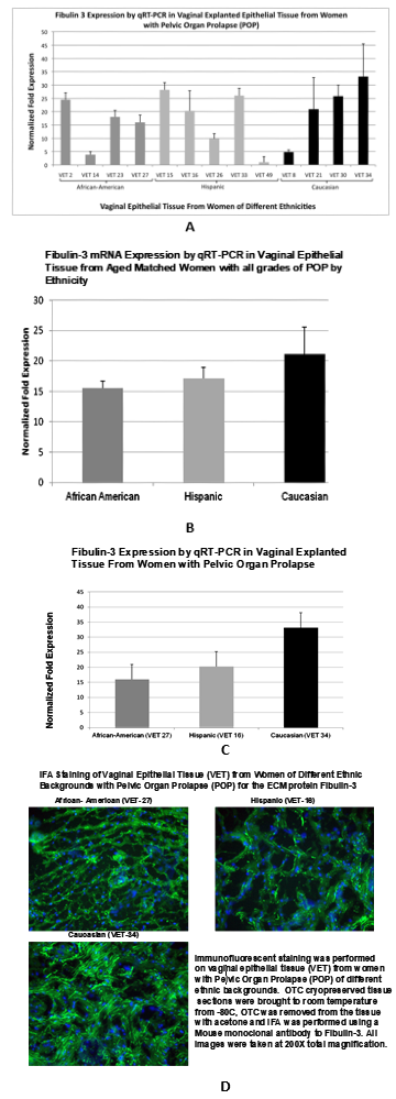
Figure 4. (A) qRT-PCR results of fibulin-3 expression in vaginal epithelial tissue from women with pelvic organ prolapse. (A) mRNA extracted from vaginal tissue explants from African American, Hispanic and Caucasian women were utilized for qRT-PCR to determine fibulin-3 transcriptional expression. Expression values were normalized to GAPDH. (B) Shows fibulin-3 expression levels by qRT-PCR in vaginal epithelial explanted tissue from aged-matched women with all grades POP by ethnicity. (C) Shows Fibulin-3 expression by qRT-PCR in vaginal explanted epithelial tissue from an African American woman (VET-27), a Hispanic woman (VET-16) and a Caucasian woman (VET 34) all with POP closely matched for age, parity and degree of prolapse. (D) Shown are immunofluorescent staining patterns of VET tissue from the African American woman (VET-27), a Hispanic woman (VET-16) and a Caucasian woman (VET 34) using an antibody to fibulin-3. FITC stained cells are green and DAPI was used to stain nuclei blue
The transcriptional expression levels of HOXA11observed in vaginal epithelium from the African women and Hispanic were higher than Caucasian women
HOXA11 transcriptional expression levels were generally higher in vaginal epithelium of African American and Hispanic women with POP when compared to Caucasian women with POP (Figure 5A). HOXA11 expression by qRT-PCR in VET from women with all grades of POP reveal that Hispanic expressed the highest levels of HOXA11 followed by African American women and Caucasian women expressed the lowest levels HOXA11. (Figure 5B). HOA11 expression by RT PCR in vaginal epithelial explanted tissue from women with POP can be observed showing similar levels of expression based the high standard error observed in VET16 from a Hispanic patient (Figure 5C). HOXA11 protein expression in vaginal epithelial explants from the African American female showed the highest degree of HOXA11 organization by protein staining patterns among the 3 women with POP (Figure 5D). The most disorganized and aberrant HOXA11 staining patterns were observed in the vaginal epithelium of the Caucasian female. An intermediate pattern of staining intensity and organization was observed in vaginal tissue of the Hispanic female (Figure 5D).
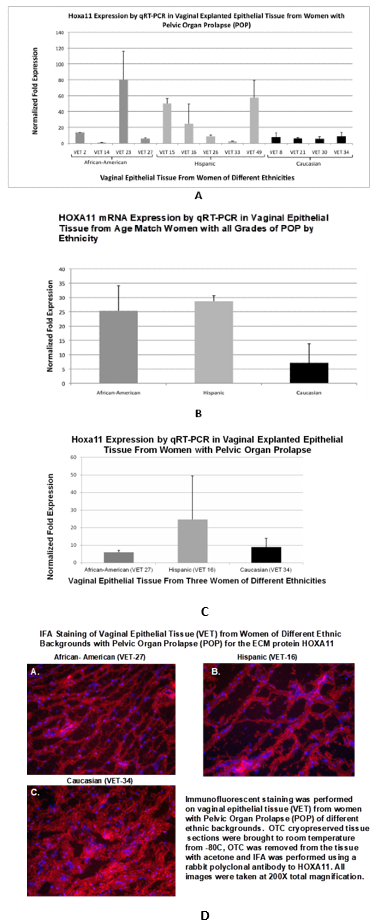
Figure 5. qRT-PCR of HOXA11 expression in vaginal epithelial tissue from women with pelvic organ prolapse. (A) Shows transcriptional expression of fibulin-3 in vaginal epithelial explanted tissue from African American, Hispanic and Caucasian women by qRT-PCR. Expression values were normalized to GAPDH. (B) Shows HOXA11 expression levels by qRT-PCR in vaginal epithelial explanted tissue from aged-matched women with all grades POP by ethnicity. (C) HOA11 expression by RT PCR in vaginal epithelial explanted tissue from women with POP. (D) Shown are immunofluorescent staining patterns of VET tissue from the African American woman (VET-27), a Hispanic woman (VET-16) and a Caucasian woman (VET 34) using an antibody to HOXA11. RITC stained cells are red and DAPI was used to stain nuclei blue
MMP2 is upregulated in vaginal epithelium African American women with POP
Because of a lack of control specimens for Caucasian and Hispanic women we examine the levels of matrix metalloproteinase-2 in vaginal epithelium of African women with and without POP. We observed a statistically significant increase in MM2 transcriptional expression among African women with POP compared to control tissue (Figure 6).
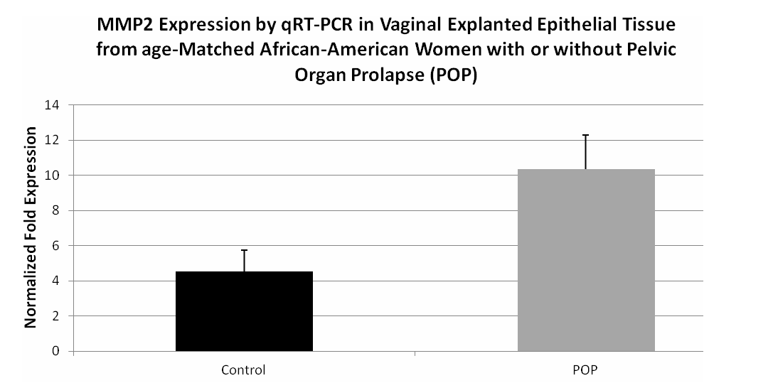
Figure 6. qRT-PCR of MMP2 expression in vaginal epithelial tissue from age-matched African American women with and without POP. (A) Shown are qRT-PCR data from vaginal tissue explants from aged matched African American women with and without POP MMP2 transcriptional expression. Expression values were normalized to GAPDH
Discussion
There are significant racial differences in prolapse prevalence, symptoms and management. This study would help address pelvic organ prolapse in women of different ethnic backgrounds, particularly Caucasian women who are disproportionately affected by POP and may provide information to identify reasons why they have a higher incidence of POP compared to other major ethnic groups. Studying the underlying causes of POP with regard to the vaginal epithelium extra-cellular matrix protein changes will allow us to better understand the mechanisms involved in this elusive medical condition and help to provide treatment alternatives other than surgery.
The significance of our most recent findings shows that the Caucasian woman with POP have a VET/ ECM staining profile for fibulin-5, fibulin-3 and HOXA11 that is disorganized and shows aberrant staining patterns compared to the African American and Hispanic female. Even more, fibulin-5 transcription was shown to be lowest in VET from the Caucasian female examined. Fibulin-5 total protein levels were shown to be largely lower among Caucasian women compared to African American women. Aberrations in expression of Fibulin-3 mRNA and protein may play less of a roll in development of prolapse in humans. While the current study shows that there may be an increase in expression of Fibulin-3 in Caucasian women in comparison to African American the study by Takacs P, et al. [25], found a similar Fibulin-3 mRNA expression and staining intensity in the uterosacral ligaments (USL) of women with and without uterine prolapse [24]. They did, however, find a significant decrease in Fibulin-5 mRNA and staining intensity in prolapsed USLs [24]. This further suggests that aberrations in fibulin-3 may not be important in the development of prolapse in women.
Conclusion
These findings may help to explain why Caucasian women are more likely to develop POP than African American women. Recent studies report Hispanic and Caucasian women have a four to five times higher risk of symptomatic prolapse compared to African American women. These preliminary data are also in support of our original hypothesis. We proposed that the vaginal epithelium undergoes a specific pattern of changes in extracellular matrix proteins (fibulin-5, fibulin-3, and HOXA11) in women with pelvic organ prolapse and the specific pattern of change varies with ethnicity. This may help to explain the higher prevalence of POP in Caucasian women and supports the notion that some women may have a predisposition for developing POP.
Acknowledgement
We thank Dr. Kevin Osteen from Vanderbilt University for his serving as a co-mentor for A.S. and his advice in the preparation of this manuscript.
Funding
A.S. was supported by a grant from Vanderbilt University Medical Scholars Program (NIH CTSA grant UL1 RR 024975); D.J.A. was supported by pilot grants from Meharry Medical College Translational Research Center (MeTRC, NIH grant U54RR026140), The Vanderbilt Meharry Center for AIDS Research (CFAR) (National Institutes of Health (NIH) grant P30AI054999), the Meharry Medical College, and Center for Clinical Research (NIH grant P20RR011792).
Conflict of interest
There are no conflicts of interest.
References
- Burrows LJ, Meyn LA, Walters MD, Weber AM (2004) Pelvic symptoms in women with pelvic organ prolapse. Obstet Gynecol 104: 982-988. [Crossref]
- Fritel X, Varnoux N, Zins M, Breart G, Ringa V (2009) Symptomatic pelvic organ prolapse at midlife, quality of life, and risk factors. Obstet Gynecol 113: 609-616. [Crossref]
- Fialkow MF, Newton KM, Lentz GM, Weiss NS (2008) Lifetime risk of surgical management for pelvic organ prolapse or urinary incontinence. Int Urogynecol J Pelvic Floor Dysfunct 19: 437-440. [Crossref]
- Subak LL, Waetjen LE, van den Eeden S, Thom DH, Vittinghoff E, et al. (2001) Cost of pelvic organ prolapse surgery in the United States. Obstet Gynecol 98: 646-651. [Crossref]
- Wu JM, Hundley AF, Fulton RG, Myers ER (2009) Forecasting the prevalence of pelvic floor disorders in U.S. Women: 2010 to 2050. Obstet Gynecol 114: 127-183. [Crossref]
- Sung VW, Hamptom BS (2009) Epidemiology of pelvic floor dysfunction. Obstet Gynecol Clin North Am 36: 421-443. [Crossref]
- Olsen AL, Smith VJ, Bergstrom JO, Colling JC, Clark AL (1997) Epidemiology of surgically managed pelvic organ prolapsed and urinary incontinence. Obstet Gynecol 89: 501-506. [Crossref]
- Smith FJ, Holman CD, Moorin RE, Tsokos N (2010) Lifetime risk of undergoing surgery for pelvic organ prolapse. Obstet Gynecol 116: 1096-1100. [Crossref]
- Nygaard I, Barber MD, Burgio KL, Kenton K, Meikle S, et al. (2008) Prevalence of symptomatic pelvic floor disorders in US women. JAMA 300: 1311-1316. [Crossref]
- Luber KM, Boero S, Choe JY (2001) The demographics of pelvic floor disorders: Current observations and future projections. Am J Obstet Gynecol 184: 1496-1501. [Crossref]
- Paladini D, Di Spiezio Sardo A, Mandato VD, Guerra G, Bifulco G, et al. (2007) Association of cutis laxa and genital prolapse: A case report. Int Urogynecol J Pelvic Floor Dysfunct 18: 1367-1370. [Crossref]
- Carley ME, Schaffer J (2000) Urinary incontinence and pelvic organ prolapse in women with Marfan or Ehlers Danlos Syndrome. Am J Obstet Gynecol 182: 1021-1023. [Crossref]
- Lind J, Wallenburg HCS (2008) Pregnancy and the Ehlers-Danlos syndrome: A retrospective study in a Dutch population. Acta Obstet Gynecol Scand 81: 293-300. [Crossref]
- Zong W, Stein SE, Starcher B, Meyn LA, Moalli PA (2010) Alteration of vaginal elastin metabolism in women with pelvic organ prolapsed. Obstet Gynecol 115: 953-961. [Crossref]
- Moalli PA, Shand SH, Zyczynski HM, Gordy SC, Meyn LA (2005) Remodeling of vaginal connective tissue in patients with prolapse. Obstet Gynecol 106: 953-963. [Crossref]
- Zhao B, Zhou J (2012) Decreased expression of elastin, fibulin-5 and lysyl oxidase-like 1 in the uterosacral ligaments of postmenopausal women with pelvic organ prolapsed. J Obstet Gynaecol Res 38: 925-931. [Crossref]
- Yanagisawa H, Davis EC, Starcher BC, Ouchi T, Yanagisawa M, et al. (2002) Fibulin-5 is an elastin-binding protein essential for elastic fibre development in vivo. Nature 415: 168-171. [Crossref]
- Nakamura T, Lozano PR, Ikeda Y, Iwanaga Y, Hinek A, et al. (2002) Fibulin-5/DANCE is essential for elastogenesis in vivo. Nature 415: 171-175. [Crossref]
- Katsuta Y, Ogura Y, Iriyama S, Goetinck PF, Klement JF, et al. (2008) Fibulin-5 accelerates elastic fibre assembly in human skin fibroblasts. Exp Dermatol 17: 837-842. [Crossref]
- McLaughlin PJ, Bakall B, Choi J, Liu Z, Sasaki T, et al. (2007) Lack of fibulin-3 causes early aging and herniation, but not macular degeneration in mice. Hum Mol Genet 16: 3059-3070. [Crossref]
- Rahn DD, Acevedo JF, Roshanravan S, Keller PW, Davis EC, et al. (2009) Failure of pelvic organ support in mice deficient in fibulin-3. Am J Pathol 174: 206-215. [Crossref]
- Drewes PG, Yanagisawa H, Starcher B, Hornstra I, Csiszar K, et al. (2007) Pelvic organ prolapse in fibulin-5 knockout mice: Pregnancy-induced changes in elastic fiber homeostasis in mouse vagina. Am J Pathol 170: 578-589. [Crossref]
- Wieslander CK, Rahn DD, McIntire DD, Acevedo JF, Drewes PG, et al. (2009) Quantification of pelvic organ prolapse in mice: Vaginal protease activity precedes increased MOPQ scores in fibulin 5 knockout mice. Bio Reprod 80: 407-414. [Crossref]
- Takacs P, Nassiri M, Viciana A, Condiotti K, Fornoni A, et al. (2009) Fibulin-5 expression is decreased in women with anterior vaginal wall prolapse. Int Urogynecol J Pelvic Floor Dysfunct 20: 207-211. [Crossref]
- Takacs P, Nassiri M, Candiotti K, Yang J, Yavagal S, et al. (2010) Differential expression of fibulins in the uterosacral ligaments of women with uterine prolapse. Arch Gynecol Obstet 282: 389-394. [Crossref]
- Taylor HS, Vanden Heuvel GB, Igarashi P (1997) A conserved Hox axis in the mouse and human female reproductive system: Late establishment and persistent adult expression of the Hoxa cluster genes. Biol Reprod 57: 1338-1345. [Crossref]
- Connell KA, Guess MK, Chen H, Andikyan V, Bercik R, et al. (2008) HOXA11 is critical for development and maintenance of uterosacral ligaments and deficient in pelvic prolapse. J Clin Invest 118: 1050-1055. [Crossref]
- Whitcomb EL, Rortveit G, Brown JS, Creasman JM, Thom DH, et al. (2009) Racial differences in pelvic organ prolapse. Obstet Gynecol 114: 1271-1277. [Crossref]
- Shah AD, Kohli N, Rajan SS, Hoyte L (2007) Racial characteristics of women undergoing surgery for pelvic organ prolapse in the United States. Am J Obstet Gynecol 197: 70.e1-70.e8. [Crossref]
- Rortveit G, Brown JS, Thom DH, Van Den Eeden SK, Creasman JM, et al. (2007) Symptomatic pelvic organ prolapse: Prevalence and risk factors in a population-based, racially diverse cohort. Obstet Gynecol 109: 1396-1403. [Crossref]
- Mattox TF, Bhatia NN (1996) The prevalence of urinary incontinence or prolapse among white and Hispanic women. Am J Obstet Gynecol 174: 646-648. [Crossref]
- Handa VL, Blomguist JL, Knoepp LR, Hoskey KA, McDermott KC, et al. (2011) Pelvic floor disorders 5-10 years after vaginal or cesarean childbirth. Obstet Gynecol 118: 777-784. [Crossref]