Abstract
Nowadays, Bichectomy surgery is a recurrent treatment with aesthetic or functional objectives. But despite being a surgery frequently performed, the surgeons need to have extensive anatomic and surgical knowledge in order to perform it safely.
Key words
Bichectomy, palsy, lipectomy, nerve damage
Introduction
Anatomically, the lower face contour is composed Bichat's fat pad, masseter muscle, mandibular bone, and the subcutaneous fat. If the buccal extension is excessive, patients may complain of rounded faces, excessive cheeks or “baby faces”. Bichectomy is presented as a technique to sculpt the facial angles and enhance aesthetics [1-5].
The buccal fat pad volume is, approximately, 9.6ml and weight of 9.3 g, and its removal should be limited to 2/3 of this volume [3]. The objective is the resection of the buccal adipose body corresponding to 30 to 40% of structure [1].
There is extensive marketing with appeal to facial aesthetics, and the procedure disseminated as a routine [5,6].
Bichectomy is a surgery of low complexity and presents excellent results in the pattern of facial aesthetics. Bichat's fat pad shows subdivisions through the masticatory space, but only the buccal extension provides the contour of the cheek region, conferring the lateral aspect of the face [7].
The adipose tissue in the cheek has 6 extensions: masseteric, superficial temporal, deep temporal, pterygomandibular, sphenopalatine, and lower orbital regions [4].
The buccal fat pad is surrounded by a defined connective capsule with septa towards the deep level, separating groups of adipose lobules [8].
Usually, the Bichectomy is performed under local anestesia. The incision is performed in buccal mucosa at bite level. After, the buccal muscle is dissected and Bichat´s ball is exposed and, without excessive traction, this portion is excised [5].
The facial nerve paralysis is one of Bichat’s fat extraction complications which might be temporary or permanente. Facial nerve innervates the facial muscles of the mimicry. The surgeon must be careful not to cause injuries to zygomatic and buccal branches of the facial nerve [2].
Cranial nerve VII has both motor and autonomic fibers with minor somatosensory components. Damage to these fibers results in ipsilateral facial paralysis [6].
Definite paralysis is not very prevalent, due to anatomical variations, both in origin and in the number of branches of the zygomatic and buccal nerves, as well as the anastomoses that these branches make, thus enabling a rich exchange of nerve fibers, which start to supply the absence of a certain mistakenly extracted nervous segment. The rich variable and anastomotic network of the terminal branches may explain the low prevalence of facial paralysis in Bichectomy procedures. Rare cases that can trigger facial paralysis can be explained by the removal of the nerve plexus together with the Bichat´s ball [2].
Case report
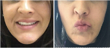
A 35-year-old female patient sought the private clinic in Maringá, Brazil, reporting an attempt of a Bichectomy surgery done a day before. The patient spoke that the dentist responsible for the surgery had great difficulty in finding Bichat´s ball and when he finally found it, couldn´t remove the entire buccal portion, despite the excessive traction used. This description is related to the left side, because the patient did not give permission to the surgeon to proceed the surgery also on the right side. She was claiming extreme pain. Through the clinical examination, it was possible to diagnose the superior labial, lower eyelid and nose wing palsy on the left side of the face (Figure 1).

Figure 1. Patient showing assimetric smile and limited movements on the left side of the face
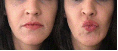
The patient was medicated with Etna ®, 2 capsules 2 times per day, for 10 days. The medication helped, but its wasn´t resolutive (Figure 2).

Figure 2. Visible ptoses of the lower eyelid and persistent limitation movements on the left side of the face
After 6 months, the patient was still showing paralysis. And, how the muscle tonicity was altered because of the non-stimulation, some ptosis was observed. The treatment of choice, at that time, was the use of botulinum toxin to create a better harmony. After one year, the patient fully recovered the facial movements. It means that, probably, the nerve lesion was incomplete, a distension of the nerve caused by the excessive force of traction, or the branches affected were terminals and its functions could be restored by anastomoses.
Conclusion
Despite being a secure surgery, Bichectomy requires great anatomy knowledge and surgery experience to keep safe nerves, vessels, glands and ducts in intimate relationship with the buccal fat pad. Besides that, the capsule that involves the Bichat´s ball, continues through all extensions, and if it's fordec by traction can compress anatomic structures, such as the nerves, and cause palsy.
References
- Junior RM, Gontijo G, Guerreiro TC, Moreira R, Sousa NL (2018) Bichectomia, a simple and fast surgery: case report. Rev Odontol Bras Central 27: 98-100.
- Porto L, Nazer M, Piazza J (2020) Anatomical Correlation among Bichat’s fat pad with the Facial Nerve Terminal Branches. Brazilian Journal of Oral and Maxillofacial Surgery 20: 12-15.
- Klüppel L, Marcos RB, Shimizu IA, Silva RD (2018) Complications associated with the bichectomy surgery. Rev Gaúch Odontol 66: 278- 284.
- Faria CA, Dias RC, Campos AC, Daher JC, Costa RS, et al. (2018) Bichectomy and its contribution to facial harmony. Rev Bras Cir Plást 33: 446-452.
- Moura LB, Spin JR, Neto SR, Filho VAP (2018) Buccal fat pad removal to improve facial aesthetics: an established technique? Med Oral Patol Oral Cir Bucal 23: 478-484. [Crossref]
- Sonne J, Lopez-Ojeda W. Neuroanatomy, Cranial Nerve. 2020.
- Santos IK, Matos JD, Barros JD, Medeiros CR, Cariri TF, et al. (2017) Bichectomy as an alternative treatment for facial harmonization – case report. Int J Adv Res 5: 1495-1502.
- Kahn JL, Wolfram-Gabel R, Bourjat P (2000) Anatomy and imaging of the deep fat of the face. Clin Anat 13: 373-382. [Crossref]


