The Philosophy of this implant is to respect as much as it is technically possible the bone physiology. The surgical theorical concept is based on three parameters: The respect of the upper femoral metaphysis elasticity. The periosteum property. The respect of the medullary stem canal [1]. The cancellous bone of both long bone’s extremities is the most important element of their elasticity and needs compression [2]. The periosteum is acting during all day life. It produces bone apposition even when or/and over 100 years [4]. The medullary canal vascularised 2/3 of the femoral shaft cortex [2], provides blood cells and interferes in bone’s remodelling [5]. At last the femoral component must reproduce the compression strain acting on the medial part of the femoral upper metaphysis in order to avoid resorption of the calcar [6-7]. For these biological facts, it is important to use them or to minimise either their destruction or their removal. The principle is to remove the least possible cancellous bone in the upper femoral metaphysis, to use the perisoteum bone apposition and do not enter in the medullary canal.
Figure 1 shows as the first femoral component was in Chrome Cobalt alloy. (Vitallium). The second is in Titanium.
The femoral component is screwed on the lateral femoral shaft. The screw diameter is 5 mms (c) to fix the plate on the femoral shaft and 7 mms (a) to maintain the greater trochanter. Two teeths (b) prevent gluteus medius muscular action. The hole’s plate and screw’s design are according to the Meyrues and Cazenave’s experimental work [8].
Eccentricity of the right metallic femoral head 0.7 cm loosening of polyethylene in 30 Years No calcar resorption, no screws fracture, periosteum inamovible apposition. Harris score: 100.
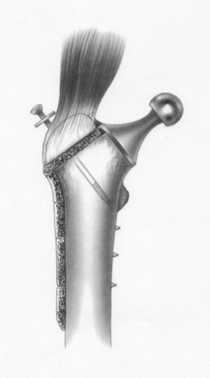
Figure 1. The femoral stem
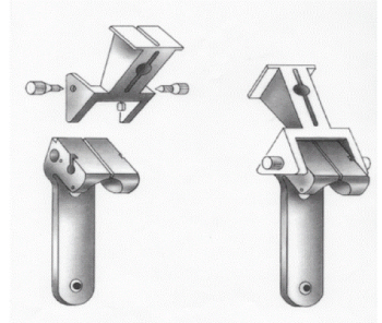
Figure 2. The ancillary instrumentation

Figure 3. The greater trochanteric cut
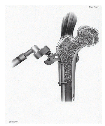
Figure 4. Femoral neck section
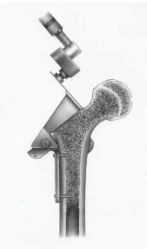
Figure 5. Fitting cuts
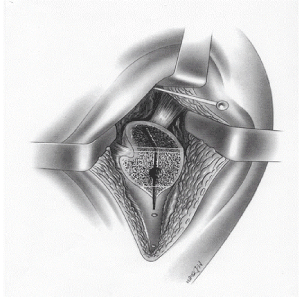
Figure 6. Presentation of setting up the implant
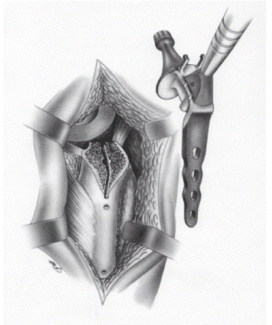
Figure 7. Implant screwed in place
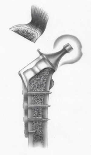
Figure 8. Greater trochanter screwed in place
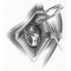
Figure 9. Aerial view of the greater trochanter in place
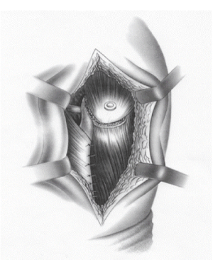
Figure 10. Results after 30 years
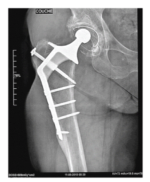
Figure 11. 0.7 cm loosening of polyethylene in 30 Years
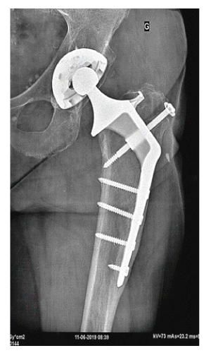
Figure 12. Twenty-six years after surgery
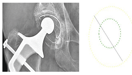
Figure 13. 30 years follow up excentration of the head: 0.7 cm and ≠0.02 mm per year
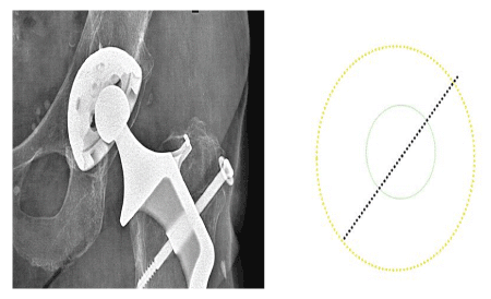
Figure 14. Polyethylene loosening 26 Years follow-up and ≠0.015 mms per year (0.015 x 26 = 0.39)
- Modifications morphologiques de la métaphyse fémorale supérieure chez l’homme atteint de la maladie ostéoporotique. Y. Cirotteau; Académie des Sciences. Mai 1999, III.
- Vico L, Collet P, Guignandon A, Lafage-Proust M-H, Thomas T, Rehailia M, Alexandre C (2000) Effects of long-term microgravity exposure on cancellous and cortical weight-bearing bones of cosmonauts. THE LANCET 355.
- Teot L, Vidal J, Dossa J (1989) Le tissu osseux; Sauramps Medical, Montpellier.
- Kibbin MB (1978) The biology of fracture healings in long bones. J Bone and Joint Surg 60B: 150-161.
- Anne Devulder. Approche micro-mécanique du remodelage osseux. Ecole Centrale Paris, Thèse: 2009. NNT: 2009ECAP0020
- Dumbleton J. Bushelow M (1990) A strain gauge analysis of the Cirotteau's hip prosthesis. Avril1990
- Pelisse J (1990) Étude sur plateau de marche de la prothèse de Y. Cirotteau. CRA.
- Etude théorique et physique expérimentale des contraintes dans les vis d’ostéosynthèse: Meyrueis JP, Bonnet, de Bazelaire, Zimmerman R, Cazenave A, Couve A. Rev. chir. orthop. 1979, Suppl.II, 65.