Abstract
Fibrous dysplasia is a rare bone disease caused by mutations in GNAS gene. The normal bone is replaced with fibrous tissue and abnormal (woven) bone. Monostotic disease occurs in one bone while polyostotic disease occurs in multiple bones [1]. It is most common in ribs (28%) and occurs in craniofacial bones in 10 to 25 % of cases [2]. Craniofacial fibrous dysplasia (CFD) lesions are detected early, but can stay silent until deformity or growth occurs. 90% of CFD lesions were detected before the age of four [3]. The differential diagnosis of CFD includes benign fibro-osseous lesions of the jaws, giant cell tumors, aneurysmal and simple bone cysts. They have common histologic findings such as hyperproliferation of fibrous material mixed with bony structures and irregular bone [4].
Key words
fibrous dysplasia, right maxillary
Case presentation
A 11-year-old girl complaining of facial asymmetry and right maxillary prominence was referred to our practice. She had no history of previous trauma or intervention.
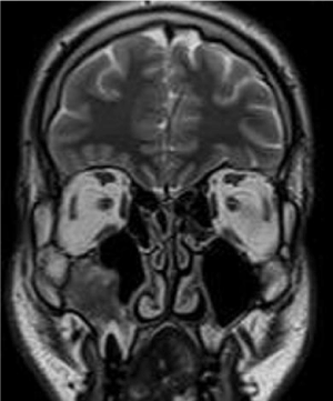
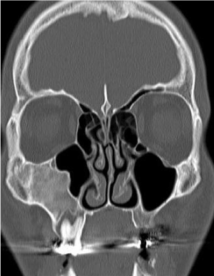
MRI examination of the facial bones was requested for this girl. The examination was performed using Toshiba MRI scanner (Toshiba Medical System, New York, USA) and the usual routine sequences were obtained. The hallmark of the examination was partial opacification of the right maxillary sinus by virtue of ill defined intensity eliciting heterogeneous intermediate T1 and predominantly low T2 signals (figure 1). Complementary CT scan was then performed using Aquillion One CT scanner (Toshiba Medical System, New York, USA), with tube potential set at 110 Kv, current at 320 mA, collimation at 1 mm and table movement at 1 mm/s. An attached workstation and software were utilized to reconstruct axial source images into 2D multiplanar coronal images. The previously thought to be partial opacification of the sinus appeared to be expansion of the right maxillary bone itself with secondary encroachment on the sinus. The right maxillary bone was expanded showing ground glass opacities, however the overlying cortex was completely intact with no evidence of fracture or breaching (figure 2).

Figure 1. Coronal T2 WI of the frontal lobes, orbits, paranasal sinuses and maxillary bones. The right maxillary sinus appears partially opacified by virtue of ill-defined intensity eliciting heterogeneous predominantly low T2 signal.

Figure 2. CT scan, Coronal 2D multiplanar image in bone window. The right maxillary bone is expanded showing ground glass opacities and intact overlying cortex with no evidence of fracture or breaching with secondary encroachment on the sinus.
The diagnosis of fibrous dysplasia was made based on CT criteria of the lesion and was confirmed lately by histopathological examination. The current case shows to what extent MRI findings could be deceiving in interpretation of bony lesions particularly in facial region and confirm the utmost importance of CT scan in assessment of craniofacial bony diseases.
Conflicts of interest
The authors declare no conflict of interests.
References
- Burke AB, Collins MT, Boyce AM (2017) Fibrous dysplasia of bone: craniofacial and dental Implications. Oral Dis 23: 697-708. [Crossref]
- Chong VF, Khoo JB, Fan YF (2002) Fibrous dysplasia involving the base of the skull. AJR Am J Roentgenol 178: 717-720.
- Hart ES, Kelly MH, Brillante B, Chen CC, Ziran N, et al. (2007) Onset, progression, and plateau of skeletal lesions in fibrous dysplasia and the relationship to functional outcome. J Bone Miner Res 22: 1468-1474. [Crossref]
- Neville BW (2009) Oral and maxillofacial pathology. Saunders/Elsevier: St Louis, MO, USA.


