Case report
A 32-year-old male who underwent a straightforward laparoscopic cholecystectomy 5 months earlier, presented with 3-day history of intermittent episodes of haematmesis and melaena. He denied any history of abdominal pain and chronic ingestion of any non-steroidal anti-inflammatory analgesics.
At presentation, his vital signs were stable, and the abdominal examination was unremarkable. The routine blood tests were within the normal range. Gastroscopy showed a deformed duodenal bulb, with narrowing of the 2nd part of duodenum by a smooth convex bulge representing either an external compression or submucosal lesion (Figure 1). Abdominal ultrasound revealed a well-defined (size: 10x6x6 cm) sacculo-tubular mass in the porta hepatis region (Figure 2A). Doppler ultrasound study showed an echogenic thick wall containing a mural thrombus with turbulent arterial flow (Figures 2B). Computed tomography (CT) scan showed a large well-defined hypodense mass with relatively hyperdense periphery arising from the hepatic artery and extending caudally (Figures 2C). A 3D CT-Angiography reconstructed images showed the presence of a huge aneurysm arising from the right hepatic artery (Figure 2D).
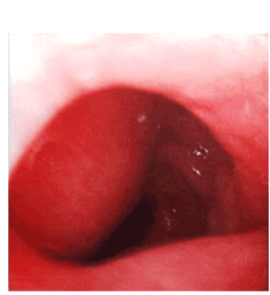
Upper GI endoscopy showing the large convex bulge projecting at the inferior wall of second part of duodenum; the mass did not exhibit visible pulsation.
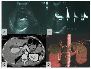
Figure 2. Ultrasound scan of the liver showing a well-defined mass at the porta hepatic with echogenic surrounding thick wall and an echoic lumen (2A). The color Doppler ultrasound showed blood flows within the lesion with turbulence (2B). The axial contrast-enhanced CT scan showed marked enhancement of the lumen of the aneurysm along the course of the hepatic artery. The non-enhancing portion of the lesion was suggestive of partial thrombosis (2C). Figure 2D shows the reconstructed image of the giant hepatic pseudo-aneurysm.
The patient was offered selective angiography and embolization, but he refused. He was discharged against medical advice and therefore lost to follow-up.
Discussion
The hepatic artery is the fourth most common site of intra-abdominal aneurysm after infra-renal aorta, iliac artery and the splenic artery. Recently, hepatic artery aneurysms are becoming the most common, due to the explosive increase in the use of percutaneous and laparoscopic hepato-biliary procedures [1]. More than one half of all detected hepatic artery aneurysms are pseudoaneurysms and the extrahepatic artery aneurysms have higher incidence of rupture than the intra-hepatic ones.
Generally, hepatic artery pseudoaneurysm (HAP) is rare (reported incidence is 0.6%) [2] but can lead to serious life-threatening complications. It was first reported after laparoscopic cholecystectomy (LC) in 1994 and attributed to iatrogenic vascular injury [1]. The injury occurs either in association with bile duct injury during LC or in isolation as a result of direct trauma or thermal injury [3]. The diagnosis is often made weeks or months after the laparoscopic procedure [4].
HAP commonly presents with gastrointestinal tract bleeding; commonly haemobilia if the pseudoaneurysm communicates with the bile duct. Also, it may present with obstructive jaundice due to the compression of the biliary tract by the expanding aneurysmal haematoma [5]. It may also present urgently with massive intraabdominal bleeding which necessitates emergency surgical intervention [6]. Contrast CT scan or dynamic CT angiography and Doppler ultrasound can ‘clinch’ the diagnosis. However, it is best made by selective angiography. The first line of treatment is non-operative in the form of selective embolization which offers reasonably good long-term results [7]. However, coil embolization carries the risk of aneurysmal rupture, hepatic necrosis and delayed bile duct stricture due to ischaemia. Sudden rupture of HAP with massive intraabdominal bleeding carries high mortality rate and hence, necessitates prompt emergency surgical intervention with ligation of the artery.
Conclusion
- Hepatic artery pseudoaneurysm (HAP) may develop weeks or months after laparoscopic cholecystectomy.
- HAP commonly present with intermittent GI bleeding (haemtemesis or melaena). If left untreated, it ruptures with massive intraabdominal bleeding which carries high mortality rate.
- HAP should be suspected in any patient presenting with GI bleeding and has a recent history of laparoscopic cholecystectomy.
References
- Pistorius GA, Walter P, Hildebrandt U, Defreyne L (1994) [Pseudo-aneurysm of the hepatic artery. A rare complication after laparoscopic cholecystectomy]. Langenbecks Arch Chir 379: 291-293. [Crossref]
- Christensen T, Matsuoka L, Heestand G, Palmer S, Mateo R, et al. (2006) Iatrogenic Pseudoaneurysms of the Extrahepatic Arterial Vasculature: Management and Outcome. HPB (Oxford) 8: 458-464. [Crossref]
- Yelle JD, Fairfull-Smith R, Rasuli P, Lorimer JW (1996) Hemobilia complicating elective laparoscopic cholecystectomy: a case report. Can J Surg 39: 240-242. [Crossref]
- Milburn JA, Hussey JK, Bachoo P, Gunn IG (2007) Right Hepatic Artery Pseudoaneurysm Thirteen Months Following Laparoscopic Cholecystectomy. Eur J Vasc Endovasc Surg 13: 1-3.
- Abdalla S, Thome A, Reslinger V, Atanasiu C, Pellerin O, et al. (2015) Compressive hematoma due to pseudoaneurysm of the right hepatic artery: a rare cause of obstructive jaundice after single-port cholecystectomy. Surg Laparosc Endosc Percutan Tech 25: e42-e444. [Crossref]
- Madanur MA, Battula N, Sethi H, Deshpande R, Heaton N, et al. (2007) Pseudoaneurysm following laparoscopic cholecystectomy. Hepatobiliary Pancreat Dis Int 6: 294-298. [Crossref]
- Rivitz SM, Waltman AC, Kelsey PB (1996) Embolization of hepatic artery pseudoaneurysm following laparoscopic cholecystectomy. Cardiovasc Intervent Radiol 19: 43-6. [Crossref]