Chronic lymphocytic leukemia / small lymphocytic lymphoma (CLL / SLL) are low grade lymphoproliferative disorders of mature B lymphocytes characterized with lymphocytosis. About 3-15% of the cases transform into a more aggressive lymphoid malignancy. This transformation is known as Richter Syndrome (RS) [1-3] CLL / SLL progression is mostly observed as prolymphocytes or immunoblasts, Hodgkin's lymphoma (HL), diffuse large B-cell lymphoma (DLBCL), T-cell non-Hodgkin's lymphoma, multiple myeloma, Burkitt's lymphoma and rarely as B-lymphoblastic lymphoma / leukemia (B-ALL) [1,4,5].
With its morphological and immunohistochemical features, Hodgkin's variant of Richter's syndrome, to be more precise, the transformation of CLL/SLL into Hodgkin's lymphoma is similar to classical HL [6]. The patients receive aggressive treatment, and they have poor prognosis [2]. We would like to present a rare case of Hodgkin’s lymphoma transformation of CLL/SLL with the review of literature.
The 69-year-old male patient was admitted to Department of Hematology with the complaints of fatigue and weakness. The patient did not have any complaints of night sweat, fever or weight loss. Facial pallor, splenomegaly and lymph node enlargements in the neck and inguinal region were observed in the physical examination. The largest of the lymph nodes was 2x1 cm. His hemogram revealed leukocytosis, and 80% mature lymphocytes and "smudge" cells were observed in the peripheral blood smear. In 2011, bone marrow aspiration and bone marrow biopsy were performed with CLL / SLL prediagnosis. In flow cytometry, CD5, CD20 and CD23 were 69%, 92% and 63% respectively. Bone marrow biopsy sample revealed lymphoid cells that diffusely infiltrated the bone marrow. They had monotonous, small, condensed chromatin and stained positive for CD20 (Clone L26, BioSB), CD79a (JCB 117, Bio SB) and CD5(Clone 1G12, Leica). The patient was diagnosed with CLL / SLL through flow cytometry and morphological and histopathological examinations. In the cytogenetic study, 46xy and 17 p were negative. In the follow-ups of the patient, an increase was detected in the lymph nodes, and his hemoglobin was 8.9 g / dl (13.5-17). Thus, rituximab (375 mg / m2) for 1 day and bendamustine (90 mg / m2) for 2 days were delivered. The tomography performed after 6 cycles of chemotherapy revealed significant regression. In the follow-up in 2015, cervical lymph node excisional biopsy was performed due to the increase detected in the number of lymph nodes and in the size of the spleen.
In the histopathological examination of the lymph node, the lymph node exhibited a diffuse infiltrate of lymphoid cells with monotonous small-medium-sized condensed chromatin (Figure 1). These cells stained positive for CD20 (Clone L26, BioSB), CD79a (JCB 117, Bio SB), PAX5 (Cat.No.RB-9406-p, Thermo), CD5 (Clone 1G12, Leica), bcl2 8Clone bcl-2/100/D5, Leica) and CD23 (Clone SP23, Thermo) in the immunohistochemical study (Figure 2). In the focuses outside CLL / SLL areas, large Hodgkin and Reed-Sternberg (H-RS) cells with prominent nucleoli were observed in inflammatory background composed of heterogeneous cell population of rare eosinophils, plasma cells and fibrohistiocytic cells (Figure 3). These cells stained positive for CD 30 (Clone RBT-CD5, BioSB) and CD 15 (SPM 119, BioSB) but negative for CD 20(Clone L26, BioSB) (Figure 4).
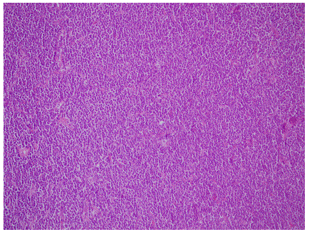
Figure 1. Lymph node with a diffuse population of small monomorphous CLL/SLL (HEX200).
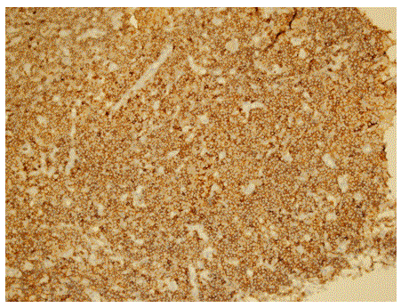
Figure 2. Lymph node with a diffuse population of small monomorphous CLL/SLL (DABXCD20).
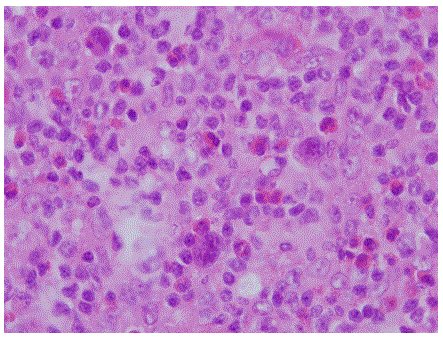
Figure 3. Reed sternberg cell in a typical polymorphous, inflamatory backround (HEX1000).
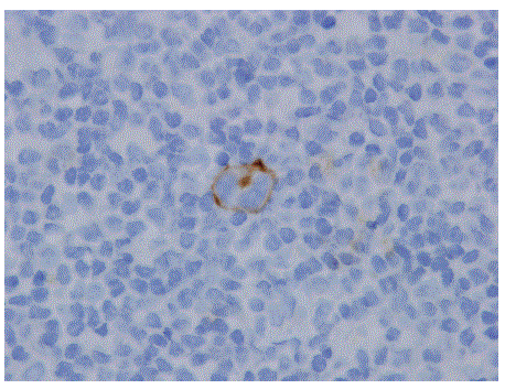
Figure 4. CD 30 expression in a Reed Sternberg cell (DABX1000).
Depending on the morphological and immunophenotypic findings, Hodgkin's lymphoma transformation of chronic lymphocytic leukemia/small lymphocytic lymphoma diagnosis was made. The patient died due to respiratory and cardiac arrest while he was being treated in the intensive care unit with the diagnoses of hypervolemia and pneumonia.
Richter's syndrome was first described by Maurice N. Richter in 1928 in a patient with rapidly fatal and diffusive lymphadenopathy and hepatosplenomegaly associated with CLL/SLL [7]. Later, other lymphoid malignancies such as prolymphocytic leukemia, HL, lymphoblastic lymphoma and "hairy cell" leukemia have also been reported in CLL/SLL patients [8]. Furthermore, it has been reported that the development frequency of secondary cancers such as basal cell carcinoma, squamous cell carcinoma, breast cancer, prostate cancer or malignant melanoma is higher in these patients [1,6].
In the studies in literature, HL transformation is encountered in the 0.4% of the CLL/SLL patients [6,8]. Bockorny et al. identified 86 cases in the literature between 1975 and 2011, including the series of Tsimberideou et al. [4]. Ages, HL transformation times and survivals of the CLL/SLL patients reported in the literature up to the date are given in Table 1. The mean HL transformation time of CLL/SLL in the series of Tsimberideou et al. was 4.6 years [6]. The patients are mostly elderly males diagnosed with the B-symptoms; lymphadenopathy and hepatosplenomegaly [1,8]. In our case, the patient was in the 7th decade, and he had lymphadenopathy and splenomegaly both in the initial symptoms and in the recurrence.
Table 1. Patients with HL transformation of CLL/SLL.
Referance (year) |
No of patient |
Age (year) |
Time for HL transformation
(year) |
Survival |
Bockorny et al. [4]
(Tsimberideou et al., (2006) [6] |
86 (18) |
65.7 (34-85) |
4.3 (0-26) |
1.7 years (0-14) |
Krause et al., (2013) [2] |
1 |
44 |
1 |
4.5 months |
Kaz´mierczak et al., (2014) [1] |
2 |
48.39 |
1 |
2years 5months |
Tadmor et al., (2014) [9] |
16 |
58 |
5.9(0,8-11,9) |
3.3 years |
Parikh et al., (2015) [10] |
26 |
67(45-88) |
6.2(0-24.5) |
3.9 years |
Janjetovic et al., (2016) [11] |
1 |
70 |
0.17 |
still alive |
Etiopathogenesis of the disease is not fully known. EBV infection has been identified in some patients. Additionally, it has been argued that immunosuppressive therapy, especially fludarabine, may increase the risk of transformation. It has been considered that elongated and significant reduction of CD 4+ and CD8+ T lymphocytes in EBV-positive B cells due to proliferation and accumulation may cause the development of a high-grade lymphoma [1,4,8].
Two morphological types of Hodgkin's lymphoma transformation of CLL/SLL have been identified. Type 1 is characterized by H-RS cells scattered over a surface containing CLL cells. In Type 2, H-RS cells are separate from the CLL cells in a typical polymorphic inflammatory cells backroun. It has been argued that Type 1 transformation occurs due to the progression of the underlying CLL cells - especially when the H-RS cells expresses the B cell markers and Type 2 develops as 2 different disorders [1,8]. The origin of H-RS cells encountered in Type 2 is different from that of CLL cells. In three of the four patients with Hodgkin's lymphoma transformation of CLL/SLL, the relation between the clones of CLL and H-RS cells has been identified by using DNA sequencing and polymerase chain reaction (PCR) [3,4,8]. On the other hand, clone has also been reported in unrelated cases, and EBV and chemotherapy are supposed to play a significant role in the development of the disease [4]. In our case, CLL / SLL and Hodgkin's cells were observed separately in the histopathological study, and H-RS cells were not stained with B-cell markers. Thus, morphologically and immunohistochemically, it was considered to be Type 2.
As in our case, the most common histological subtype encountered in the literature is the mixed cellular type. It is followed by nodular sclerosing, lymphocyte-poor and lymphocyte rich types [1,4].
The most effective treatment is not known yet. HL-targeted, multiple chemotherapy protocols are employed. In general, the response to the treatment and clinical course are poor [1,4,6].
Hodgkin’s transformation of CLL / SLL is a rare disorder with a poor clinical course and requires different treatment approaches. Its definitive diagnosis is made with histopathological study. Molecular genetic studies have a significant role in understanding its etiopathogenesis, which will make the development of specific treatments for the disease possible. When a disease progression in follow-up patients with CLL/SLL is identified clinically, histopathological evaluation of the diagnosis is significant to detect the possible comorbid transformations. It is emphasized through this case that in patients with the above mentioned clinical features, the transformation may occur not only towards the B-cell lymphomas but also towards the leukemias or Hodgkin's disease.
- Maciej K, Renata KB, Andrzej B, El-zbieta C, Lidia G, et al. (2014) Hodgkin lymphoma transformation of chronic lymphocytic leukemia: cases report and discussion. Med Oncol 31: 800. [Crossref]
- John RK, Lee CD, Latoya CK (2013) Hodgkin lymphoma transformation of chronic lymphocytic leukemia/small lymphocytic lymphoma. Bayl Univ Med Cent 26: 16–18. [Crossref]
- Toshiyuki O, Bassam NS, Dennis DW, Randy DG, Steven DH, et al. (1998) Origin of the Hodgkin/Reed-Sternberg Cells in Chronic Lymphocytic Leukemia With ‘‘Hodgkin’s Transformation’’. Blood 91: 1757-1761. [Crossref]
- Bruno B, Ion C, Constantin AD (2012) Hodgkin lymphoma as Richter transformation in chronic lymphocytic leukaemia: a retrospective analysis of world literature. Br J Haematol 156: 50-66. [Crossref]
- Wojciech G (2014) Chronic lymphocytic leukemia/small lymphocytic lymphoma and B-cell prolymphocytic leukemia. In: Wojciech Gorczyca. Atlas of differential diagnosis in neoplastic hematopathology, 3th edn. CRC pres. Boca Raton. 211- 234.
- Tsimberidou AM, O'Brien S, Kantarjian HM, Koller C, Hagemeister FB, et al. (2006) Hodgkin Transformation of Chronic Lymphocytic Leukemia. The M. D. Anderson Cancer Center Experience. Cancer 107: 1294-302. [Crossref]
- Richter MN (1928) Generalized Reticular Cell S2021 Copyright OAT. All rights reserv Lymphatic Leukemia. Am J Pathol 4: 285-292. [Crossref]
- Tsimberidou AM, Keating MJ (2005) Richter syndrome: biology, incidence, and therapeutic strategies. Cancer 103: 216-228. [Crossref]
- Tadmor T, Shvidel L, Goldschmidt N, Ruchlemer R, Fineman R, et al. (2014) Hodgkin's variant of Richter transformation in chronic lymphocytic leukemia; a retrospective study from the Israeli CLL study group. Anticancer Res 34: 785-790.
- Parikh SA, Habermann TM, Chaffee KG, Call TG, Ding W, et al. (2015) Hodgkin transformation of chronic lymphocytic leukemia: Incidence, outcomes, and comparison to de novoHodgkin lymphoma. Am J Hematol 90: 334-338. [Crossref]
- Janjetovic S, Bernd HW, Bokemeyer C, Fiedler W (2016) Hodgkin's lymphoma as a rare variant of Richter's transformation in chronic lymphocytic leukemia: A case report and review of the literature. Mol Clin Oncol 4: 390-392. [Crossref]