Abstract
Congenital heart disease complicated with brain abscess is mostly seen in children, adults are rare. Patients often have no typical clinical symptoms at the beginning of the disease, which can be misdiagnosed. At this time, we need to use imaging examinations and related medical history to diagnose. Here we report a case of adult congenital heart disease complicated with cerebral abscess.
Key words
brain abscess, ;congenital heart disease, adult
Introduction
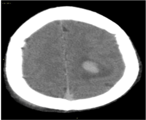
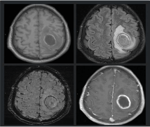
Male patient, 24 years old. He had no apparent cause for dizziness and unconsciousness two days ago. Family members complained of foaming at the mouth, on the way to the hospital, patient’s consciousness gradually cleared and had a headache, dizziness. Right upper limb was weak. Physical examination: T36.2?, the heart rate was 89 beats per minute, breathe was 20 times per minute, Bp115/63 mmHg, conscious, and it’s a chronic ill. The valve could be heard ?-? grade systolic murmur. The two pupils, such as the circle, etc., were sensitive to the light reflection, no resistance to the neck, the right nasal lip groove was a little shallow, the right upper limb was numb, the muscle strength of the right upper limb was 4 levels, the residual muscle was normal. The emergency Computed Tomography(CT) of brain (Figure 1) showed. There is a ovoid high-density shadow in the left parietal lobe, about 2.3 cm × 1.5 cm in size, the CT numerical value is about 63 HU.The boundary is clear, there is a large area of edema around the lesion, and intracranial arteriovenous density increases significantly, considering the hematoma and subarachnoid hemorrhage in the left parietal lobe. He was admitted to the hospital emergency department of cerebral hemorrhage, the Neurology Department. To give him furosemide to promote diuresis, xingnaojing injection and edaravone to nourish his brain, sodium valproate to relieve spasm, continuous oxygen therapy and gastric nursing, etc. No improvement in symptoms after a week. The Magnetic Resonance Imaging (MRI) showed (Figure 2). A circular shadow in the left frontal and parietal, about 3.5 cm × 2.6 cm × 3.4 cm, the lesions with annular high TIWI and low Flair signal, internal T1WI is uneven and Flair signal is slightly high, with lamellar and irregular edema around the nidus, Diffusion Weighted Imaging (DWI) with limited dispersion performance inside. Susceptibility Weighted Imaging (SWI) showed the low signal on the edge of lesions. Enhanced scan assumes the circinate and uniform reinforcement, but it’s inner isn’t enhanced. Midline structure slightly shifts to the right, the subcallosal slightly downward shifts, considering brain abscess.

Figure1. A ovoid high-density shadow in the left parietal lobe, about 2.3 cm x 1.5 cm in size, the CT numerical value is 63HU. The boundary is clear, there is a large area of edema around the lesion, and intracranial arteriovenous density increased significantly.

Figure 2. A circular shadow in the left frontal and parietal about 3.5 cm × 2.6 cm × 3.4 cm, the lesions with annular high TIWI and low Flair signal, internal T1WI is uneven and Flair signal is slightly high, with lamellar and irregular edema around the nidus. Susceptibility Weighted Imaging(SWI) showed the low signal on the edge of lesions. Enhanced scan assumes the circinate and uniform reinforcement, but it’s inner isn’t enhanced. Midline structure slightly shifts to the right, the subcallosal slightly downward shifts.
Echocardiographic examination: Complex congenital heart disease: pulmonary atresia, aortic valvular ventricular septal defect, central atrial septal defect (ASD), right aortic arch. Combined with the related. examinations, considering it’s the combination of congenital heart disease and cerebral abscess.
We gave him meropenem and linezolid to make him more resistant to infection, along with dehydration therapy. Patient had a headache obviously, vomited frequently after strengthening dehydration, poor mental and physical strength, the temperature was up to 40 degrees centigrade, got gradual right hemiplegia. the mind was clear, the right limb muscle strength grade was 2-3, the right Babinski was positive. Planning surgical treatment, he was transferred to neurosurgery. The red blood cells(RBC) was 7.29 T/L, hemoglobin concentration (HGB) reached 204 g/L, hematocrit (HCT) was about 65.7%. The function of liver and kidney, ANCA, the BNP, STD and ENA were basically normal, aspartate amino transferase was about 19 u/L, CK - MB0.5 ng/mL.
The department recommends surgery:
1. Craniotomy, with a great risk, could be life-threatening with cardiopulmonary failure.
2. Drilling drainage, also has a bigger risk, abscess wall is unresectable, may get recurrence.
After repeated instructions to the patient's father, the families choose drilling drainage operation. Drilling drainage tube placement reached the site at the depth of 4cm saw clearly frustrated feeling, extracted about 40ml of viscous liquid. The operation went well, and the patient's clinical symptoms were markedly improved.
Discussion and conclusion
Cerebral abscess is a rare and serious complication of congenital heart disease, which occurs in patients over two years old of non-corrected congenital heart disease [1]. Adult congenital heart disease is rare. Because the blood of this type of patient is from right to left, without the filtration of capillaries, there are more opportunities for patients with asymptomatic bacteraemia to be invaded by bacteria. And because the brain tissue is in a state of chronic hypoxia, the disease caused by erythrocytosis, haemal viscosity heighten causes cerebral blood stream to be slow, and to get the cerebral vascular thrombosis and cerebral infarction, and softening the brain, all of these are more conducive to bacterial growth and brain abscess [2]. The brain abscess is divided into otogenic and rhinogenous cerebral abscess, haematogenous, traumatic and cryptogenic cerebral abscess. The combination of cerebral abscess and cerebral abscess belongs to haematogenous obscess. Pathology can be divided into acute inflammatory stage, suppurative necrosis stage and abscess formation stage.
CT and MRI diagnosis of the disease is extremely helpful, they can show the size, shape, number of abscess, wall thickness and multiple cavity, it also can display the edema of brain abscess, whether there is a placeholder effect and whether there are complications [3]. Brain abscesses are generally single, CT scan is generally characterized by round or irregular low-density shadow, along with slightly high density ring around the legion, this patient of brain abscess in CT scan, characterized by high density, is due to cyanotic type congenital heart disease caused by oxygen for a long time, not enough blood to carry oxygen, resulting methemoglobin hematic disease. Longer time it can be changed in the cerebral vasculature. There are also some scholars believing that cyanotic congenital heart disease can be combined with endocarditis, and the inflammatory embolus can deposit slowly in the blood vessels, which can also lead to higher density of blood vessels and abscesses in the brain. The patient had a sudden onset, and CT density in the intracranial vascular increased widely, and a high-density shadow in the left parietal lobe, were more likely misdiagnosed as cerebral hemorrhage, so we should carefully ask patients’ medical history before diagnosis. The patients who have brain hemorrhage always have typical symptoms, the majority of patients have a history of trauma. Head CT scan shows vascular density with cyanotic congenital heart disease are often higher than the average density of brain abscesses. Patients with severe cases of the disease are similar to the injection contrast agent, which can be misdiagnosed as cerebral hemorrhage, subarachnoid hemorrhage and venous sinus thrombosis and so on [4]. Therefore, the diagnosis of cerebral abscess should be suspected if the CT examination of the congenital heart disease is high in the intracranial density. The main reason which affects the CT value in human blood is the visible protein in RBC and plasma, especially the hemoglobin containing iron, it takes up most of the protein in the blood, also is the main cause of increased blood CT density [5]. Many researchers believe that the CT value of intracranial vessels is directly proportional to the number of HB, that is, the blood vessel density increases when the concentration of HB in the blood increases. Congenital heart disease with brain abscess is treated with antibiotic resistance to infection in the early stage. When the capsule forms, a craniotomy can remove the pus cavity completely, but the risk is very high, and it can cause cardiac dysfunction. Drilling drainage are the preferred method in the treatment of congenital heart disease with brain abscesses, patients can obviously relieve, when the abscess is cured, it should be done as early as possible to avoid the recurrence of brain abscess.
References
- Paixão A, de Andrade FF, Sampayo F (1989) Congenital cardiopathy and cerebral abscess. Acta Medica Portuguesa 2: 73.
- Matson DD, Salam M (1961) Brain abscess in congenital heart disease. Pediatrics 27: 772-789.
- Sun ZQ, Luo LM, Huang WC (2009) Imaging Diagnosis of Brain Abscess in Patients with Congenital Heart Diseases. Clinical Journal of Medical Officers 37: 94-96.
- Liu H, Zhang XT, Wang J, Min T, Yuping H, et al. (2012) CT features on increased cerebral vascular density and its pathological mechanism in patients with cyanotic congenital heart disease. Chinese J Radiol 46: 300-303.
- Yin WW, Xu HZ, Chen W (2008) CT findings in hyperhemoglobinemi a mimicking a contrast-enhanced study. Chinese J Radiol 42: 1265.


