Black/African Americans (AAs) experience inordinate COVID-19 mortality in major cities across the United States (US) compared to other racial ethnic groups [1]. In Chicago for example, although AAs comprise just 30% of the city population, they bear the burden of deaths at 70%. While this striking demographic imbalance is often ascribed to inequalities in health care and insurance coverage, and other social determinants (structural racism, socio-economic status), the biological implications that may also play a role remain incompletely understood. To explain this health disparity, we hypothesize that the current serologic and molecular test kits for SARS-CoV-2 do not account for adaptive viral mutations occurring in a host sector which is demographically distinguishable. This rationale is based on evidence that specialized mutations could in theory impinge on antibody and viral RNA testing consistency in AAs so as to systematically reduce opportunities for prompt clinical interventions. Hence, it is paramount to investigate whether the current COVID test kits are molecularly optimal to confidently detect SARS-CoV-2 in the AA demographic.
We base our hypothesis on the following: for AAs, reliability of present FDA-approved COVID-19 tests may be ineffective due to 1) the high susceptibility of SARS-CoV-2 to error-prone RNA-dependent RNA- polymerases (RNA polymerase) of RNA viruses, yielding mutation reservoirs on which AA demographic selective pressures may act, and from 2) the vulnerability of serologic and viral genome tests to consequent probe-sequence mismatch against the heterogeneous targets queried, increasing false negatives. Relatedly, RNA viruses including coronavirus [2-5], HIV-1 [6-10] etc., via myriad mutations, and over many infection cycles, generate sequentially diverging “quasispecies”, stemming from this faulty proofreading by the viral polymerase [11,12]. Thus, each AA infectee's full array of variant virus would be structurally and temporally unrepeatable [13] but may consistently feature a number of demographically specific virion types. We surmise that in the context of host pressures, underlying inflammatory disease phenotypes (e.g., hypertension, type 2 diabetes) could potentially result in increased error-prone RNA polymerase and viral regulatory gene changes due to elevated virus replicability; this could increase the pool of quasispecies. These sequential divergences could not only produce genetically favored variants (i.e., more pathogenic species, though not the subject of our discussion here), but also escape detection by molecular and serologic screens. Importantly, the production of the pathophysiologically diverse quasispecies contributes to many biomedically-relevant phenomenon, including immune system evasion, vaccine and antiviral inefficacies, failures in virulence, cell tropism and host range restrictions [14-19]. These factors lend urgency to molecular optimization of COVID-19 tests to minimize polymorphism-associated false negative diagnoses in minority populations.
Molecular test
The FDA approved molecular test for SARS-CoV-2 infection which employs reverse transcription (RT) followed by polymerase chain reaction (PCR) is based on the amplification of a selected region of the virus nucleocapsid (N) gene using oligonucleotide primers, whose extension reduces probes conjugated with a reporter dye. In the process, the probe, annealed to a specific target sequence located between the forward and reverse primers, will be degraded in the extension phase of the PCR cycle by the 5’ nuclease activity of Taq polymerase, which causes the reporter dye to separate from the quencher dye, generating a fluorescence signal. As the cycling progresses, the reporter dye molecules are increasingly cleaved from their respective probes, raising fluorescence intensity proportional to virus infection loads (i.e., more viral RNA). Fluorescence intensity is monitored at each PCR cycle by the Applied Biosystems 7500 Fast Dx Real-Time PCR System with SDS version 1.4 software (coronavirus RT-PCR kits – CDC2019-novel coronavirus-FDA). The main concern around use of qPCR-based sensitivity and accuracy, both of which can be affected by mutations or polymorphisms in the primers/probe binding sites. In the context of the SARS- CoV-2 assay, what would happen if the genomic RNA derived from the infected AAs does not perfectly match the primer/probe sequences in the RT-PCR kit due to viral gene mutations? Moreover, mismatch between the 3‘ end of the primer -- where extension initiates -- and the target viral sequence will be especially fatal to amplification, greatly diminishing fluorescence and precluding a positive readout of the infection. Acquired RNA mutations in either primer and/or probe binding sites will affect the accuracy and sensitivity of the diagnostic test; if these mutations are selectively exacerbated in the AA population, then sensitivity of the assay may be compromised and detection of SARS-CoV-2 infection may be missed. This may (1) increase the opportunity for further spread of the virus and (2) negatively affect disease course by delaying treatment.
Serologic test
The serologic test using antibody/antigen suffers an equivalent vulnerability due to the short length of the epitope, where mutations in the viral genomic sequences encoding the target epitope could greatly affect the specificity and sensitivity of serologic (or antibody) test. Serologic tests are designed to detect antibodies that are in serum or plasma components of blood, that are in response to SARS-CoV-2 infection, and that interact with purified SARS-CoV-2 spike (S) protein as antigen (designed by the Vaccine Research Center at the National Institutes of Health). As with the PCR primers, the amino acid length of the epitope to elicit antibody against SARS-CoV-2 is short, making the test vulnerable to blunted resolution by single nucleotide mismatch. Specifically, the length of the epitope presented on the MHC class I is typically 8 to 11 amino acids [20-23], corresponding to the 24 to 33 nucleotides (or 8 to 11 codons), and thus a mutation in one out of 24 to 33 nucleotides, if non-synonymous, would appreciably reduce affinity between an AA’s antibody raised against a distinctive epitope and the antigen probe based on the non-AA span. The identical
failure in outcome could result if the amino acid sequence motif in antigen recognizing antibody were otherwise sufficiently changed by genetic mutation.
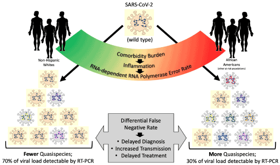
Taking into consideration these potential flaws in diagnostic test design, given the inherent mutational events of SARS-CoV-2, it is imperative to characterize SARS-CoV-2 quasispecies of AAs and examine whether molecular motifs essential for RT-PCR and epitope probes differ at noticeable rates between non-AAs and AAs, ultimately improving the diagnostic accuracy and sensitivity of the serologic and molecular assays. Moreover, meaningful polymorphisms may be incorporated as subpopulation markers to augment subject inclusivity and nationwide diagnostic confidence for the molecular tests. Taken together, investigation of racially divergent viral quasispecies distributions in SARS-CoV-2 pathobiology studies will rectify COVID-19 testing flaws that are believed to aggravate pandemic mortality among AAs (Figure 1).

Figure 1. Representation of the Hypothetical Impact of Comorbidity on SARS-CoV-2 Quasispecies Generation and Testing. Wild type SARS-CoV-2 (the most common form, represented as a yellow virion), serves as the reference RNA sequence for development of a diagnostic RT-PCR assay. However, it is clear that SARS-CoV-2’s RNA polymerase is error prone and is capable of generating quasispecies upon replication (shown as grey virions with various RNA colors). Upon mutation in PCR-targeted sequence(s), the assay’s sensitivity and accuracy of diagnosis is compromised. Here, we propose the possibility that SARS-CoV-2 may exhibit a higher RNA mutation/error rate in response to comorbid conditions and their resultant inflammatory phenotypes. If so, it clearly follows that vulnerable, at-risk populations such as African Americans who endure a heavier burden of comorbid disease (e.g., hypertension) would be adversely affected by the resulting false negative rates. This could be a cryptic contributor to the observed disparity in COVID-19 mortality rates in such populations
- Yancy CW (2020) COVID-19 and African Americans. JAMA [Crossref].
- Rodríguez AK, Garzaro DJ, Loureiro CL, Gutiérrez CR, Ameli G, et al. (2014) HIV-1 and GBV-C co- infection in Venezuela. The Journal of Infection in Developing Countries 8: 863-868.
- Tang JW, Cheung JL, Chu IM, Sung JJ, Peiris M, et al. (2006) The large 386-nt deletion in SARS-associated coronavirus: evidence for quasispecies? J Infect Dis 194: 808-813. [Crossref]
- Xu D, Zhang Z, Wang FS (2004) SARS-associated coronavirus quasispecies in individual patients. N Engl J Med 350: 1366-1367. [Crossref]
- Briese T, Mishra N, Jain K (2014) Middle East respiratory syndrome coronavirus quasispecies that include homologues of human isolates revealed through whole-genome analysis and virus cultured from dromedary camels in Saudi Arabia. mBio 5: e01146-14 [Crossref]
- Yu F, Wen Y, Wang J, Gong Y, Feng K, et al. (2018) The transmission and evolution of HIV-1 quasispecies within one couple: a follow-up study based on next-generation sequencing. Sci Rep 8: 1-8. [Croosref]
- Collins KR, Quiñones-Mateu ME, Wu M, Luzze H, Johnson JL, et al. (2002) Human immunodeficiency virus type 1 (HIV-1) quasispecies at the sites of Mycobacterium tuberculosis infection contribute to systemic HIV-1 heterogeneity. J Virol 76: 1697-1706.
- Dampier W, Nonnemacher MR, Mell J, Earl J, Ehrlich GD, et al. (2016) HIV-1 genetic variation resulting in the development of new quasispecies continues to be encountered in the peripheral blood of well-suppressed patients. PloS one 11: e0155382. [Crossref]
- Liu Y, Jia L, Su B, Li H, Li Z, et al. (2020) The genetic diversity of HIV-1 quasispecies within primary infected individuals. AIDS Res Hum Retroviruses 36: 440-449. [Crossref]
- de Azevedo SS, Caetano DG, Côrtes FH, Teixeira SL, dos Santos Silva K, et al. (2017) Highly divergent patterns of genetic diversity and evolution in proviral quasispecies from HIV controllers. Retrovirology 14: 1-13.
- Steinhauer DA, Domingo E, Holland JJ (1992) Lack of evidence for proofreading mechanisms associated with an RNA virus polymerase. Gene 122: 281-288. [Crossref]
- Bernad A, Blanco L, Lázaro J, Martin G, Salas M (1989) A conserved 3′→ 5′ exonuclease active site in prokaryotic and eukaryotic DNA polymerases. Cell 59: 219-228. [Crossref]
- Holland JJd, De La Torre J, Steinhauer D (1992) RNA virus populations as quasispecies. Genetic diversity of RNA viruses: Springer pp:1-20.
- Domingo E, Perales C (2019) Viral quasispecies. Plos Genetics 15: e1008271.
- Spindler K, Horodyski F, Grabau E, Nichol S, Vandepol S (1982) Rapid evolution of RNA genomes. Science 215: 1577-1585. [Crossref]
- Holland J (2006) Transitions in understanding of RNA viruses: a historical perspective. Quasispecies: Concept and implications for virology: Springer. pp: 371-401.
- Domingo E, Perales C (2018) Quasispecies and virus. European Biophysics Journal 47: 443-457.
- Domingo E (1989) RNA virus evolution and the control of viral disease. Prog Drug Res 33: 93-133. [Crossref]
- Briones C, Domingo E (2008) Minority report: hidden memory genomes in HIV-1 quasispecies and possible clinical implications. AIDS Rev 10: 93-109. [Crossref]
- Craig L, Sanschagrin PC, Rozek A, Lackie S, Kuhn LA, et al. (1998) The role of structure in antibody cross-reactivity between peptides and folded proteins. J Mol Biol 281: 183-201.
- Steers NJ, Currier JR, Jobe O, Tovanabutra S, Ratto-Kim S, et al. (2014) Designing the epitope flanking regions for optimal generation of CTL epitopes. Vaccine 32: 3509-3516.
- Trolle T, McMurtrey CP, Sidney J, Bardet W, Osborn SC, et al. (2016) The length distribution of class I–restricted T cell epitopes is determined by both peptide supply and MHC allele–specific binding preference. J Immunol 196: 1480-1487. [Crossref]
- Lundegaard C, Lund O, Buus S, Nielsen M (2010) Major histocompatibility complex class I binding predictions as a tool in epitope discovery. Immunology 130: 309-318. [Crossref]

