Introduction: Trismus or lock jaw is one of the frustrating problems not only for patients but also for the treating doctors. Various treatment options available for trismus only have temporary results. In this original article, nasolabial flap was used for covering the raw area of buccal mucosa and pterygomandibular raphe after releasing the trismus.
Methodology: Total of six patients (4 male, 2 female) with juxta-articular trismus were selected and trismus release was done transorally along with covering of the raw area of buccal mucosa with inferiorly pedicled nasolabial flap. Postoperatively all the patients were kept on regular follow up for 6 months thorough physiotherapy.
Results: Mouth opening improved in all the patients. Mouth opening improved from 8.5 mm (range 7-9 mm) preoperatively to 23.5mm (20 to 26 mm) at 6 months postoperative follow up. No intraoperative, postoperative complications were recorded in the series.
Conclusion: Nasolabial flap is a versatile, easily available, well vascularised flap which needs little surgical dexterity. When used for trismus, it gives good results not only in terms of mouth opening but also replacing all the diseased buccal mucosa in submucosal fibrosis which is a niche for malignant transformation there by improving patient’s quality of life.
nasolabial flap, juxta articular, trismus, pterygomandibular raphe, submucous fibrosis
First description of nasolabial flap was in Sushrutha’s book, Sushrutha Samhita which is one of the ancient books in medicine [1]. Initial descriptions of nasolabial flaps were primarily superiorly based for nasal alar reconstruction and dorsum of nose [2]. Essar in early nineteenth century described the use of inferiorly based nasolabial artery to close palatal fistula. Superiorly based flap receives random blood supply from infraorbital artery and transverse facial artery whereas inferior based flap receives blood supply from superolabial and alar branches of facial artery [3]. Inferiorly based nasolabial flap is a pedicled, locally available, reliable and economical option for single time treatment of non-articular causes of trismus especially secondary to trauma and oral submucosal fibrosis.
Study design
A cross sectional study done between august 2016 to June 2018 in a single multidisciplinary teaching hospital from South India after obtaining clearance from the institute review board.
Inclusion criteria
A total of six patients were selected with trismus secondary to trauma (iatrogenic or accidental) and oral submucosal fibrosis.
Exclusion criteria
Trismus secondary to Temporomandibular joint involvement by doing a clinical evaluation from oromaxillofacial team and Orthopantomogram.
A total of six patients were surgically treated after obtaining informed written consent and due permission from the institutional ethics committee. Patients’ demographics and preoperative maximum inter incisior distance (Figure 1a) were illustrated in table 1. All the patients were clinically evaluated by Oromaxillofacial team and Digital Orthopantomogram was taken to rule out intra-articular causes of trismus (Figure 1b).
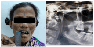
Figure 1. A) Preoperative maximum interincisor distance. B) Preoperative orthopantomogram
Table 1. Illustration of patient demographics
Patient |
Age/Sex |
Etiology |
Side |
Preoperative IID (in mm) |
Case 1 |
40 / F |
Dental abscess, post dental surgery |
Unilateral |
7 |
Case 2 |
24/M |
Oral submucous fibrosis |
Bilateral |
9 |
Case 3 |
26/M |
Oral submucous fibrosis |
Bilateral |
8 |
Case 4 |
28/F |
Trauma to RMT during childhood |
Unilateral |
7 |
Case 5 |
24/M |
Corrosive ingestion in childhood |
Unilateral |
9 |
Case 6 |
25/M |
Oral submucosal fibrosis |
Bilateral |
8 |
IID- Inter Incisor Distance, mm- millimeters
Preoperative Doppler was used in all cases to record the flow in the facial artery at the mandibular margin (Figure 2a). Patients were followed up to 6 months postoperatively and maximum interincisior distance measured with a measuring scale in millimetres.
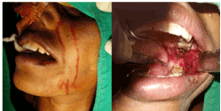
Figure 2. A) Marking of nasolabial flap with pedicle. B) After ablation of scar tissue in the buccal space and retromolar trigone
All cases were taken under general anaesthesia with flexible laryngoscopic guided nasotracheal intubation. Inferiorly based nasolabial flap was used in all the cases. The scar tissue in the buccal mucosa, fibrous bands and pterygomandibular raphe are ablated in all cases with radiofrequency ablater and hemostasis achieved with bipolar diathermy as shown in figure 2b. Intraoperative maximum interincisor distance was measured in all patients after release of trismus. Skin marking for nasolabial flap and corresponding facial artery were made as shown in figure 2a. The nasolabial flap was raised above the plane of facial musculature with cold instruments and cautery is avoided to decrease thermal damage to the flap (Figure 3a). An optimum sized tunnel is made behind the posterior margin of the orbicularis oris to communicate with buccal space (Figure 3b). Nasolabial flap is brought into the buccal space through this tunnel (Figure 3b). The proximal most skin at the base of the flap trimmed meticulously avoiding injury to the underlying pedicle. This segment is undermined, and donor site primarily closed. The margins of the buccal mucosa and flap are approximated and closed primarily. Physiotherapy was started from seventh postoperative day and asked to continue for 6 months to avoid scar contracture. All the patients were followed up to 6 months and their maximum interincisor distance is measured. Figure 4 showing 6 months follow up after surgery in case 1.
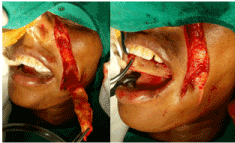
Figure 3. A) Nasolabial flap ready to be tunneled. B) Flap tunnel into the buccal space behind the orbicularis oris muscle
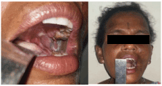
Figure 4. Six months postoperative follow up picture showing well healed flap at retromolar trigone and minimal visible scar on the face
Mouth opening improved in all the patients. No intraoperative and perioperative complications were encountered in any of them. The average preoperative IID noted in the series is 8.6 mm (7-9 mm). All the patients have an IID more than 35 mm intra operatively after releasing the trismus bands. One-month postoperative average IID noted was 25.2 mm (20-28 mm) and at 6 months, average IID was 23.5 mm (20 to 26 mm). The comparison of the preoperative, one and six-month postoperative interincisor distance is illustrated in table 2. Three out of four male patients had scarce intraoral hair growth on the flap which was not troublesome according to the patient.
Table 2. Comparison of preoperative and postoperative IID
Patient |
Preoperative IID (in mm) |
Postoperative IID at 1 month (in mm) |
Postopertive IID at 6 months (in mm) |
Case 1 |
7 |
25 |
23 |
Case 2 |
9 |
25 |
23 |
Case 3 |
8 |
28 |
26 |
Case 4 |
7 |
20 |
20 |
Case 5 |
9 |
26 |
25 |
Case 6 |
8 |
27 |
24 |
IID- Inter Incisor Distance, mm-millimeters
Trismus is limitation in opening of mouth due to reduced mandibular mobility. Trismus can be secondary to intra-articular causes like temperomandibular ankylosis, arthritis and meniscal pathology. Most common extraarticular causes are oral submucosal fibrosis, scarring secondary to dental infections, dental surgeries and post-traumatic causes [4]. The cause of trismus in the present series was oral submucosal fibrosis in 60% (3/5), post traumatic in 33.3% (2/6) and iatrogenic in 16.6% (1/6) of patients. Most of the treatment options for trismus are conservative. Most common conservative measures include mouth opening exercises using serial incremental use of ice cream sticks, therabite systems, dynasplint trismus systems, where the later options are not always suitable for low socioeconomic groups [5,6]. There are also minimal invasive measures like various injections like steroids, hyaluronic acid and placental extracts into the scarred retromolar trigone region to slacken the fibrous bands. Although these injections produce temporary relief, they may further worsen the condition due to repeated trauma [7]. Various surgical procedures have been proposed for trismus ranging from simple release of fibrous bands to reconstruction by pedicled free flaps. Most commonly performed procedure for early trismus due to oral submucosal fibrosis is release of fibrous bands followed by aggressive mouth opening exercises which will have high recurrence due to rebound fibrosis during healing [8]. Khanna et al. [9] proposed a technique of palatal island flap for trisums. However, he mentioned the main limitations of this flap include less available donor tissue along with limited reach of the pedicle. Tepan et al. [10] used tongue flaps for covering the raw area in trismus due to submucosal fibrosis with limitations include dysphasia, dehiscence in postoperative period, and chance of aspiration. Free radial forearm flaps are also used in trismus but because of its complexity, high cost, long duration of surgery and donor site morbidity this flap is not a practical one all the time [12].
Nasolabial flaps are versatile, quick, can easily be harvested and locally available flaps. Moreover, the donor site can easily be closed without need for complex reconstruction work. The skin over the nasolabial fold is nourished by multiple feeders from the superior labial artery, infraorbital artery and near the dorsum of nose from the angular artery. The inferior based nasolabial flap with facial artery as its pedicle can be used to reconstruct floor of mouth, buccal mucosa and retromolar trigone area [2]. Mohit et al. [12] also used extended nasolabial flap for covering the raw area after removal of fibrous bands secondary to oral submucous fibrosis in 28 patients. The interincisor distance improved from 11 mm before surgery to 39mm at the end of 6 months. In our series, we noted an improvement from 8.6 mm to 23.5 mm at the end of 6 months. As stated by others, all our patients also received aggressive physiotherapy for 6 months [12,13].
In patients with oral submucous fibrosis, improvement in mouth opening helps in regular examination and early detection of malignant transformation. Posterior buccal mucosa is the most common site for malignant transformation in oral submucous fibrosis as it has chronic irritation from the misaligned maxillary molars. As nasolabial flaps replace this part of the buccal mucosa the chances of this area to get involved in malignant transformation is less [12]. Take in time for nasolabial flaps is excellent with minimal flap failure because of robust blood supply. Growth of intraoral hair which was reported by other authors is also seen in our series at the end of 3 months. As the flap is not sensate it has not troubled the patients and the hair has become scarce at 6 months follow up visit. Postoperative complications like palatal perforation, partial flap necrosis and are not seen in our series as noted by others [12].
Nasolabial flap is the best option for trismus secondary to extra-articular causes. This flap can be easily adaptable, harvested with minimal surgical dexterity unlike other regional and free flaps. The take in time for this flap is excellent with very less complication rate owing to the robust blood supply. It is a cost-effective option for developing countries like India even in modern day practice.
- Pers M (1967) Cheek flaps in partial rhinoplasty: a new variation: the in-and-out flap. Scand J Plast Reconstr Surg 1: 37-44.
- Schmidt BL, Dierks EJ (2003) The nasolabial flap. Oral Maxillofac Surg Clin 15: 487-495. [Crossref]
- Barthelemy I, Paoli JR, Boutault F, Fabie M (1996) A superiorly pedicled nasobuccal flap. Its value in the reconstruction of posterior-superior loss of substance of the oral mucosa. Rev Stomatol Chir Maxillofac 99: 217-220. [Crossref]
- Dhanrajani PJ, Jonaidel O (2002) Trismus: aetiology, differential diagnosis and treatment. Dent Update 29: 88-94. [Crossref]
- Kamstra JI, Roodenburg JLN, Beurskens CHG, Reintsema H, Dijkstra PU (2013) Thera Bite exercises to treat trismus secondary to head and neck cancer. Support Care Cancer 21: 951-957. [Crossref]
- Kamstra JI, Reintsema H, Roodenburg JLN, Dijkstra PU (2016) Dynasplint Trismus System exercises for trismus secondary to head and neck cancer: a prospective explorative study. Support Care Cancer 24: 3315-3323. [Crossref]
- Borle RM, Borle SR (1991) Management of oral submucous fibrosis: a conservative approach. J Oral Maxillofac Surg 49: 788-791. [Crossref]
- Kamath VV (2015) Surgical interventions in oral submucous fibrosis: a systematic analysis of the literature. J Maxillofac Oral Surg 14: 521-531. [Crossref]
- Khanna JN, Andrade NN (1995) Oral submucous fibrosis: a new concept in surgical management: report of 100 cases. Int J Oral Maxillofac Surg 24: 433-439. [Crossref]
- Tepan MG, Saigal GS, Tilak SB (1986) Use of tongue flap in submucous palatal fibrosis. J Laryngol Otol 100: 455-460. [Crossref]
- Wei FC, Chang YM, Kildal M, Tsang WS, Chen HC (2001) Bilateral small radial forearm flaps for the reconstruction of buccal mucosa after surgical release of submucosal fibrosis: a new reliable approach. Plast Reconstr Surg 107: 1679-1683. [Crossref]
- Agarwal M, Gupta DK, Tiwari AD (2011) Extended nasolabial flaps in the management of oral submucous fibrosis. J Maxillofac Oral Surg 10: 216-219. [Crossref]
- Thakur S, Deepthi SH, Gupta K, Randhawa G (2016) The Nasolabial flap in oral submucosal fibrosis- a case series. Int J Prevent Clinical Dental Res 3: 228-331.
Editorial Information
Editor-in-Chief
Chin-Lung Kuo
Taoyuan Armed Forces General Hospital
Taiwan
Article Type
Research Article
Publication history
Received date: June 19, 2020
Accepted date: July 07, 2020
Published date: July 10, 2020
Copyright
©2020 Kumar L. This is an open-access article distributed under the terms of the Creative Commons Attribution License, which permits unrestricted use, distribution, and reproduction in any medium, provided the original author and source are credited.
Citation
Kumar L, Bhasker P, Pol SA (2020) Inferiorly pedicled nasolabial flap for extra-articular trismus. Otorhinolaryngol Head Neck Surg 5: doi: 10.15761/OHNS.1000247
Corresponding author
Shashikant Anil Pol
Department of ENT, JIPMER, Puducherry, UT, India
E-mail : bhuvaneswari.bibleraaj@uhsm.nhs.uk
Figure 1. A) Preoperative maximum interincisor distance. B) Preoperative orthopantomogram
Figure 2. A) Marking of nasolabial flap with pedicle. B) After ablation of scar tissue in the buccal space and retromolar trigone
Figure 3. A) Nasolabial flap ready to be tunneled. B) Flap tunnel into the buccal space behind the orbicularis oris muscle
Figure 4. Six months postoperative follow up picture showing well healed flap at retromolar trigone and minimal visible scar on the face
Table 1. Illustration of patient demographics
Patient |
Age/Sex |
Etiology |
Side |
Preoperative IID (in mm) |
Case 1 |
40 / F |
Dental abscess, post dental surgery |
Unilateral |
7 |
Case 2 |
24/M |
Oral submucous fibrosis |
Bilateral |
9 |
Case 3 |
26/M |
Oral submucous fibrosis |
Bilateral |
8 |
Case 4 |
28/F |
Trauma to RMT during childhood |
Unilateral |
7 |
Case 5 |
24/M |
Corrosive ingestion in childhood |
Unilateral |
9 |
Case 6 |
25/M |
Oral submucosal fibrosis |
Bilateral |
8 |
IID- Inter Incisor Distance, mm- millimeters
Table 2. Comparison of preoperative and postoperative IID
Patient |
Preoperative IID (in mm) |
Postoperative IID at 1 month (in mm) |
Postopertive IID at 6 months (in mm) |
Case 1 |
7 |
25 |
23 |
Case 2 |
9 |
25 |
23 |
Case 3 |
8 |
28 |
26 |
Case 4 |
7 |
20 |
20 |
Case 5 |
9 |
26 |
25 |
Case 6 |
8 |
27 |
24 |
IID- Inter Incisor Distance, mm-millimeters