Abstract
More than 10% of cirrhotic patients will require surgeries during the last two years of life. Among the causes of morbidity and mortality, most are derived from portal hypertension. The Transjugular Intrahepatic PortoSystemic Shunt (TIPS) has been used before abdominal surgeries to reduce portal hypertension, including the gastroesophageal junction (GEJ), the most common site of collateral circulation. The case of a patient with portal hypertension and esophageal-gastric varices who underwent installation of TIPS as a bridge therapy to total gastrectomy is presented. The results show a reduction of esophagogastric varices, allowing esophagojejunoanastomosis to be performed without bleeding and in tissue not damaged by endoscopic ligatures.
This study proposes using TIPS as temporary therapy in cirrhotic patients with complications associated with portal hypertension (PH) prior to abdominal surgeries that include GEJ. Since this anatomical site is the most affected by varicose disease and the management of this pathology with endoscopic therapy causes alterations in the anatomy and histological characteristics of the distal esophagus, which impoverishes the quality of the results and is associated with a higher rate of complications. While TIPS therapy is a minimally invasive procedure, it would reduce both intraoperative and postoperative complications: bleeding, anastomosis dehiscence, infections, prolonged surgical time, among others. In this way, it would increase the spectrum of patients who are candidates for surgical resolution, the rate of successful procedures, and reduce associated mortality.
The presentation or exacerbation of complications such as encephalopathy and liver dysfunction do not currently position it as the therapy of choice, so despite the favorable results, more experience and larger cohorts are required to estimate the adequate performance of this therapy accurately.
key words
portal hypertension, esophageal varices, systemic porto shunt
Introduction
More than 10% of cirrhotic patients will require surgeries during the last two years of life, sometimes highly complex abdominal surgeries. However, high surgical mortality rates have been reported in this type of patient [1-4]. The mortality of cirrhotic patients electively undergoing extrahepatic abdominal surgeries has been estimated to be between 10% and 57%. Consequently, numerous potentially curative surgical procedures are contraindicated in cirrhotic patients [5,6]. On the other hand, Child A cirrhotic patients have a 10% postsurgical mortality, Child B 30% - 31%, and 76 - 82% for Child C cirrhotic [7].
The complications that most frequently determine mortality are perioperative hemorrhage, sepsis, operative wound complications, coagulopathy, and kidney failure. Each of these derives in its entirety or an essential part from a common pathophysiological phenomenon, portal hypertension [2,6,8].
Portal hypertension (PH) is defined as an increase in the pressure of the portal venous system above five mmHg, which can cause numerous complications [9]. Among them are the promotion of the development of collateral circulation within the peritoneal cavity, varicose veins, and splenomegaly. The above, together with the coagulation disorders secondary to hypoprothrombinemia and thrombocytopenia present in these patients, cause an increased risk of perioperative bleeding [3,5,8]. On the other hand, due to its hypervolemic state and splanchnic vasodilation, PH produces ascites, frequently contributing to operative wound infections and sepsis. In addition, increasing intra-abdominal pressure promotes the dehiscence of operative wounds [4,6].
There is a growing interest in effective preoperative methods that lower portal pressure in cirrhotic patients and reduce associated mortality. Given the effectiveness of portal pressure reduction through preoperative portosystemic shunts and the reduction of perioperative complications of cirrhotic patients, it has postulated TIPS as a possible portal decompression tool to its minimally invasive nature [3,6,10].
In the literature, TIPS have been described before abdominal surgeries of various parenchyma, including the stomach. However, there are no records of surgeries that technically involve the GEJ [2,5,6,8], this being the most common site for the development of collateral circulation [9,10]. Therefore, it makes us wonder if the use of TIPS can go beyond the indications described so far. Thus, the objective of this study is to present the case of a patient with PH and esophageal-gastric varices who underwent the installation of TIPS as bridging and neoadjuvant therapy to total gastrectomy with subsequent reconstruction with esophagus jejunum anastomosis.
Case report
A 52-year-old male patient with a history of chronic liver damage due to alcohol, classified as Child A and MELD 8, consulted for upper gastrointestinal bleeding. Upper gastrointestinal endoscopy was performed, which observed numerous varices at the level of the gastroesophageal junction (GEJ) (Figure 1). Endoscopic ligation was managed to control the bleeding; however, multiple lesions were observed in the fundus and gastric body during the procedure, compatible with a multifocal neuroendocrine tumor (Figure 2). The histopathological study concluded the presence of a neuroendocrine tumor with Ki67 greater than 5%.
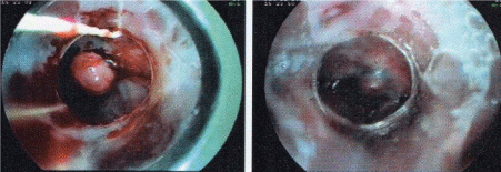
Figure 1. Endoscopy shows the presence of high-risk esophageal varices and ligation in situ in the distal esophagus
Subsequently, the etiological and dissemination study is carried out. Computed tomography (CT) of the abdomen and pelvis showed numerous peri gastric adenopathy (the largest of 13 mm). Also, simple liver cysts in segments 2 and 4, PHT, and splenomegaly were detected. Chest CT did not reveal alterations, and serological studies for HBV and HCV were negative.
Total gastrectomy with curative intent is indicated based on the pathological findings, upper gastrointestinal endoscopy, and negative dissemination study. However, its performance implies a high risk of variceal bleeding, putting the patient’s life at risk. On the other hand, despite its effectiveness, endoscopic treatment of gastroesophageal varices is not an adequate strategy in this patient because it causes anatomical distortion, and complex surgical management in the tissues esophagojejunanastomosis will be performed. In this context, and to achieve curative treatment, we decided to decrease portal pressure prior to surgery by installing TIPS (Figure 3). Specifically, we proceeded to install TIPS portocava, which passed without incident.
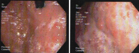
Figure 2. Multiple multifocal nodular lesions in the body and gastric fundus
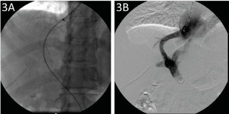
Figure 3. Radiological evidence of TIPS installation. (A) Fluoroscopy in which the release of a self-expanding metallic stent 10 mm x 8 cm is visualized with its ends in the right suprahepatic vein and the right portal vein (white arrow). The access with a 5 French sheath in the right portal vein branch is also identified. (B) Fluoroscopy with subtraction showing permeability of the TIPS, well-positioned. A metallic stent is observed, with an in situ measurement system and access with a 5 French sheath in the right portal vein branch
Results
It was possible to reduce the portal pressure from 22 to 11 mmHg, while the pressure in the vena cava showed an increase from 0 to 6 mmHg after the shunt. In sum, a total portosystemic gradient of 22 - 5 was obtained. Despite the promising results, the patient developed grade 3 encephalopathy, a known complication of this procedure, so this did not prevent the planned surgical intervention from being carried out.
Total gastrectomy with D2 lymphadenectomy was performed on the fifth day without incident. During the intraoperative period, a significant decrease in the number and size of the esophagogastric varices was observed, which allowed safe handling of the operative area during the esophageal section. Esophageal tissue with ligature scars and fibrosing was identified that favored subsequent anastomosis. This anastomosis was performed with a 25-mm circular mechanical suture without incidents (Figures 4 and 5).
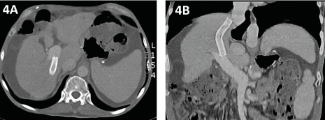
Figure 4. (A) Recent postsurgical axial section images in portal phase, identifying patent TIPS, postgastrectomy postoperative changes, and moderate ascites. (B) Coronal MIP reconstruction showing TIPS with ends in the right suprahepatic vein - inferior vena cava and right portal vein. Coronal MIP reconstruction is well located and permeable
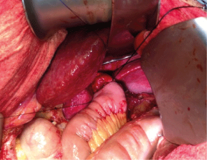
Figure 5. Intraoperative process: the findings highlight the left liver lobe with signs of chronic liver damage. In addition, the esophagus-jejunal anastomosis is visualized
The patient evolved favorably, with no anastomotic leakage or other complications during the postoperative period. The patient was discharged on the eighth postoperative day. At the last outpatient review, the patient tolerated feeding well and showed no signs of hepatic encephalopathy.
Discussion
In 1981, Schwartz demonstrated a decrease in intraoperative bleeding in cirrhotic patients with portal hypertension by performing, concomitant to the primary surgery, a Porto-cavus shunt [3]. Similarly, Sandblom describes using a Porto-cavus shunt, this time prior to surgery, in four patients with ascites who required a biliodigestive anastomosis. Once the "pre-surgical" Porto-cavus shunt had been performed and the ascites controlled, the surgery was performed without complications [5].
TIPS or transjugular hepatic portosystemic shunt is the procedure by which the portal vein is decompressed by creating a low resistance channel, usually between the right hepatic vein and the right portal vein through a fenestrated metal implant, which may or may not be covered, which crosses the cirrhotic liver parenchyma. Angiography-guided endovascular route is installed, which avoids performing a laparotomy, takes approximately one to two hours, and is technically successful in most cases with low complication rates [9].
Currently, TIPS has an established and widely accepted role as temporary therapy for patients awaiting liver transplantation and as a treatment for some complications of portal hypertension [6,9]. Specifically, prevention of recurrent variceal bleeding and salvage therapy in patients with acute variceal bleeding when pharmacological and endoscopic therapy has failed [6,8,9,11]. However, the indications have been increasing due to the evidence of its effectiveness in various pathologies. Other indications for TIPS that show promise are refractory ascites, Budd Chiari syndrome, severe hypertensive portal gastropathy, hepatorenal syndrome, refractory hepatic hydrothorax, and early therapy in cirrhotic patients with esophageal varices prior to the first episode of HDA [6, 11,12].
In the literature included in this review, there are few reports and case series in which the use of TIPS is described as part of the pre-surgical preparation in extrahepatic abdominal surgeries. However, there are descriptions of TIPS prior to cardiac, esophageal, renal, ureteral, pancreatic, colonic, hernioplasty, retroperitoneal, abdominal, gallbladder, orthopedic, rectal, aortic, pelvic, ovarian, and two gastric surgeries (in which performed a distal subtotal gastrectomy, which technically does not involve the gastroesophageal junction) 2. There are no reported cases of TIPS use within these series before total gastrectomy [2,5,6,8].
Through this report, we present the case of a patient with DHC who, in the context of variceal UGH, detected numerous gastric lesions compatible with neuroendocrine neoplasms. Due to the finding, the need arises for total gastrectomy for curative oncological purposes. However, due to numerous large varices in the GEJ, where the esophagojejunanastomosis is performed, surgery is relatively contraindicated due to a high risk of bleeding.
In the same way, as the case presented, all the cases reported in the literature on using TIPS as a preoperative preparation were performed in patients whose main contraindication to extrahepatic abdominal surgery was portal hypertension refractory to medical treatment. In these cases, the literature is evident in recommending endoscopic variceal ligation prior to surgery to reduce the risk of bleeding. During this, meticulous handling is recommended since varicose veins are very thin-walled vessels subjected to high pressure, which in minor lacerations can cause massive bleeding [13]. In our case, where the surgical technique involves esophagogastric section and subsequent esophagojejunal anastomosis, variceal treatments using EDA cause anatomical distortion and local tissue damage that are not recommended.
In this specific clinical context, the indication for using TIPS as an alternative method of reducing portal pressure and treating esophagogastric varices is based and justified. The above is under the premise of the minimally invasive nature that does not increase the risk of bleeding [6] and, in turn, does not distort the anatomy of one of the key points to be manipulated during a total gastrectomy, the distal esophagus.
TIPS stent installation was performed five days before total gastrectomy, an uneventful procedure. Then, surgical intervention was carried out, which showed a significant decrease in collateral circulation and gastroesophageal varices. This surgical intervention allowed the esophagogastric section to be carried out without incident, with minimal bleeding, and to perform the esophagojejunanastomosis in the adequate tissue, thus reducing the possibility of bleeding, dehydration, and anastomotic leakage, achieving a total gastrectomy plus lymphadenectomy with free surgical margins. Regarding the results in the literature, the series has shown improvements in the various indicators of PH and pre-surgical prognostic factors of mortality [5,6,8,14]. Significant improvements have been reported in the Child score, MELD, a decrease in the level of ascites, and a lower rate of operative wound complications [13]. In addition, some series have measured the decrease in portosystemic gradients after TIPS, which have decreased abruptly, an average of 18 mmHg [6].
Conclusion
We postulate TIPS as an effective and safe neoadjuvant therapy for total gastrectomies. Like other neoadjuvant treatments, it allows us to offer curative treatments simplified and at a lower rate of complications to those who previously only offered palliative therapy. Always bear in mind that it is a case presentation and literature review.
Due to the limited number of reported cases, there is considerable variability in the management and treatment protocol of these patients, with significant variations still existing between types of patients, comorbidities, clinical contexts, the time between pre-surgical TIPS and main surgery, type of surgery, among other. Therefore, we believe that more experience and larger cohorts are required to accurately estimate the proper performance of this therapy, optimize it, and generate unique treatment protocols.
Conflict of interest
The authors declare that they have no conflict of interest.
References
- Probst A, Probst T (1995) Prognosis and life expectancy in chronic disease. Dig Dis Sci 40: 1805-1815.
- John J Kim, Narasimham L Dasika, Esther Yu, Robert J Fontana (2009) Cirrhotic patients with a transjugular intrahepatic portosystemic shunt undergoing major extrahepatic surgery. J Clin Gastroenterol 43: 574-579. [Crossref]
- Schwartz SI (1981) Biliary tract surgery and cirrhosis: a critical combination. Surgery 90: 577-583.
- Aranha GV, Sontag SJ, Greenlee HB (1982) Cholecystectomy in cirrhotic patients: a formidable operation. Am J Surg 143: 55-60. [Crossref]
- Daniel Azoulay, Fernando Buabse, Ivana Damiano, Alaoua Smail, Philippe Ichai, et al. (2001) Neoadjuvant transjugular intrahepatic portosystemic shunt: A solution for extrahepatic abdominal operation in cirrhotic patients with severe portal hypertension. J Am Coll Surg 193: 46-51.
- A Gila, F Martínez-Regueiraa, J L Hernández-Lizoaina, F Pardoa, JM Oleaa, et al. (2004) The role of transjugular intrahepatic portosystemic shunt prior to abdominal tumoral surgery in cirrhotic patients with portal hypertension. EJSO 30: 46-52. [Crossref]
- Garrison RN, Cryer HM, Howard DA, Polk HC Jr (1984) Clarification of risk factors for abdominal operations in patients with hepatic cirrhosis. Ann Surg 199: 648-655.
- Christine Schlenker, Stephen Johnson, James F Trotter (2009) Preoperative transjugular intrahepatic portosystemic shunt (TIPS) for cirrhotic patients undergoing abdominal and pelvic surgeries. Surg Endosc 23: 1594-1598.
- Bilbao JI, Quiroga J, Herrero JI, Benito A (2002) Transjugular intrahepatic portosystemic shunt (TIPS): current status and future possibilities. Cardiovasc Intervent Radiol 25: 251-269.
- Rossle M, Ochs A, Gulberg V (2000) A comparison of paracentesis and transjugular intrahepatic portosystemic shunting in patients with ascites. N Engl J Med 342: 1701-1707. [Crossref]
- Rosado B, Kamath PS (2003) Transjugular intrahepatic portosystemic shunts: an update. Liver Transpl 9: 207-217.
- Juan Carlos García-Pagán, Karel Caca, Christophe Bureau, Wim Laleman, Beate Appenrodt, et al. (2010) Early use of TIPS in patients with cirrhosis and variceal bleeding. N Engl J Med 362: 2370-2379. [Crossref]
- Gene Y Im, Nir Lubezky, Marcelo E Facciuto, Thomas D Schiano (2014) Surgery in patients with portal hypertension a preoperative checklist and strategies for attenuating risk. Clin Liver Dis 18: 477-505. [Crossref]
- Moulin G, Champsaur P, Bartoli JM, Chagnaud C, Rousseau H, et al. (1995) TIPS for portal decompression to allow palliative treatment of adenocarcinoma of the esophagus. Cardiovasc Intervent Radiol 18: 186-188.


