Objective: Brachytherapy is an essential component in the definitive treatment of locally advanced cervical cancer to improve local control (LC) and overall survival (OS). This technique requires the placement of the intrauterine tandem through the cervical orifice, which can lead to perforation uterine. The objective of this study is to show the role and benefits of intraoperative ultrasound guidance in cervical brachytherapy.
Materials and methods: A prospective study conducted on 67 patients with locally advanced cervical cancer treated with concurrent chemoradiation followed by intracavitary brachytherapy using ultrasound for real-time assessment of tandem placement in 152 insertions.
Results: The median age of the patients was 52.6 years (33-77). Among the 152 insertions, 3 perforations were detected with a rate of 1.9%. One was on the anterior wall of the uterus, one on the lateral wall, and the last one on the uterine fundus.
Conclusion: intraoperative ultrasound guiding the application is an easy method to provide sure and efficient data to reduce the risk of uterine perforations and the wrong position of the tandem.
cervical cancer, intracavitary brachytherapy, intraoperative ultrasound, tandem, perforation.
CCRT: concurrent chemoradiotherapy; CT: computed tomography; FIGO: international federation of gynecology and obstetrics; Gy: gray; Hb: hemoglobin; HDR: High Dose Rate; HPV: human papillomavirus; ICBT: intra-cavitary brachytherapy; LC: local control; MRI: magnetic resonance imaging; OS: overall survival
Cervical cancer is a major health problem. It is common in developing countries, which HPV infection is the main risk factor. The diagnosis is often made at an advanced stage. Definitive Cisplatin-based concurrent chemoradiotherapy (CCRT) followed by intra-cavitary brachytherapy (ICBT) is standard treatment [1]. The technique of ICBT consists of placing a tandem in the uterine cavity often blindly advancing it until feeling the uterine fundus. Uterine perforation is the main peroperative complication of this technique with rates ranging from 2 to 14% [2-4], which may alter local control of the tumor. Data suggest that the routine use of intraoperative ultra-sound facilitates ideal tandem placement and decreases the risk of uterine perforation, thereby diminishing an underappreciated source of toxicity while optimizing disease control [5,6]. Granai et al. [7] reported on routine intraoperative ultra-sound for 72 patients and noted no clinically evident perforations. Rotmensch et al. [8] investigated the use of intraoperative ultrasound for applicator placement in 20 implants. Unsatisfactory placement was detected in nine implants (45%) including six (30%) perforations. These complications were unknown to the clinician inserting the applicators. Rotmensch et al. [8] concluded that the use of intraoperative ultrasound was helpful when difficulty was encountered in the placement of the applicator. Potential complications could be identified early without resorting to more invasive corrective procedures.
The objective of this study in the reviews the role of preoperative ultra-sound guidance in gynecologic brachytherapy, in terms of reducing the risk of uterine perforation and minimizing complications.
The current study is a retrospective study of 67 patients who received ICBT guided by intraoperative ultrasound. Over a period of five-months, from December 2019 to April 2020 at the National Institute of Oncology Rabat. Clinical and radiologic data were gathered from the medical record of patients. The selection criteria for the present study included all cervical cancer who had undergone tandem placement under real-time intraoperative imaging ultrasound guidance. Twenty patients were excluded because no records of ultra-sound utilization were available, or the use of applicator within a tandem (cylinder).
All patients received whole pelvis irradiation to the primary tumor and pelvis lymph nodes to a dose of 46 Gy in 2 Gy per fraction. A parametrial boost (10 Gy additional in 2 Gy per fraction) was provided if parametrial infiltration is still persistent. A lymph node boost (14-20 Gy additional in 2 Gy per fraction) was provided if lymph node enlargement was diagnosed by CT.
Concurrent chemotherapy is based in cisplatin at a dose of 40 mg/m² per week (maximum dose of 70 mg/m²). Complete blood count, blood urea nitrogen, and serum creatinine were evaluated before prescription of the chemotherapy protocol and weekly. Contraindication of cisplatin-based chemotherapy are creatinine clearance< 60 ml/min, anemia with Hb< 8g/dl, absolute neutrophil count less than 500/mm3, the platelet count less than 100000/mm3.
Brachytherapy is usually scheduled in the last week of external radiotherapy. The brachytherapy protocols adopted in our service are 4x7Gy (two insertions per week with one-week interval), 3x8Gy (a weekly insertion) or 2x9Gy (a weekly insertion).
The application of brachytherapy in the operating room includes several successive stages: a pelvic examination of the patient under spinal anesthesia to assess the residual tumor and parametrial involvement. The placement of a urinary catheter with a 120-400 ml bladder filling of saline solution for better uterine visualization and move up the uterine body. Real-time intraoperative transabdominal ultrasound scanning was done with BK medical machine (Philips Healthcare, Amsterdam, The Netherlands) with curvilinear probe.
Ultrasonographic scanning with sagittal and or transverse sections allows verification of the uterine height already measured on the initial MRI, endometrial echogenicity, cervical-uterine angle and, uterine position (acutely anteverted or retroverted).
The applicator type is chosen beforehand according to the patient’s anatomy and tumor residue. After cervical orifice dilatation, the uterine tandem was gently inserted through the orifice into the uterine cavity and positioning was evaluated during the procedure by real-time ultrasound guidance. The tandem can be repositioned if it is shorter or more advanced, or stacked against the lateral, anterior or posterior walls of the uterus. The vaginal tandem of the applicator was threaded onto the uterine tandem and then inserted into the vagina before solidarization of the whole and vaginal packing. CT scanning to rule out any uterine perforation evaluates the application. All patients were treated by the High Dose Rate ICBT machine with the Oncentra planning system (Nucletron).
If a perforation was detected, the applicator was removed, and the treatment will be staggered for a week. Analysis was performed using SPSS version 16.0.
67 patients were included in this study and 152 insertions for ICBT were performed with US guidance.
The median age was 52.6 years with a range of 33-77 years. 54.6% of patients were at stage IIB by FIGO classification. The median initial tumor size was 4.61 cm and the median tumor size at the time of ICBT was 1.98 cm. 91% of patients were having an anteflexion uterine position. The patient characteristics are detailed in the attached table (Table 1).
Table 1. patient characteristics
Variable |
Number des patients |
% |
Total of patients: |
66 |
100 |
Median age (year)
<40 years
>40 years |
52.6
58
8 |
87.87
12.13 |
Genital activity :
The number of pregnancies (median) :
The number of parities (median) : |
5.06
4.33 |
|
The notion of intra-uterine device : |
1 |
1.5 |
The notion of sexually transmitted infection : |
17 |
25.7 |
Fibroma : |
3 |
4.5 |
histological type :
Squamous cell carcinoma
Adenocarcinoma
Adenosquamous carcinoma
Trabecular carcinoma |
54
10
1
1 |
81.8
15.2
1.5
1.5 |
Tumor Stage:
IA
IB
IIA
IIB
IIIA
IIIB
IIIC1
IIIC2
IVA |
3
3
8
36
2
1
10
2
1 |
4.5
4.5
1.2
54.6
3
1.5
15.2
3
1.5 |
Initial tumor size
Median (cm)
<4 cm
>4 cm |
4.61
32
34 |
48.5
51.5
|
Residue tumor size after CCRT :
Median (cm)
<2 cm
>2 cm
|
1.98
42
24
|
63.6
36.4 |
uterine position:
Anteflexion :
Retro flexion :
Latero-deviation : |
60
3
3 |
91
4.5
4.5 |
Only 3 of those insertions had a uterine perforation with a rate 1.9 %. The perforation sites were the anterior wall, uterine fundus and lateral wall (Table 2). The first case of perforation was in a patient treated for stage IIIA squamous cell carcinoma of the cervix, the tumor residue after CCRT was one cm, during the application of brachytherapy the uterus was retroflexed and whose retroflexion manoeuvers resulted in perforation at the anterior wall. the second case was a patient treated for a squamous cell carcinoma of the cervix stage IIB, the perforation was I the lateral wall because of the latero deviated position of the uterus during the application. The last case was among a stage IIIC2 squamous cell carcinoma of the cervix, the tumor residue was one cm after CCRT, the cause of the perforation was an overestimation of uterine height in an ante flexed uterine and the perforation was in the uterine fundus.
Table 2. perforation characteristics
The characteristics of perforations (n=3) : |
Number of cases |
|
3 (1.9%) |
Sites perforations :
Anterior wall :
Lateral wall :
Uterine fundus : |
1
1
1 |
Uterine position during perforation:
Ante flexion :
Retro flexion :
Latero-deviation : |
1
1
1 |
Initial stage :
IIB
IIIA
IIIC2 |
1
1
1 |
Residue tumor size (cm) :
1
2
4 |
1
1
1 |
Catheterizable orifice:
Yes : |
3 |
Cause of the perforation:
Retroflexed uterus with ante flexion failure:
Overestimation of uterine height:
Lateral-deviated uterus: |
1
1
1 |
In all cases, the orifice of the cervix was catheterizable. No major complication such as a bowel or bladder perforation occurred.
After each, the application was removed, and symptomatic treatment was administrated. A second tentative was performed one week after and was successfully confirmed on the CT-scan following applicator placement.
Brachytherapy is a fundamental part of radiotherapy treatment for locally advanced cervical cancer. Indeed, extremely high doses are delivered at the level of the tumor and its immediate environment, while normal tissues are spared. Appropriate placement of tandem and ovoids, in conjunction with standard source loading, creates a pear-shaped isodose distribution as seen in the ante-posterior view (Figure 1) and a banana-shaped distribution in the lateral view (Figure 1). These therapeutic characteristics are linked to the high dose gradient correlated with the physical properties of the dose distribution in the order of 10%/mm. It is therefore logical that optimal placement of the brachytherapy tandem applicator is strongly associated with superior outcomes. In addition, treating with a tandem that has perforated the uterus is associated with significant gastrointestinal toxicity [9]. For these reasons, optimal applicator placement, including the intra-uterine placement of the tandem is an integral component of optimal therapy for carcinoma of the cervix.
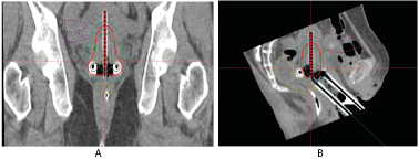
Figure 1. Dose distribution in brachytherapy. Anteroposterior (A) and profile (B) Gray line: Isodose 150%. Red line: Isodose 100%. Green line: Isodose 50%
Researchers at Fox Chase Cancer Center [10] evaluated data from patients treated for cervical cancer. They showed that patients with the ideal or adequate application (symmetry and equidistance between the tandem and the ovoid) are associated with a significantly high local control rate (68 vs 34%) with improved survival (60 vs 40%). Therefore, optimal placement of the applicator is strongly recommended, combined with much better results in terms of overall survival and local control [11,12].
One of the major intraoperative complications of intracavitary brachytherapy is uterine perforation. Previous studies have shown perforation rates ranging from 2% to 14% if applied without intraoperative ultrasound. In another study carried out at the National Institute of Oncology in Morocco between January 2014 and February 2016 including 270 patients with 570 insertions revealed a perforation rate of 5.8 % of insertions.
The uterine perforation usually occurs at the posterior wall, but also at the uterine fundus (Figure 2), therefore a good understanding with a clear visualization of the position, size, and flexion of the uterus is necessary to avoid such a complication [13]. Uterine perforation can also lead to direct traumatism to adjacent organs such as the bladder and small bowel which can lead to an increased dose at their level. This perforation may result in the patient a discomfort or abdominal pain. After a uterine perforation, a second visit to the operating room is necessary, the application must be removed and a second general or spinal anesthesia then a second insertion should be provided, as well as a treatment delay that may extend overall treatment time and compromise central control rates in the long term [14].
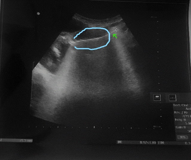
Figure 2. Ultrasound image showing uterine perforation (blue: uterus boundary, green arrow: exiting of the tandem)
The upper uterine perforations can however be treated by turning off the source in the last stop positions of the tandem which goes beyond the uterine fundus. A change in treatment time is shown to significantly influence the dosimetric parameters of brachytherapy [15].
The use of ultrasound to guide insertion dates back to the 1980s. Real-time ultrasound permits the radiotherapist to correct the inadequate length of the uterine tandem or penetration at the myometer and then, reduce the risk of uterine perforation (Figure 3).
Matsuyama et al. [16] reported a rate of 9.8% uterine perforation without ultrasound guidance. Whereas with the routine use of ultrasound when placing the uterine tandem, Watkins et al. [17] and Schaner et al. [18] reported a uterine perforation rate of 1.4%. Thus, the rates found in our study join those of the literature with a rate of 1.9%.
Historically, the perforation rate reported in the series that did not consistently use post-implant CT or MRI for the evaluation of the application and detection the uterine perforation was significantly lower than the series that used it [19-21]. However, the severity of the complications related to these events was higher (including death), which was possibly linked to that reduced sensibility of clinical evaluation for detecting perforations [22]. Other than the obvious complications of peritoneal infection secondary to uterine perforation, it is reasonable to relate part of the late toxicity events, such as bowel obstruction or necrosis, to the activation of sources outside the uterine cavity, which could occur in an unnoticed perforation.
From 1999 to mid-2007, treatment planning was performed via fluoroscopy, using orthogonal images. Before mid-2007, computed tomography CT of the pelvis was performed to confirm applicator positioning only in cases where insertion was difficult or perforation was suspected. Since mid-2007, routine CT imaging has been performed on all HDR brachytherapy procedures for treatment planning purposes. And therefore, the increased use of three-dimensional planning for brachytherapy allowed increased verification of the tandem position after insertion, earlier diagnosis of the perforation, and a window for the possibility of reinserting the applicator or adapt the treatment to avoid the activation of the source positions outside the uterine cavity. In our study, all applications have been evaluated by a postimplant CT.
Ultrasound is also useful in the context of a difficult application in a population with high-risk factors of perforation: cervical stenosis, history of perforation, or improperly positioned uterus. May et al. [23] evaluated the placement of the applicator with ultrasound guidance in case of the retroverted uterus, 33 insertions were realized to dilate the cervix and reposition the uterus, the anteversion was obtained in all applications without perforation. May et al concluded that the use of ultrasound showed positive results without complications in a population with a high risk of perforation.
Other modalities for verification of applicator placement have been used as well. Irvin et al. [24] described direct endoscopic visualization to provide irrefutable evidence of tandem location; however, this procedure is both time-consuming and expensive.
The use of ultrasound also permits to reduce the time of the application since it allows verification of the positioning of the applicator before fixing and solidarizing all the different parts of the applicator and thus minimizing the risk of a repeated surgical procedure. Davidson et al. [25] realized the value of ultrasound on 35 applications, based on their experience the ultrasound reduces the risk of perforation and reduces the time required for the application.
Intracavitary brachytherapy is integral to the success of definitive radiotherapy for cervical cancer, but technical challenges can limit successful applicator placement. Data for series (spanning 1996-2004), ICBT was not possible in 44 patients, and 73% of these were limited by technical considerations [26]. The most common reason cited was the inability to cannulate the cervical orifice. In our study, no patients have been unable to receive ICBT secondary to technical limitations surrounding applicator placement. Intraoperative ultrasound guidance accounts for the high success rate of applicator placement. Real-tile feed-back and device visualization are useful in the context of an effaced or distorted cervical orifice and allow for aggressive sounding and dilatation.
Limitation of this retrospective study that it does not compare insertions guided by intraoperative ultra-sound directly with blind tandem insertions. A randomized trial addresses this. Thus, despite these limitations, we feel that the present study is reverent and important to current clinical practice.
Ultrasound guidance is an accessible, innocuous inexpensive and fast radiological device that can easily be incorporated into gynecological brachytherapy centers, even in developing countries. Intraoperative ultrasound is an essential tool for optimizing the placement of the uterine tandem and reducing the rate of uterine perforation. Proper training of staff is necessary to ensure safe and optimal use. Improvement brachytherapy technology contributes to improving local control, survival and quality of care, and reduced patient morbidity.
We thank the Radiotherapy department and our radiotherapist-‘s colleagues at the National Institute of Oncology of Rabat who provided care and support for the patient.
The authors report no conflict of interest concerning the case in this paper.
- National Comprehensive Cancer Network. Cervical Cancer (2015) http://www.nccn.org/professionals/physician_gls/pdf/cervical.pdf. Accessed on November 10, 2015.
- Barnes EA, Thomas G, Ackerman I, Barbera L, Letourneau D (2007) Prospective comparison of clinical and computed tomography assessment in detecting uterine perforation with intracavitary brachytherapy for carcinoma of the cervix. Int J Gynecol Cancer 17: 821-826. [Crossref]
- Jhingran A, Eifel PJ (2000) Perioperative and postoperative complications of intracavitary radiation for FIGO stage I-III carcinoma of the cervix. Int J Radiat Oncol Biol Phys 46: 1177-1183.
- Kim RY, Levy DS, Brascho DJ, Hatch KD (1983) Uterine perforation during intracavitary application. Radiology 147: 249-251.
- Small W, Strauss JB, Hwang CS (2011) Should uterine tandem applicators ever be placed without ultrasound guidance? No: a brief report and review of the literature. Int J Gynecol Cancer 21: 941-944.
- Davidson M, Yuen J, D’Souza D (2008) Optimization of high-dose-rate cervix brachytherapy applicator placement: the benefits of intraoperative ultrasound guidance. Brachytherapy 7: 248-253. [Crossref]
- Granai CO, Doherty F, Allee P (1990) Ultrasound for diagnosing and preventing malplacement of intrauterine tandems. Obstet Gynecol 75: 110-113.
- Rotmensch J, Waggoner SE, Quiet C (1994) Ultrasound guidance for placement of difficult intracavitary implants. Gynecol Oncol 54: 159e162.
- Unningham DE, Stryker JA, Velkley DE (1981) Routine clinical estimation of rectal, rectosigmoidal, and bladder doses from intracavitary brachytherapy in the treatment of carcinoma of the cervix. Int J Radiat Oncol Biol Phys 7: 653-660.
- Corn BW, Hanlon AL, Pajak TF, Owen J, Hanks GE (1994) Technically accurate intracavitary insertions improve pelvic control and survival among patients with locally advanced carcinoma of the uterine cervix. Gynecol Oncol 53: 294-300.
- Corn BW, Shaktman BD, Lanciano RM (1997) Intra- and perioperative complications associated with tandem and colpostat application for cervix cancer. Gynecol Oncol 64: 224-229. [Crossref]
- Viswanathan AN, Moughan J, Small W (2009) Quality of cervical cancer brachytherapy implantation in RTOG prospective trials. Int J Radiat Oncol Biol Phys 75: 86-87.
- Viswanathan AN, Thomadsen B (2012) American brachytherapy society cervical cancer recommendations committee; American Brachytherapy Society. American Brachytherapy Society consensus guidelines for locally advanced carcinoma of the cervix. Part I: general principles. Brachytherapy 11: 33.
- Perez CA, Grigsby PW, Castro-Vita H (1995) Carcinoma of the uterine cervix. I. Impact of prolongation of overall treatment time and timing of brachytherapy on outcome of radiation therapy. Int J Radiat Oncol Biol Phys 32: 1275-1288.
- Ibhade OR, Oyeyemi OE, Idayat AB (2015) Tandem-ring dwell time ratio in Nigeria: dose comparisons of two lading patterns in standard high-dose-rate brachytherapy planning for cervical cancer. J Contemp Brachytherapy 7: 161-170.
- Matsuyama T, Tsukamoto N, Matsukuma K (1986) Uterine perforation at the time of brachytherapy for the carcinoma of the uterine cervix. Gynecol Oncol 23: 205-211.
- Viswanathan AN, Erickson BA (2010) Three-dimensional imaging in gynecologic brachytherapy: a survey of the American Brachytherapy Society. Int J Radiat Oncol Biol Phys 76: 104-109.
- Schaner PE, Caudell JJ, De Los Santos JF (2013) Intraoperative ultrasound guidance during intracavitary brachytherapy applicator placement in cervical cancer: the University of Alabama at Birmingham experience. Int J Gynecol Cancer 23: 559-566.
- Villasanta U (1970) Radium and external irradiation versus radium and operation for early invasive carcinoma of the uterine cervix. Am J Obstet Gynecol 106: 498-505.
- Volterrani F, Lombardi F (1980) Long termresults of radium therapy in cervical cancer. Int J Radiat Oncol Biol Phys 6: 565-570.
- Kim RY, Levy DS, Brascho DJ, Hatch KD (1983) Uterine perforation during intracavitary application. Prognostic significance in carcinoma of the cervix. Radiology 147: 249-251.
- Barnes EA, Thomas G, Ackerman I (2007) Prospective comparison of clinical and computed tomography assessment in detecting uterine perforation with intracavitary brachytherapy for carcinoma of the cervix, Int J Gynecol Cancer 17: 821-826.
- Corn BW, Hanlon AL, Pajak TF (1994) Technically accurate intracavitary insertions improve pelvic control and survival among patients with locally advanced carcinoma of the uterine cervix. Gynecol Onco 53: 294e300.
- Irvin W, Rice L, Taylor P (2003) Uterine perforation at the time of brachytherapy for carcinoma of the cervix. Gynecol Oncol 90: 113-122.
- Davidson MT, Yuen J, D’Souza DP (2008) Optimization of high-dose-rate cervix brachytherapy applicator placement: the benefits of intraoperative ultrasound guidance. Brachytherapy 7: 248-253.
- Barraclough LH, Swindell R, Livsey JE (2008) External beam boost for cancer of the cervix uteri when intracavitary therapy cannot be performed. Int J Radiat Oncol Biol Phys 71: 772-778. [Crossref]
Editorial Information
Editor-in-Chief
Prof. Hamid Yahya Husain
Dubai Health Authority, Dubai
Article Type
Research Article
Publication history
Received date: May 10, 2021
Accepted date: May 22, 2021
Published date: May 31, 2021
Copyright
©2021 Imane Mbarki. This is an open-access article distributed under the terms of the Creative Commons Attribution License, which permits unrestricted use, distribution, and reproduction in any medium, provided the original author and source are credited.
Citation
Imane Mbarki, Samia Hajar Touimi, Hanane Elkacemi, Ayeb.Kebdani, Sanae Elmajjaoui, et al. (2021) Intraoperative ultrasound guidance during intra-cavitary brachytherapy of cervical cancer. Med Case Rep Rev, 2021 DOI: 10.15761/MCRR.1000170