Background and purpose: The central facial palsy (cFP) after stroke is associated with a deficit of the voluntary movements of the lower face contralateral to the lesioned hemisphere. The facial expression is reduced, and the communication is limited. Emotional interaction, muscular control and the quality of life are negatively affected. Therefore, the question arises whether the number of cFP after ischemic stroke is a relevant size that we have to deal with.
Method: To the best of our knowledge, there is no study that reported the prevalence of cFP after first ever ischemic stroke. Therefore, in this prospective study we calculated the 1-year prevalence of cFP after ischemic stroke in 2019.
Results: With the consideration of the exclusion and inclusion criteria from 416 patients 155 patients showed cFP (prevalence of 37.3%). 78.7% of patients with cFP had a stroke within the supply area of the middle cerebral artery.
Conclusion: With such a high prevalence number, it is important for physicians and therapists to consider the clinical consequence of cFP. Evidence-based studies concerning a rehabilitation strategy to improve cFP are still lacking. Consequently, for the future, studies are necessary to effectively support the recovery process of cFP.
ischemic stroke, prevalence, central facial palsy
ACA: Anterior Cerebral Artery; cFP: The Central Facial Palsy; MCA: The Middle Cerebral Territory; MT: Multiple Territories; PCA: The Posterior Territory; PR: Prevalence Rate.
After ischemic stroke, the central facial palsy (cFP) is accompanied by a deficit of the voluntary movements of the lower face contralateral to the lesioned hemisphere while a limited mobility of the upper hemi-face is usually absent. The classic explanatory model describes bilateral corticonuclear projections from each primary motor cortex to the motoneurons innervating the upper facial muscles and unique contralateral projections to the motoneurons innervating the lower facial muscles.
With cFP the facial muscular activity is reduced and consequently, communication, facial expression, daily food intake and control of saliva could be compromised [1]. With reduced facial feedback the related neural activity of emotions is also abnormally modulated [2,3]. These circumstances have a negative influence on the quality of life [4,5]. So, there are enough reasons to investigate in detail the number of cFP as a relevant variable. To the best of our knowledge, there are no studies that reported the prevalence of cFP after first ever ischemic stroke. This is the first study in Germany that investigates the prevalence of cFP after supranuclear lesion. With a high prevalence value of cFP, there is a subsequent necessity to provide all patients suffering from cFP with additional specific rehabilitative therapy.
Study population
Patients after first ever ischemic stroke, admitted to the Moritz Klinik, were included from 1 January 2019 to 31 December 2019. The Moritz Klinik is an inpatient rehabilitation center of neurology, located in Thuringia (middle-east of Germany). In the specified period each patient with a stroke diagnosis was included in the first stage of this prospective study. Exclusion criteria were: hemorrhage, previous ischemic stroke, stroke longer than six months ago, previous peripheral and / or cFP, lack of compliance with the examination, vigilance dysfunction, other concomitant disease which affects the central nervous system, e.g. Multiple Sclerosis, traumatic brain injury etc. During the one-year registration 479 stroke patients in total were included. Considering inclusion and exclusion criteria 63 patients were excluded from the total number (n = 41 because of concomitant diseases, n = 11 due to vigilance dysfunction, and n = 10 lack of compliance to the examination and one patient with confused information regarding stroke localization that was not fit to the side of hemiparesis). Consequently, 416 patients (161 females, 255 males; age: 19-92 years) fulfilled all criteria and were included in the final analysis.
Stroke localization
Diagnosis of Stroke was made based on the criteria of guidelines of the German Society of Neurology (www.dgn.org/leitlinien). According to the stroke localization obtained by CCT (n = 231), MRI (n = 185) or both (n = 298), we divided stroke patients into five categories: 1. the anterior territory (ACA; brain regions which are supplied by the anterior cerebral artery), 2. supply area of the middle cerebral artery (the middle cerebral territory, MCA), 3. the posterior territory (PCA; brain regions in relation to vertebral artery, basilar artery, posterior inferior cerebellar artery and posterior cerebral artery), 4. multiple territories (MT) referring to patients with multilocular infarction and 5. patients without stroke demarcation in neuroimaging (NO).
Diagnosis of cFP
Each patient who met the related exclusion and inclusion criteria was tested. The following examinations were performed: patients were asked to look forward without any facial movement to evaluate if there is a facial asymmetry in rest. Patients, who indicated a former asymmetry not due to pathological causes were compared with their German National Identity Card (ID) to rule out a false positive diagnosis of a cFP in case of a natural innate facial asymmetry. With accordance and normal voluntary facial movements as described below, patients were labeled as clinically unremarkable due to cFP.
To assess the voluntary facial movement patients were requested to perform the following movements: to close their eyes gently, to squint their eyes, to raise the eyebrows, to knit the eyebrows, to wrinkle the nose, to smile with closed lips, to show teeth (to snarl), to bring the lips forward, to pull the corners of their mouth downward. We assessed facial asymmetry at rest and of single facial movements by comparing both sides of face. cFP was diagnosed when there was a difference between both sides.
Prevalence of cFP
During the one-year registration 479 stroke patients in total were included. Considering inclusion and exclusion criteria 63 patients were excluded from the total number (n = 41 because of concomitant diseases, n = 11 due to vigilance dysfunction, and n = 10 lack of compliance to the examination and one patient with confused information regarding stroke localization that was not fit to the side of hemiparesis). Consequently, 416 patients (161 females, 255 males; age: 19-92 years) fulfilled all criteria and were included in the final analysis.
260 patients did not show any paresis of facial movement. From these patients 22 had initially a false positive diagnosis (8.5%), because considering their IDs their lower face asymmetry already existed before the stroke. These patients were considered as patients with a natural innate facial asymmetry.
155 (61 females, 94 males) demonstrated a cFP (37.3%). Their time between stroke and evaluation of cFP was between seven- and 135-days post onset of stroke symptoms, with a mean of 24 days.
The number of patients with cFP was most after stroke in the MCA group (78.7%). Age distribution of stroke patients without and with cFP were similar. For stroke localization and age distribution, please see Figure 1.
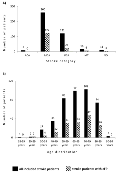
Figure 1. A) Distribution of patients in total and those with cFP in relation to stroke territories. B) Categorization of age with the related distribution of patients in total and those with cFP
All acronyms are explained in the section methods.
One patient showed a peripheral facial paresis after a brain stem and cerebellar lesion (4.2% of all patients within PCA category) and were excluded from cFP cluster.
cFP after ischemic stroke is a relevant number with a prevalence of 37.3%. To the best of our knowledge, there is no prospective study that reported the prevalence of cFP after first ever ischemic stroke.
A previous study reported 28 out of 47 stroke patients had cFP (59.6%). This study, however, focused on analyzing the association between pattern of facial movement and their stroke localization (6).
Some studies investigated the prevalence of pure isolated facial paresis-dysarthria syndrome and found 11 out of 2,000 (0.55%) stroke patients showed corresponding clinical symptoms (7). Another study found a prevalence of 0.4% by searching a database with more than 2,000 stroke patients (8). All our cFP patients had additional clinical symptoms (e.g. hemiparesis) and we did not find an isolated facial paresis-dysarthria syndrome. This discrepancy might be due to the lower number of patients we included. In our prospective study, we included 416 patients in one year, while other studies analyzed retrospectively their database over the past six and four years.
It must be considered that from clinical aspects cFP recovers fast during the first days after stroke. We investigated our patients within 7 days up to 135 days post onset. Therefore, an analysis at the earlier time point would probably increase the prevalence of cFP.
cFP and territory lesion
Previous studies reported that there are different cortical motor regions responsible for the facial muscle movements. For the lower face, it is represented in MCA territory and for the upper face movement brain regions within the anterior circulation is responsible [6,9]. In our population we had nine patients with an anterior circulation lesion. None of them showed any disturbance of face movement, most patients (78.7%) with cFP were after MCA territory lesion (compared to 15,5% of the group of posterior territory).
Consequences of cFP
Patients with facial movement disturbance have reduced facial expression and simultaneously their emotional expression are weakened. With less facial movement, the related cortical feedback is reduced and therefore the connectivity to emotion-specific network is limited [10-12] and abnormally modulated [2,3]. These circumstances, as a matter of fact, reduce the quality of life as a whole [4,5]. Therefore, it is important to be aware of emotional consequences of each patient with cFP after supranuclear lesion.
From the clinical point of view, it is important to be aware of cFP because more than one-third of stroke patients showed cFP. Considering sequelae of cFP for the future, we need multimodal evidence-based rehabilitation strategies to force its recovery process.
We are grateful to Anke Oertel for her support of study organization and Kristin Busch, Susanne Georgiew, Conny Großer, Katharina Herzog, Lisa-Marie Jacob, Patricia Klöpfel, Kati Matterne, Diana Rosner for their assistance of patients’ selection.
- Konecny P, Elfmark M, Urbanek K (2011) Facial paresis after stroke and its impact on patients' facial movement and mental status. J Rehabil Med 43: 73-5. [Crossref]
- Hennenlotter A, Dresel C, Castrop F, Ceballos-Baumann AO, Wohlschläger AM, et al. (2009) The link between facial feedback and neural activity within central circuitries of emotion--new insights from botulinum toxin-induced denervation of frown muscles. Cereb cortex 19: 537-42. [Crossref]
- Kheirkhah M, Brodoehl S, Leistritz L, Götz T, Baumbach P, et al. (2020) Abnormal Emotional Processing and Emotional Experience in Patients with Peripheral Facial Nerve Paralysis: A MEG Study. Brain Sci 10: 147. [Crossref]
- Konecny P, Elfmark M, Horak S, Pastucha D, Krobot A, et al. (2014) Central facial paresis and its impact on mimicry, psyche and quality of life in patients after stroke. Biomed Pap Med Fac Univ Palacky Olomouc Czech Repub 158: 133-7. [Crossref]
- Schimmel M, Ono T, Lam OL, Muller F (2017) Oro-facial impairment in stroke patients. J Oral Rehabil 44: 313-26. [Crossref]
- Cattaneo L, Saccani E, De Giampaulis P, Crisi G, Pavesi G (2010) Central facial palsy revisited: a clinical-radiological study. Ann Neurol 68: 404-8. [Crossref]
- Celebisoy M, Tokucoglu F, Basoglu M (2005) Isolated dysarthria-facial paresis syndrome: a rare clinical entity which is usually overlooked. Neurol India 53: 183-5. [Crossref]
- Sands KA, Shahripour RB, Kumar G, Barlinn K, Lyerly MJ, et al. (2016) Acute Isolated Central Facial Palsy as Manifestation of Middle Cerebral Artery Ischemia. J Neuroimaging 26: 499-502. [Crossref]
- Morecraft RJ, Louie JL, Herrick JL, Stilwell-Morecraft KS (2001) Cortical innervation of the facial nucleus in the non-human primate: a new interpretation of the effects of stroke and related subtotal brain trauma on the muscles of facial expression. Brain 124: 176-208. [Crossref]
- Klingner CM, Volk GF, Maertin A, Brodoehl S, Burmeister HP, et al. (2011) Cortical reorganization in Bell's palsy. Restor Neurol Neurosci 29: 203-14. [Crossref]
- Klingner CM, Volk GF, Brodoehl S, Witte OW, Guntinas-Lichius O (2014) The effects of deefferentation without deafferentation on functional connectivity in patients with facial palsy. Neuroimage Clin 6: 26-31. [Crossref]
- Gothard KM. The amygdalo-motor pathways and the control of facial expressions. Front Neurosci 8: 43. [Crossref]