Background The exercise test is widely used in diagnosis and prognosis of coronary artery disease. It’s routinely performed in stable and asymptomatic patients despite the low level of evidence for this indication, according to the guidelines.
The objective was to analyse whether these tests had prognostic value and determine the impact of a positive test on the cardiologist's attitude. Also, we wanted to study its value in diabetics.
Methods The sample group consisted of all exercise tests performed in one year in our cardiology department. The cases were the routine tests and controls were the prognostic tests.
Results We analysed 597 exercise tests on patients with coronary disease. More than half were routine. In more than half of routine positive tests did not change the clinical attitude. The prognosis was significantly better in cases, even when the result was positive, and the clinical attitude was not changed. Same trend was observed in diabetics.
Conclusion Routine exercise testing in patients with coronary artery disease, asymptomatic, stable, diabetic or not, has a very limited prognostic value. The long-term prognosis of asymptomatic patients with a positive test is excellent. We advocate following clinical guidelines and not doing exercise tests in asymptomatic with stable coronary disease.
Exercise test, Coronary artery disease, Prognostic
The exercise test (ET) remains the most widely used and technically available test to diagnose and study the presence and severity of coronary artery disease (CAD). It is a crucial test in the management of patients with CAD. The most accepted indications for an ET in patients with known CAD are, in brief: to determine the likelihood of significant anatomical or functional CAD that may be of prognostic importance, to identify its functional class and to evaluate the effects of therapy [1].
In the cardiology clinic, routine ET is often performed in the course of monitoring asymptomatic patients with CAD, despite being a controversial indication. In fact, in clinical guidelines [1], the indication in asymptomatic patients is considered class IIb, and in revascularized [2] patients it is only class III.
For this study, we hypothesized that routine ET in asymptomatic and stable patients with CAD, whether or not they were diabetic, had no added value in their follow-up compared with ET made for prognostic purposes in risk stratification.
The main objective of the study was to analyse whether routine ET in stable, asymptomatic patients with CAD had prognostic value. Secondary objectives were: 1) to ask the same question in a diabetic subgroup and 2) study any changes in the clinical approach following a positive result in the routine ET.
We performed a case-control study, comparing routine against prognostic ET, using as a sample all the ET performed during one year in our cardiology department.
Inclusion criteria were: 1) cases: routine ET in patients with CAD, stable and asymptomatic, 2) controls: prognostic ET in patients with CAD and clinical changes or acute coronary syndrome (ACS).
The variables studied were:
- ET type: 1) routine: conducted in patients with CAD, asymptomatic and stable, meaning no hospitalization from cardiovascular causes in the last 6 months, or 2) prognostic: for risk stratification and prognostic evaluation after a clinical change (onset of angina or change in occurrence threshold) or after ACS in any form or presentation. All ET were performed on a treadmill-type ergometer according to Bruce’s protocol [3].
- Results of the ET: positive criteria that were used in routine practice - a) clinical criteria: typical chest pain suggestive of angina; b) electrical criteria: ST depression of 0.1 mV or more for at least 80 ms, in at least 2 consecutive leads.
- Clinical attitude to a positive result on the ET: 1) no change in attitude, 2) modification of antianginal drug therapy (increase or addition of new drugs) or 3) performance and outcome of coronary angiography - percutaneous coronary intervention (PCI), coronary artery bypass graft (CABG) or no intervention.
- Diagnosis of diabetes mellitus [4]: HbA1c ≥6.5%, fasting plasma glucose ≥126 mg/dL plasma glucose ≥200 mg/dl after 2 hours of a glucose tolerance test; classic symptoms of hyperglycemia and random plasmatic glucose ≥200 mg/dl.
- Time of follow-up: months until the last visit in clinic or until the presentation of a clinical event.
- Clinical events in follow-up: ACS in any form - ACS without ST-elevation, myocardial infarction without ST-elevation, ACS with ST elevation and ST-elevation acute myocardial infarction; we applied the common criteria for ACS used in clinical practice [5,6], but always with objective data - chest pain suggestive of myocardial ischemia plus electrocardiographic changes (T wave alterations or up/down transient or persistent ST) and/or significant elevation of myocardial necrosis biomarkers (troponin I of 0.04 ng/ml with typical ascending positive curve).
Statistical analysis: 1) We used the Chi2 test for qualitative comparisons; for values less than 5, the Fisher’s exact test was used. 2) Quantitative variables were expressed as mean +/- standard deviation and median. 3) To analyse the prognosis we studied the ACS-free interval, performing an univariate analysis of survival according to the product limit method of Kaplan-Meier.
During one year, 1833 ETs were conducted, of which 597 were performed in patients with CAD. Of these 332 (55.6%) were routine (cases) and 265 (44.4%) were prognostic (controls).
The results of the ETs can be seen in Table 1, where the percentage of negative routine tests was higher than the percentage of negative prognostic tests.
Table 1. Exercise test results.
Total sample (N=597) |
|
Positive result |
Negative result |
p |
Routine (cases) (n = 332) |
82 (20) |
264 (80) |
0.003 |
Prognostic (controls) (n = 265) |
68 (31) |
183 (69) |
Diabetics (n=168) |
|
Positive result |
Negative result |
p |
Routine (cases) (n = 87) |
17 (19.5) |
70 (80.5) |
0.018 |
Prognostic (controls) (n = 81) |
29 (36) |
52 (64) |
Value (%)
We further analysed what the attitude of the cardiologist was to positive results in patients with routine positive ET (n = 150). Over half decided not to modify the clinical regimen, despite the positive result, compared with 13% of controls. Of all patients with positive ET, regimen was changed in 102. Table 2 shows the changes made; it can easily be seen that in both groups the greatest changes were in pharmacological treatment.
Table 2. Clinical approach following a positive exercise test result (n = 150).
|
Routine |
Prognostic |
p |
Total |
68 (45.3) |
82 (54.7) |
|
No changes |
37 (54.4) |
11 (13.4) |
0.000 |
Pharmacologic changes |
20 (29.4) |
31 (37.8) |
0.123 |
Coronary angiography |
7 (10.3) |
7 (8,5) |
1 |
Pharmacologic and Coronary angiography |
4 (15.9) |
33 (40.3) |
0.000 |
Value (%)
The percentage of patients with modified antianginal therapy who also underwent coronary angiography was much higher in the control group (prognostic tests).
Taking all the cases, 5 patients (7.3% of all positive routine ETs) underwent PCI compared with 24 in the control group (29% of total positive prognostic ETs) (p = 0.001). With respect to CABG, 3 cases (4.4% of total positive routine ETs) were operated on against 5 controls (6.1% of total positive prognostic ETs) (p = 0.73).
The diabetic subgroup was 168 patients (28.1% of total). As in the global sample, Table 1 shows that the percentage of negative routine tests was higher than the percentage of negative prognostics. In 53% of diabetic patients with routine ET, the clinical approach to a positive test result did not change.
Follow-up
Follow-up (available in 557 patients, 93% of total) was 14.4 months on average (± 10.4), with a median of 21 months.
Outcome: ACS free interval
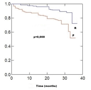
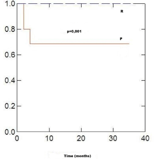
Kaplan-Meier graphs show that the ACS-free interval was higher in asymptomatic patients who underwent routine ET (Figure 1). At follow-up, 30 cases (9%) experienced an event compared with 57 controls (21.5%). In the follow-up of patients with positive ET, neither group with routine ET suffered any acute event, compared with 19 (23%) of the symptomatic group (p = 0.000). Asymptomatic patients with positive results that did not prompt any change in clinical approach (n = 48), as seen in Figure 2, still had an excellent prognosis (0 events versus 3 (6.2%) in controls).

Figure 1. ACS-free interval: total sample
R: routine exercise tests; P: prognostic exercise tests

Figure 2. ACS-free interval: positive exercise test without changes in clinical approach
R: routine exercise tests;P: prognostic exercise tests
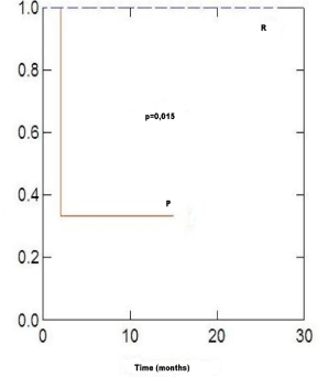
This fact was duplicated in the diabetic subgroup with positive results and no clinical changes (n = 12), as shown in Figure 3, with a marked separation of the lines from the first month of follow-up; in this subgroup, 2 events occurred in the control group (67%).

Figure 3. ACS-free interval: positive exercise test in diabetics without changes in clinical approach
R: routine exercise tests; P: prognostic exercise tests
Revascularized patients (n = 344, 57.6% of the total) presented the same trend. Of 41 revascularized patients with positive ET, 29 received no modified treatment. In the follow-up, 3 controls (of 22) with positive ET and no clinical changes presented ACS (p = 0.003).
The ET is essential in the diagnosis and prognosis of patients with CAD. Despite its relatively limited sensitivity and specificity, both in the diagnosis of CAD (68% and 77%) [7], and multi-vessel CAD (81% and 66%) [8], it is a test that provides clinically relevant data. In these patients, the primary objective is to objectify the presence of inducible ischemia, both from the clinical and electrocardiographic standpoint, but the clinician may also be interested in other variables such as blood pressure response to exercise, chronotropism, inotropism, arrhythmic inducibility and functional class.
As mentioned in the Introduction, there is widespread routine use of ET in the clinical management of patients with CAD. Clinical guidelines don’t agree, because the recommendation has a low level of evidence, and even in revascularized patients it is contraindicated [2]. For this reason, we performed this study to try to answer this question of its value and consider whether, despite the concensus recommendation, it is worth performing routine ET.
As discussed in Results, the percentage of routine ETs with negative results was greater than the percentage of negatives in the prognostic group, which is logical, since patients with routine tests were asymptomatic and in stable condition.
When analysing the attitude of the cardiologist when faced with the ET results, it was observed that with more than half the patients with positive routine tests it was decided no to change the clinical approach. It seems paradoxical to find prognostic factors, such as silent ischemia, and theoretically find them in more than half the asymptomatic patients, and not absolutely modify the therapeutic management, either pharmacological or interventional.
Among the 150 patients with positive results in which the clinical approach was changed, the majority took the form of changes in drug therapy, not statistically different between groups. Significant differences were seen in the percentage of combined changes in medical therapy and coronary angiography, which were much higher in the group of prognostic tests, which is again logical since these are patients presenting with ACS in any form.
Although the methodology is different, our results are similar to those in the study of Casella G, et al. [9], which retrospectively reviewed 766 patients with a remote history of myocardial infarction with Q wave, stable, asymptomatic and adequately treated, who underwent a routine ET. They noted that none of the predictors of risk of reaching the primary endpoint (cardiac death or nonfatal reinfarction) was related to the ET; they only found relationships with older age, FC ≥90 bpm and ST depression in resting electrocardiogram. They concluded that the value of ET in identifying patients with the above profile at risk of cardiovascular events is insignificant. Certainly, the cases in our sample are not identical, since one inclusion requirement was a history of CAD and chronic infarction; however, the results are in essential agreement, which is that the discriminative ability of the ET is very low in stable and asymptomatic coronary patients.
The reason to perform routine ET is clearly to prevent events and change the prognosis. In the Kaplan-Meier ACS free interval graphics, we can see that the prognosis is better for patients with routine ET. This fact is easily assumed, as compared with patients who have just suffered an ACS. However, we were surprised that no patient had any ischemic event. In principle, one might think that the group of patients with positive routine ET where the cardiologist does not change the clinical approach may have a worse prognosis, precisely because of the positive result.
When we analysed the subgroup of diabetic patients, we found the results of the ETs were very similar to those of the global sample. The diabetic group certainly generates more questions about silent ischemia in clinical practice; however, when we try to "get ahead" of events, in our study we found many in the positive ET group were not offered any therapeutic changes. In a review in the late '90s on studies using multivariate analysis of clinical data and ET’s ability to predict the presence of angiographic CAD, it was seen that diabetes mellitus was a good predictor of the presence of CAD in 4 of 11 studies considered, in 3 of which the prevalence of diabetes was 19%, while in the other 7, in which diabetes was not a predictor, it was less than 16%. When analysing extension studies of CAD, diabetes was also a good predictor. The authors concluded that whether a variable is a good clinical predictor may depend on the frequency of that variable in the population [10]. However, in our sample the frequency of diabetes was higher (28.1%), despite which no significant differences in prognosis were seen, whether or not the ET was positive. Clearly, we are not comparing the same studies, since we didn’t conduct a multivariate analysis and all of our patients (cases and controls) already had CAD. Studies such as DIAD (Detection of Ischemia in Asymptomatic Diabetics study) have shown that ischemia resolves in most asymptomatic type 2 diabetics after 3 years of intensive treatment for cardiovascular risk factors with drugs such as aspirin, statins, and angiotensin converting enzyme (ACE) inhibitors [11], although the "DIAD patients", unlike the diabetics in our series, had no known CAD.
Califf RM, et al. [12] studied ischemia prognostic parameters in 5886 patients with CAD in order to develop a score (Duke Angina Score) that included only clinical parameters related to the type and/or frequency of angina, and ischemic electrocardiographic changes at rest, all with prognostic value. Thus, if a patient with 3-vessel disease, normal left ventricle and a score of 0, had an infarction-free survival at 2 years of 90%, this was reduced to 68% if the score was equal to or greater than 9. This is consistent with the importance of the clinical prognosis of angina, as we show in our study.
It is known that vulnerable atherosclerotic plaques which can trigger an ACS have a characteristic composition, a thin fibrous capsule, large lipid cores, fewer smooth muscle cells, more macrophages and less collagen, than stable plaques, and exhibit eccentric remodelling of the arterial wall, therefore causing less coronary lumen stenosis [13,14]. These vulnerable plaques rarely cause significant stenosis before rupture and onset of ACS. However, stable plaques have a thicker fibrous capsule, a smaller lipid core, more smooth muscle cells, fewer macrophages, more collagen, and provoke concentric remodelling causing narrowing of the coronary lumen. These lesions cause ischemia and anginal symptoms but are less likely to trigger an ACS [15,16]. Furthermore, significant lesions are those that can be detected by the ET, because they limit blood flow under conditions of increased oxygen demand during exercise. However, lesions that can trigger an ACS themselves don’t limit coronary flow and aren’t detected by the ET. This pathophysiological mechanism explains the limitation of ET in detecting vulnerable plaques, potentially triggering ACS.
Routinely performing ET consumes resources, generates waiting lists in the saturated public health system and is associated with economic costs. Moreover, we cannot ignore the anxiety generated in the patient over a positive ET result, which as a routine test is presented as a predictor of the status of their disease, then in most cases does not actually change the clinical approach. And, a very important aspect, we see that the prognosis is not changed when faced with a positive test. For these reasons, we advocate that routine ET not be performed in patients with stable and asymptomatic CAD.
This is a retrospective study and therefore there may be a selection bias in the studied group of patients (cases). However, this is unlikely, since our department, at that time, was conducting an annual ET for risk stratification in this kind of patient.
Among the cases that were included, some patients had a history of acute myocardial infarction and other did not; however, all could be considered low risk, given their stability and the absence of clinical symptoms.
The Kaplan-Meier univariate is also a limitation, since it does not include variables that could explain some of the effect.
Moreover, the decision to change the regimen depends on the clinical cardiologist, who is not subject to established criteria in a research protocol; therefore, it is influenced by criteria (uncontrollable) decided upon by each cardiologist and the patient, whose opinion always counts, but who will be especially sensible to invasive procedures determined by the outcome of a non-invasive test such as the ET.
Routine ET in asymptomatic patients, stable, with CAD, has very limited prognostic value versus that performed against clinical changes. Neither adds value in diabetics. The long-term prognosis of these asymptomatic patients with positive ET is excellent compared to symptomatic patients. We advocate following clinical guidelines and performing ET in patients with CAD, but only for prognostic purposes, depending on clinical indications.
- Fox KM, Garcia MA, Ardissino D, Buszman P, Camici PG, et al. (2006) Guidelines on the management of stable angina pectoris: executive summary: The Task Force on the Management of Stable Angina Pectoris of the European Society of Cardiology. Eur Heart J 27: 1341–81. [Crossref]
- Gibbons RJ, Balady GJ, Chaitman BR, Fletcher GF, et al. (2002) ACC/AHA 2002 guideline update for exercise testing: summary article: a report of the American College of Cardiology/American Heart Association Task Force on Practice Guidelines (Committee to Update the 1997 Exercise Testing Guidelines). J Am Coll Cardiol 40: 1531-1540. [Crossref]
- Bruce RA (1956) Evaluation of functional capacity and exercise tolerance of cardiac patients. Mod Concepts Cardiovasc Disease 25: 321-326. [Crossref]
- American Diabetes Association (2018) 2. Classification and Diagnosis of Diabetes: Standards of Medical Care in Diabetes-2018. Diabetes Care 41: S13-S27. [Crossref]
- Roffi M, Patrono C, Collet JP, Mueller C, Valgimigli M, et al. (2016) 2015 ESC Guidelines for the management of acute coronary syndromes in patients presenting without persistent ST-segment elevation: Task Force for the Management of Acute Coronary Syndromes in Patients Presenting without Persistent ST-Segment Elevation of the European Society of Cardiology (ESC). Eur Heart J 37: 267-315. [Crossref]
- Van de Werf F, Bax J, Betriu A, Blomstrom-Lundqvist C, Crea F, et al. (2008) Management of acute myocardial infarction in patients presenting with persistent ST-segment elevation. Eur Heart J 29 :2909-2945. [Crossref]
- Gianrossi R, Detrano R, Mulvihill D, Lehmann K, Dubach P, et al. (1989) Exercise-induced ST depression in the diagnosis of coronary artery disease. A meta-analysis. Circulation 80: 87-98. [Crossref]
- Detrano R, Gianrossi R, Mulvihill D, Lehmann K, Dubach P, et al. (1989) Exercise-induced ST segment depression in the diagnosis of multivessel coronary disease: a meta analysis. J Am Coll Cardiol 14: 1501-1508. [Crossref]
- Casella G, Pavesi PC, Niro MD, Bracchetti D (2001) Negative and positive predictive values of routine exercise testing in stable, medically-treated patients several years following a Q-wave myocardial infarction. Ital Heart J 2: 271-279. [Crossref]
- Yamada H, Do D, Morise A, Atwood JE, Froelicher V (1997) Review of studies using multivariable analysis of clinical and exercise test data to predict angiographic coronary artery disease. Prog Cardiovasc Dis 39: 457-481.
- Wackers FJT, Chyun DA, Young LH, Heller GV, Iskandrian AE, et al. (2007) Resolution of Asymptomatic Myocardial Ischemia in Patients With Type 2 Diabetes in the Detection of Ischemia in Asymptomatic Diabetics (DIAD) Study. Diabetes Care 30: 2892–2898. [Crossref]
- Califf RM, Mark DB, Harrell FE Jr, Hlatky MA, Lee KL, et al. (1988) Importance of clinical measures of ischemia in the prognosis of patients with documented coronary artery disease. J Am Coll Cardiol 11: 20-26. [Crossref]
- Naghavi M, Libby P, Falk E, Casscells SW, Litovsky S, et al. (2003) From vulnerable plaque to vulnerable patient: a call for new definitions and risk assessment strategies. Part I. Circulation 108: 1664-1672. [Crossref]
- Naghavi M, Libby P, Falk E, Casscells SW, Litovsky S, et al. (2003) From vulnerable plaque to vulnerable patient: a call for new definitions and risk assessment strategies. Part II. Circulation 108: 1772-1778.
- Ambrose JA, Tannenbaum MA, Alexopoulos D, Hjemdahl-Monsen CE, Leavy J, et al. (1988) Angiographic progression of coronary artery disease and the development of myocardial infarction. J Am Coll Cardiol 12: 56-62. [Crossref]
- Little WC, Constantinescu M, Applegate RJ, Kutcher MA, Burrows MT, et al. (1988) Can coronary angiography predict the site of a subsequent myocardial infarction in patients with mild-to moderate coronary artery disease? Circulation 78: 1157-1166. [Crossref]


