Abstract
Oculo-facio-cardio-dental (OFCD) syndrome is a gene disorder characterized by congenital cataracts, facial anomalies, congenital heart disease, and a number of different dental anomalies. Here we report a case involving a young girl with OFCD syndrome showing dental anomalies in the permanent tooth exchange process from the time of primary tooth eruption. We conducted regular dental checkups for this patient for over 12 years. Her dental anomalies included delayed eruption and prolonged retention of primary teeth, delayed eruption of permanent teeth, radiculomegaly of primary and permanent teeth, supernumerary teeth, and bulky fused teeth. Roots of primary anterior teeth and cuspids were remarkably long, and root resorption was inhibited. Using extracted pulp cells from the primary tooth, which was extracted to promote eruption of permanent teeth, we evaluated osteoclast differentiation; no differentiation was observed. After extraction of the primary teeth, the succedaneous teeth began to erupt immediately; however, absorption of the primary teeth was incomplete due to OFCD syndrome, which was caused by the lack of/inhibited osteoclast differentiation at the appropriate time. Additionally, this suggests that subsequent eruption of the permanent teeth will be delayed. To our knowledge, this is the first report of a case of OFCD syndrome involving radiculomegaly of primary teeth to be described in detail.
Key words
congenital cataracts, congenital heart disease, dental anomalies, facial anomalies, OFCD syndrome, radiculomegaly
Introduction
Oculo-facio-cardio-dental (OFCD) syndrome is a rare gene disorder caused by a mutation of the BCOR gene on an X chromosome [1]. It is characterized by congenital cataracts, various facial anomalies (broad nasal tip, long philtrum, etc.), congenital heart disease, and dental anomalies [2-9]. Skeletal abnormalities such as the abnormal flexure of the second and third toes, mild hearing loss, and mental delay have also been reported [3,5,7,10,11]. Radiculomegaly of the permanent teeth, is noted in most conventional cases of OFCD syndrome, but other dental anomalies, including delayed eruption, persistence of primary teeth, oligodontia, hyperdontia, and fused teeth have been reported [11]. Congenital heart disease and congenital cataract are typically diagnosed after birth, and treatments are subsequently started. It is difficult for medical specialists to diagnose OFCD syndrome because dental anomalies gradually develop from the primary tooth eruption period; many cases are diagnosed by a dentist after radiculomegaly is observed in permanent teeth [9,12]. Only genetic screening has enabled the earlier diagnosis of OFCD syndrome owing to the mutation of the BCOR gene.
The present case showed delayed eruption of primary (anterior) teeth, radiculomegaly of the primary and permanent teeth, supernumerary teeth, persistence of primary tooth dentition, delayed eruption of permanent teeth, malformations such as fused teeth, and numerous dental anomalies such as malocclusion. Here we report the first known case of OFCD syndrome involving dental anomalies that appeared during the exchange process from primary to permanent tooth dentition in detail.
Case report
The case described in the present report was 14-year-old girl who had been undergoing regular medical examinations since the age of 1 year and 7 months. She was referred to our department with the chief complaint of delayed eruption of the primary teeth (Figure 1). She was a second child born by normal delivery after 38 weeks of gestation. Her birth weight and length were 3000 g and 50 cm, respectively. She presented with a history of congenital cataracts, congenital glaucoma, tetralogy of Fallot, hammertoe, mild mental retardation, a mildly broad nasal tip, and a long philtrum (Figures 1 and 2). She underwent surgery for congenital cataracts at age 2 months and for glaucoma at 1 year and 5 months. Her cardiac diseases were repaired at age 1 year. She continues to receive treatment for her glaucoma, which was caused by her cataracts, and her right ear still shows mild hearing loss.
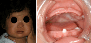
Figure 1. Facial and oral photographs of the present case at age 1 year and 7 months showing a mildly broad nasal tip and long philtrum, as well as delayed eruption of anterior teeth.
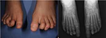
Figure 2. Hammertoe deformity of the second and third toes
An intraoral examination at the initial visit (age 1 year and 7 months) revealed that only her four first primary molars and lower right primary central incisor had erupted (Figure 1B). The eruption of her anterior teeth was slower than that of her molar teeth. Primary dentition was complete at the age of 4 years and 2 months. All of her first molars had erupted by age 8 years without any problems. She managed to maintain healthy teeth and good oral health with regular maintenance. However, no signs of the primary deciduous incisors were observed (Figure 3A). At this time, genetic screening showed a mutation of the BCOR gene.
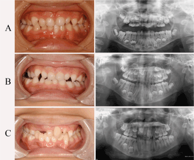
Figure 3. Oral photographs and panoramic radiograph A. at age 8 years, B. at age 10 years, and C. at age 13 years. B. Oral photograph showing persistent primary dentition/unerupted permanent dentition, and panoramic radiograph showing radiculomegaly and defective absorption of primary teeth. C. Panoramic radiograph showing radiculomegaly of permanent teeth, supernumerary teeth in the mandibular region, a maxillary anterior tooth, and fused teeth, including the upper maxillary right central and lateral incisors.
Radiographs revealed that her roots were remarkably long. All of her primary teeth were retained, and a root resorption defect was observed. No signs of eruption of permanent anterior teeth and cuspids were recognized. The radicular point of the lower canine teeth extended to the mandibular cortical plate (Figure 3A). The eruption of succedaneous teeth was noted at a periodical examination (Figure 3B). Currently, this patient is preparing for orthodontic treatment (Figure 3C).
A computerized tomography scan of her maxilla confirmed the finding described above and additionally showed that the permanent right central and lateral incisors were fused, as well the presence of a supernumerary tooth (Figure 4). This led to a diagnosis of difficult eruption of successive permanent teeth.
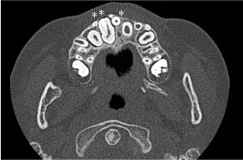
Figure 4. Computed tomography image of the maxilla at age 8 years and 11 months. A median maxillary supernumerary tooth (*). Fused teeth including the upper maxillary right central and lateral incisors (**).
In addition, the mandibular primary central incisor, lateral incisors, and primary cuspids, which showed deficient exfoliation, were extracted sequentially. All primary teeth extracted were remarkably long compared with those in Japanese controls (Figure 5, Table 1).
Table 1. Comparison of the length of primary teeth between the present case and Japanese patients
Tooth name (mandibular) |
Japanese minimum |
Japanese average |
Japanese maximum |
Patient
(mm) |
A |
12.6 |
13.59 |
15.0 |
24.4 |
B |
13.7 |
15.94 |
18.6 |
24.6 |
C |
16.0 |
17.34 |
18.8 |
27.8 |
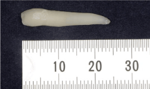
Figure 5. Extracted lower right primary central incisor at 9 years of age. The root is significantly long, and the apex of the root is sharp.
We compared osteoclast differentiation and cell-proliferating potency of the pulp cells of the patient with the pulp cells of a healthy patient. However, no osteoclast differentiation was observed in pulp cell culture of the patient (Figure 6). Moreover, the proliferation potency of the cultured cell was high compared with a control (Figure 7).
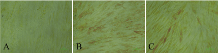
Figure 6. Comparison by tartrate-resistant acidic phosphatase (TRAP) staining after 7 days in culture with osteoclast differentiation. A. Oculo-facio-cardio-dental (OFCD) syndrome (mandibular primary cuspid): negative results by TRAP staining (no osteoclast differentiation). B. Control 1 (mandibular cuspid with normal dentition): positive results by TRAP staining (osteoclast differentiation). B. Control 2 (mandibular incisor with normal dentition): positive results by TRAP staining (osteoclast differentiation).
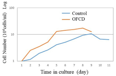
Figure 7. Cell growth curve. The pulp cells in the case with OFCD syndrome proliferated faster than those of the control. OFCD syndrome (mandibular primary canine), control (mandibular canine with normal dentition).
Discussion
The dental anomalies of most patients with OFCD syndrome exhibit delayed primary and permanent dentition, persistent primary teeth, supernumerary teeth, root radiculomegaly of permanent teeth (cuspid in particular) and fused permanent dentition. Radiculomegaly of primary teeth has not been reported in studies on eight cases of primary dentition, 30 cohort studies, or elsewhere [5,13]. However, in the present case, remarkable radiculomegaly of the primary dentition (panoramic images) was observed.
Additionally, the formation of the permanent teeth continued with no signs of root resorption of the primary teeth, and the root bottom extended to the mandibular cortical bone. In this account we performed extraction of primary teeth in a purpose by eruption of the permanent teeth instruction sequentially. The extracted primary teeth were significantly long compared with Japanese controls [14].
As for the extracted primary teeth, absorption had not started (Figure 5). The eruption of successional teeth was promoted by the extraction of the primary teeth, as expected. The first molar without the preceding primary teeth was slow but erupted without a problem. From these results, we thought that the delayed eruption of the permanent teeth in OFCD syndrome was caused by a primary tooth root resorption deficiency.
Osteoclast differentiation was not observed in the pulp cell culture obtained exodontia in this patient. Therefore, we speculated that the primary tooth root resorption deficiency found in most cases of OFCD syndrome results from osteoclasts not being able to differentiate at the time root resorption begins.
Furthermore, we compared the proliferation potency of the control’s pulp cell with the cultured pulp cell derived from the present case. As a result, we found that the proliferation potency of the pulp cell in the present case was higher compared with the control. Mesenchymal stem cells obtained from the pulp cells of patients with OFCD syndrome have been shown to enhance osteo-dentinogenic potential [15,16]. Therefore, the increased cells proliferation potency in the present case was assumed to be the result of the continuation of abnormal root formation.
Panoramic X-rays showed few abnormalities in the molar teeth compared with the front teeth and cuspids. In addition, the eruption of the anterior teeth was slower than that of the posterior teeth, and more right than left dental anomalies were found (Figure 3). Root resorption of the primary teeth and eruption of the permanent teeth on the right side were later than those on the left, and bulky fused teeth were observed (Figure 4). Furthermore, the cataracts and degree of hearing loss in the present case were more severe on the right than on the left side. Interestingly, laterality defects in OFCD syndrome have been reported and should be investigated [13].
The most challenging problem remaining in the present case remains the treatment of the fused teeth on the right side of the maxilla, which consist of the maxillary right central incisor, lateral incisor, and adjacent supernumerary teeth. Extracting these fused teeth would result in a major defect in the body of the maxilla. Therefore, we plan to perform orthodontic treatment with crown restoration after pulp treatment in coordination with an orthodontist, endodontist, and prosthetic specialist after all of the present case’s premolar teeth have erupted and treatment has begun.
Conclusion
2021 Copyright OAT. All rights reserv
The case described in the present report involved numerous dental anomalies in the cuspid and anterior primary and permanent dentition, as well as congenital heart disease, congenital cataracts, hammertoe, mild hearing loss, and a mildly broad nasal tip. OFCD syndrome is difficult to differentiate clinically from congenital rubella syndrome and several other diseases unless radiculomegaly of the permanent teeth is observed in X-rays. However, because dental anomalies become a more serious problem with age, pedodontists should be prepared for treatment. We hope that medical specialists suspect OFCD syndrome when the eruption of primary teeth (particularly anterior primary teeth) is significantly delayed around 10 months after birth in children with symptoms such as congenital heart disease and congenital cataracts and refer such cases to pedodontists.
Ethics approval
The experimental protocol of the present study was reviewed and approved by the Institutional Review Board for Ethical Issues at Nagasaki University (approval number: 140407283) and the Committee for Ethical Issues on Human Genome and Gene Analysis (approval number: 1499).
Conflicts of interest
None
References
- Hayward JR (1980) Cuspid gigantism. Oral Surg Oral Med Oral Pathl 49: 500-501
- Davoody A, Chen IP, Nanda R, Uribe F, Reichenberger EJ, et al. (2012) Oculofaciocardiodental syndrome: a rare case and review of the literature. Cleft Palate Craniofac J 49: e55-60.
- Rudrappa S, Kumar R, Kumar GS (2010) Oculo-facio-caedio-dental syndrome in a girl and her mother. Indian J Hum Genet 16: 169-171.
- Cogulu D, Ertugrul F (2008) Dental management of a patient with oculo-facio-cardio-dental syndrome. J Dent Child (Chic) 75: 306-308.
- Tsukawaki H, Tsuji M, Kawamoto T, Ohyama K (2005) Three cases of oculo-facio-cardio-dental (OFCD) syndrome. Cleft Palate Craniofac J 42: 467-476.
- Oberoi S, Winder AE, Johnston J, Vargervik K, Slavotinek AM, et al. (2005) Case reports of oculofaciocardiodental syndrome with unusual dental findings. Am J Med Genet A 136: 275-277.
- Horn D, Chyrek M, Kleier S, Lüttgen S, Bolz H, et al. (2005) Novel mutations in BCOR in three patients with oculo-facio-cardio-dental syndrome, but none in Lenz microphthalmia syndrome. Eur J Hum Genet 13: 563-569.
- Schulze BR, Horn D, Kobelt A, Tariverdian G, Stellzig A (1999) Rare dental abnormalities seen in oculo-facio-cardio-dental (OFCD) syndrome: three new cases and review of nine patients. Am J Med Genet 82: 429-435.
- Gorlin RJ, Marashi AH, Obwegeser HL (1996) Oculo-facio-cardio-dental (OFCD) syndrome. Am J Med Genet 63: 290-292.
- Verma G, Singh GK, Tandon P, Verma SL (2014) A rare syndrome with unusual dental findings: Oculo-facio-cardio-dental syndrome. J Oral Maxillofac Pathol 18: 331.
- Ng D, Thakker N, Corcoran CM, Donnai D, Perveen R, et al. (2004) Oculofaciocardiodental and Lenz microphthalmia syndromes result from distinct classes of mutations in BCOR. Nat Genet 36: 411-416.
- Kawamoto T, Motohashi N, Ohyama K (2004) A case of oculo-facio-cardio-dental syndrome with integrated orthodontic-prosthodontic treatment. Cleft Palate Craniofac J 41: 84-94.
- Hilton E, Johnston J, Whalen S, Okamoto N, Hatsukawa Y, et al. (2009) BCOR analysis in patients with OFCD and Lenz microphthalmia syndromes, mental retardation with ocular anomalies, and cardiac laterality defects. Eur J Hum Genet 17: 1325-1335.
- Onda S (1994) Primary teeth anatomy. Tokyo: Oral Health Association of Japan.
- Fan Z, Yamaza T, Lee JS, Yu J, Wang S, et al. (2009) BCOR regulates mesenchymal stem cell function by epigenetic mechanisms. Nat Cell Biol 11: 1002-1009.
- Suda N, Moriyama K (2009) Human diseases associated with abnormal tooth roots. J Oral Biosci 51: 199-204.