Abstract
Purpose: To evaluate the long term visual results, effectiveness, and safety of limbal conjunctival autograft transplantation associated with an amniotic membrane (AM) transplantation in patients with total limbal stem cell deficiency.
Methods: A retrospective study involving 22 eyes of 22 patients with unilateral limbal stem cell deficiency who attended to Oftalmosalud Instituto de Ojos between September 2009 and May 2017 and who were operated with limbal stem cell transplantation associated with cryopreserved AM transplantation. Successful transplantation and associated complications were assessed at 1 year of follow-up and in the last follow up time. Success was defined as epithelial stability, no corneal neovascularization, no conjunctival inflammation, and no symblepharon. Statistical analysis was performed using Stata 13 software.
Results: Patients included had 54.4% (12 eyes) alkali burn, 36.36% (8 eyes) acid burn, 9.09% (2 eyes) fire burn. 100% of the patients had total corneal neovascularization. Visual acuity without correction improved significantly from 2.22 LogMAR (DS: 0.81) to 0.78 LogMAR (DS: 0.39, p <.001) at 3.27 years of follow-up. 100% of patients gain 1 or more lines of vision. Transplant success was achieved in 63.63% (14/22) of the patients. The most frequent cause of failure was recurrent neovascularization. The most frequent postoperative complication was the dry eye in 81.81% (18 eyes) of the patients.
Conclusions: Limbal conjunctiva autograft transplantation associated with the amniotic membrane is effective in the treatment of total unilateral limbal stem cells deficiency, generating a significant improvement in the visual acuity of these patients.
Introduction
The corneal limbus is located in the transition zone between the cornea and the sclera and is essential for epithelial regeneration of the cornea since it contains limbal stem cells that are the progenitors of the corneal epithelium [1]. There are multiple causes of limbal stem cell deficiency, including Stevens Johnson Syndrome, chemical burns, pemphigus, etc [2,3]. Deficits of limbal stem cells leads to a loss of limbal niche homeostasis and corneal epithelium, leading to corneal neovascularization, loss of cornea transparency and finally loss of vision [4].
Great progress has been made in the transplantation of stem cells in various tissues of the human body [5]. At the ocular level since the discovery of stem cells in the corneal limbus by Schermer and coauthors in 1986 [6], many advances have also been made. The concept of limbal transplantation to improve ocular surface was first reported at the First World Congress of Cornea in 1964 by Dr. Barraquer and Strampelli [7]; Then Kenyon and Tseng [8] popularized the limbal transplantation with the CLAU technique (limbal conjunctiva autograft transplantation), and to date, authors have described promising results [9-14], however few studies report long term results [15] of this technique.
The amniotic membrane, from the amniotic sac, has factors that suppress the transforming growth factor beta (TGF-B) and the myofibroblast proliferation avoiding scar formation, in addition it counts on protease inhibiting factors, accelerating the apoptosis of the cells inflammatory, thus reducing neovascularization and inflammation. On limbal stem cells transplantation it has been used for its anti-inflammatory effects or as a basement membrane for the growth of the epithelial cells on it [16-22].
The purpose of the present study is to evaluate the long term visual results, effectiveness and safety of limbal stem cell transplantation associated with amniotic membrane transplantation in patients with total unilateral limbal deficiency.
Material and methods
A retrospective study involving 22 eyes of 22 patients who attended the Entity for presenting total unilateral limbal stem cell deficit and corneal neovascularization and were submitted to limbal stem cell transplantation associated with amniotic membrane transplantation during the period between September 2009 and May 2017 by the same surgeon (MAH) with a minimum follow-up of 1 year. The diagnosis of limbal stem cell deficits was performed using a impression cytology that confirmed the presence of globet cells on the corneal surface.
The study was developed according to the principles of the Helsinki Declaration, the Ethics Committee of the Entity approved the study and all patients signed informed consent.
Preoperative and postoperative information at 1 year and at the latest follow up included: uncorrected visual acuity (AVSC), best corrected visual acuity (BCVA), refraction, ophthalmic examination, fluorescein staining, intraocular pressure (Goldman tonometer), ocular ultrasound, Quantel, Medical) and impression cytology and complications. Corneal neovascularization was defined as the presence of anomalous blood vessels on the cornea and presence of goblet cells in the impression cytology; preoperative neovascularization was classified as total (Figure 5a) or associated with fibrosis (Figure 3a), inflammation was also classified as mild, moderate and severe, and the number and type of surgeries prior to stem cell transplantation were recorded.
The donor tissue was obtained from the contralateral eye, which should be healthy, absent from any ocular pathology except for refractive defect correctable with glasses.
Postoperative transplantation success was defined as epithelial stability (no epithelial defects), no corneal neovascularization, no inflammation, and no symptoms at one year of follow-up; In addition, the cause of transplant failure and associated side effects were recorded.
Procedures
Impression cytology
Impression cytology was performed for the diagnosis of limbal stem cell deficiency by means of the positive presence of goblet cells in the corneal epithelium. A light microscope at 40X was used. Under topical anesthesia, a strip of cellulose acetate filter paper was placed on the cornea for 5 seconds. The strips were fixed to 95% ethanol, and stained with Schiff periodic acid and haematoxylin, and then fixed to a sheet. They were evaluated under light microscopy to describe cell morphology and to identify the presence of goblet cells [23].
Amniotic membrane preparation
The amniotic membrane was obtained from planned cesarean of healthy patients (mother and fetus) who had an uncomplicated pregnancy with serological tests from the mother that were negative for various diseases (HIV, TORCH). The patients signed the informed consent and the act of donation.
Immediately after delivery, the placenta was placed in a sterile container and shipped to the Institution's Laboratory Department. There, the amnions were removed from the chorion under sterile conditions within a horizontal laminar flow cabinet (BBS_H1300, Biobase) and sterile physiological solutions were washed with supplemented with a combination of antibiotics. It was then placed on a sterile support (Polyvinyl Floride Membranes) to be cut into small pieces (4cm x 4cm) and finally stored in containers with sterile culture medium supplemented with glycerol in a 1: 1 concentration. All membranes obtained were maintained at -80 ° C until used. Figure 1 shows some of the steps in the preparation of the amniotic membrane.
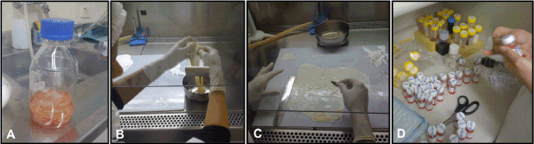
Figure 1. Amniotic membrane preparation. A: Arrival to the laboratory of the membrane in a sterile flask; B: Separation between amnion and chorion; C: Cutting the membrane; D: Packaging.
Surgical technique
The technique proposed by Kenyon and Tseng (10) and modified with the use of amniotic membrane (17) was used. All patients were operated under general anesthesia, once achieved asepsis and antisepsis in both eyes, from the donor eye (healthy eye), a 6 x 5 mm rectangular sector involving conjunctiva and limbus was taken using the vanax scissors and kept in viscoelastic (4% chondroitin sulfate) for later use. In the burn eye, a 360 peritomy was performed to remove fibrotic and neovascular tissue on the cornea. Subsequently, the graft was placed at 12 and 6 hours sutured with 10-0 nylon and then corneal, limb and proximal conjunctiva were coated with the amniotic membrane, which was sutured with 10-0 nylon spaced points.
Post-surgical treatment consisted of a topical treatment with Vigamox (Moxifloxacin hydrochloride, Novartis Pharmaceuticals) 1 drop every 4 hours and 0.4% Lagricel Often (sodium hyaluronate, Sophia RX) 1 drop every 6 hours for 8 days and Flumetol NF Often (Fluorometholone 1mg, Sophia RX) from day 8 to 30 (every 8 hours). The stitches were removed at 8 postoperative days. In cases of inflammation, custom topical and systemic corticosteroid regimens were used. Sections of neovascularization were treated with subconjunctival injections of bevacizumab (2.5 mg/0.1 mL) and triamcinolone (1 mg/0.1 mL).
Results
We analyzed 22 eyes of 22 patients; the mean age was 35.72 years (SD: 17.18). 9.09% (2) were women and 90.09% (19) were men. 63.63% (14) were workers, 27.27% (6) were students, 9.09% (2) housewife.
100% of the patients tested positive for goblet cells on impression cytology, confirming the diagnosis of limbal stem cell failure in these eyes. Table 1 shows the preoperative characteristics of the eyes under study.
Table 1. Preoperative characteristics of patients with total unilateral limbal stem cell deficiency.
Preoperative characteristics |
Mean (DS) |
Number of eyes |
22 |
Age |
35.72 (17.18) |
UCVA |
2.22(0.81) |
BCVA |
2.22 (0.81) |
Time between ocular burn and CLAU* (months) |
6.37 (3.77) |
Preoperative characteristics |
Percentage (Number) |
Causal Agent |
|
Alkali |
54.4% (12/22) |
Acid |
36.36% (8/22) |
Fire |
9.09% (2/22) |
Impression cytology (positive Globet cells) |
100% (22/22) |
Corneal Neovascularization |
3.45 (0.52) |
Total |
100% (22/22) |
Total associate to fibrosis |
45.45% (10/22) |
Inflammation |
|
Mild |
18.18% (4/22) |
Moderate |
27.27% (6/22) |
Severe |
54.54% (12/22) |
Other associate disease |
|
Yes |
0% (0/22) |
No |
100% (22/22) |
Amniotic membrane transplantation in acute stage |
|
Yes |
36.36% (8/22) |
No |
63.63% (14/22) |
Previous surgeries to CLAU* |
|
Tectonic corneal transplant (due to corneal perforation) |
18.18% (4/22) |
Symblepharon surgery |
36.36% (8/22) |
*CLAU: limbal conjunctiva autograft transplantation
At 3.27 years of follow up, 100% of patients improved one or more lines of uncorrected visual acuity, none of the eyes experienced visual acuity loss. 54.54% of patients had uncorrected visual acuity of 20/60 or better. Table 2 shows the visual results and complications related to the technique and Figure 2 shows the preoperative and postoperative pictures of some patients.
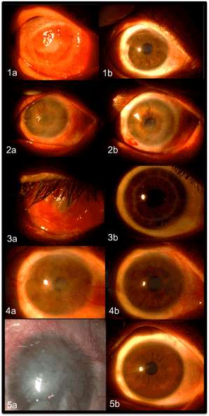
Figure 2. Preoperative (a) and postoperative (b) of 5 cases of limbal conjunctival autograft transplantation associated with an amniotic membrane in patients with unilateral total limbal deficiency.
Table 2. Clinical results of limbal conjunctival autograft transplantation associated with amniotic membrane.
Preoperative characteristics |
Percentage (N) |
Successful CLAU* |
63.63% (14/22) |
Time for corneal epithelization (days) |
8.27 (3.84) |
Cause of failure |
|
Neovascularization reached the pupil |
36.36%(8/22) |
Persistent epithelial defect |
18.18% (4/22) |
Persistent inflammation |
27.27% (6/22) |
Associated diseases |
18.18% (4/22) |
Symblepharon |
9.09% (2/22) |
Collateral Effects |
|
Partial Neo vascularization requiring injection of anti VEGT |
72.72% (16/22) |
Partial Neo vascularization requiring second procedure |
27.27% (6/22) |
Inflammation resolved with topical and systemic corticosteroids |
18.18% (4/22) |
Inflammation resolved with topical corticosteroids |
45.45% (10/22) |
Dry eye |
81.81 %(18/22) |
Mild |
63.63% (14/22) |
Moderate |
18.18% (4/22) |
Corneal opacity requiring corneal transplantation |
18.18% (4/22) |
Eyes worsened uncorrected visual acuity after 1 year |
4.54% (1/22) |
|
|
|
Mean (SD) |
UCVA postoperative at 1 year (LogMAR) |
0.72 (0.35) |
BCVA postoperative at 1 year (LogMAR) |
0.53 (0.46) |
Average follow-up time (years) |
3.27 (1.34) |
Final UCVA postoperative (LogMAR) |
0.78 (0.39) |
Final BCVA postoperative (LogMAR) |
0.58 (0.52) |
UCVA: Uncorrected visual acuity; BCVA: Best corrected visual acuity; CLAU: limbal conjunctiva autograft transplantation
95.45% (21 eyes) of the donor's eyes did not suffer any complications; only 1 eye presented a conjunctival granuloma in the immediate postoperative period (1 week) in the area of the donor tissue extraction that was resolved with its surgical excision, without affecting the visual acuity of the patient. 100% of the eyes presented adequate healing in the area where the donor tissue was extracted at the last follow up.
Discussion
Regenerative treatment with stem cells has had a breakthrough in medicine in recent years [5] and the specialty of ophthalmology has not been left behind; the international literature has demonstrated the effectiveness of limbal stem cell transplantation in the treatment of corneal neovascularization [9-15]; however, there are few studies reporting long term results of this procedure in combination with amniotic membrane use.
Corneal neovascularization is characterized by a vascularization of the corneal epithelium, chronic inflammation, epithelial defects, photophobia and loss of vision, leading to blindness where corneal transplants are ineffective because the limbal niche induces corneal rejection. The chemical burns represent one of the main causes of corneal neovascularization and unfortunately affect the economically active population, with a high social cost, in our series of cases, 90.9% of the patients were students or workers and the average age was 35.72 years.
Our results show that 100% of patients improved one or more lines of vision in postoperative visual acuity at 3.27 year of follow-up and none of the eyes experienced visual acuity loss, demonstrating that it is an effective procedure at long term. It should be noted that 72.72% of the patients had a visual acuity of hand movement or finger counts, in the latest postoperative follow up, 54.54% of the patients had best corrected visual acuity of 20/60 or better. Despite the good long-term results of this procedure, it requires very close follow-up by the specialist, in order to solve the main complications related to the disease, in our case series, 72.72% of the patients presented some type of inflammation, neovascularization or secondary disease as dry eye, which required some type of treatment or intervention that resolved favorably in all cases. Of the cases that presented limbal transplant failure that was 36.37%; it is important to note that 27.27% had a severe ocular burn in the preoperative period associated with corneal perforation and scar ulceration. Lang and coauthors [24], has described these as preoperative risk factors for transplant failure; The encouraging aspect of these cases is that despite being considered as a "failure", all these patients experienced improvement of preoperative visual acuity. Our results are very similar to those found by others authors, where failure rate after the transplant has been reported between 6 and 50% [9-16]. In addition, the present technique proved to be safe, since 100% of donor's eyes presented adequate healing and no complications in the donor eye.
In conclusions, the present study demonstrated that limbal conjunctival autograft transplantation associated with an amniotic membrane is effective and safe at long term follow up in the treatment of total unilateral limbal stem cells deficiency.
Acknowledgement
1. Tec Violeta Malaber for collaboration in the collection of the amniotic membrane.
2. Blga. Carmen Maldonado for collecting photos and technical support.
References
- Beebe DC, Masters BR (1996) Cell lineage and the differentiation of corneal epithelial cells. Invest Ophthalmol Vis Sci 37: 1815-1825. [Crossref]
- Voiculescu OB, Voinea LM, Alexandrescu C (2015) Corneal neovascularization and biological therapy. J Med Life 8: 444-448. [Crossref]
- Jongkhajornpong P, Lekhanont K, Siriyotha S, Kanokrungsee S, Chuckpaiwong V (2017) Factors Contributing to Long-Term Severe Visual Impairment in Stevens-Johnson Syndrome and Toxic Epidermal Necrolysis. J Ophthalmol 2017: 2087578. [Crossref]
- Arvelo F, Sojol F, Cotte C (2012) [Limbal deficiency syndrome]. Invest Clin 53: 205-217. [Crossref]
- Nagpal A, Choy FC, Howell S, Hillier S, Chan F, et al. (2017) Safety and effectiveness of stem cell therapies in early-phase clinical trials in stroke: a systematic review and meta-analysis. Stem Cell Res Ther 8: 191. [Crossref]
- Schermer A, Galvin S, Sun TT (1986) Differentiation-related expression of a major 64K corneal keratin in vivo and in culture suggests limbal location of corneal epithelial stem cells. J Cell Biol 103: 49-62. [Crossref]
- King JH, McTigue JW, MEDICO (Organization) (1965) International Eye Bank. The cornea world congress; papers. Washington: Butterworths.
- Kenyon KR, Tseng SC (1989) Limbal autograft transplantation for ocular surface disorders. Ophthalmology 96: 709-722. [Crossref]
- Dua HS, Saini JS, Azuara-Blanco A, Gupta P (2000) Limbal stem cell deficiency: concept, aetiology, clinical presentation, diagnosis and management. Indian J Ophthalmol 48: 83-92. [Crossref]
- Rama P, Ferrari G, Pellegrini G (2017) Cultivated limbal epithelial transplantation. Curr Opin Ophthalmol 28: 387-389. [Crossref]
- Arora R, Dokania P, Manudhane A, Goyal JL (2017) Preliminary results from the comparison of simple limbal epithelial transplantation with conjunctival limbal autologous transplantation in severe unilateral chronic ocular burns. Indian J Ophthalmol 65: 35-40. [Crossref]
- Meller D, Thomasen H (2017) [Limbal epithelial stem cell transplantation: Current state and perspectives. Ophthalmologe 114: 298-306. [Crossref]
- Moreira PB, Magalhães RS, Pereira NC, Oliveira LA, Sousa LB (2015) Limbal transplantation at a tertiary hospital in Brazil: a retrospective study. Arq Bras Oftalmol 78: 207-211. [Crossref]
- Sangwan VS, Sharp JAH (2017) Simple limbal epithelial transplantation. Curr Opin Ophthalmol 28: 382-386. [Crossref]
- Wylegala E, Dobrowolski D, Tarnawska D, Janiszewska D, Gabryel B, et al. (2008) Limbal stem cells transplantation in the reconstruction of the ocular surface: 6 years experience. Eur J Ophthalmol 18: 886-890. [Crossref]
- Kim JC, Tseng SC (1995) Transplantation of preserved human amniotic membrane for surface reconstruction in severely damaged rabbit corneas. Cornea 14: 473-484. [Crossref]
- Pires RT, Chokshi A, Tseng SC (2000) Amniotic membrane transplantation or conjunctival limbal autograft for limbal stem cell deficiency induced by 5-fluorouracil in glaucoma surgeries. Cornea 19: 284-287. [Crossref]
- Barreiro TP, Santos MS, Vieira AC, de Nadai Barros J, Hazarbassanov RM, et al. (2014) Comparative study of conjunctival limbal transplantation not associated with the use of amniotic membrane transplantation for treatment of total limbal deficiency secondary to chemical injury. Cornea 33: 716-720. [Crossref]
- Muraine M, Descargues G, Franck O, Villeroy F, Toubeau D, et al. (2001) [Amniotic membrane graft in ocular surface disease. Prospective study with 31 cases]. J Fr Ophtalmol 24: 798-812. [Crossref]
- Tseng SC, Prabhasawat P, Barton K, Gray T, Meller D (1998) Amniotic membrane transplantation with or without limbal allografts for corneal surface reconstruction in patients with limbal stem cell deficiency. Arch Ophthalmol 116: 431-441. [Crossref]
- Meller D, Dabul V, Tseng SC (2002) Expansion of conjunctival epithelial progenitor cells on amniotic membrane. Exp Eye Res 74: 537-545. [Crossref]
- Tseng SC, Li DQ, Ma X (1999) Suppression of transforming growth factor-beta isoforms, TGF-beta receptor type II, and myofibroblasts differentiation in cultured human corneal and limbal fibroblasts by amnioctic membrane matrix. J Cell Physiol 179: 325-335. [Crossref]
- Rivas L, Oroza MA, Pérez-Esteban A, Murube-del-Castillo J (1991) Topographical distribution of ocular surface cells by the use of impression cytology. Acta Ophthalmol (Copenh) 69: 371-376. [Crossref]
- Lang SJ, Böhringer D, Geerling G, Reinhard T (2017) Long-term results of allogenic penetrating limbo-keratoplasty: 20 years of experience. Eye (Lond) 31: 372-378. [Crossref]