Introduction: Calcaneal fractures can be life debilitating injuries. Bilateral involvement and presence of open fracture both contribute to poorer outcomes.
Case presentation: We present a case of a 36-year-old male who fell of a 1.5 m height and sustained a bilateral tongue-type calcaneal fractures, with the right side being a Gustilo type 2 open fracture. There was tenting of the skin on the left side which together with the open fracture element on the right side, necessitated urgent surgical intervention. Surgical management involved wound debridement on the right side and fixation of the calcaneal fractures by 2 cannulated screws on each side. Regular follow up for wound review and check x rays showed fracture healing with restoration of full weight bearing after 9 weeks and 12 weeks on his left side and right side, respectively.
Conclusion: This report shows successful surgical management and early restoration of function of a rare entity of calcaneal fractures.
Fracture, Calcaneum, Bilateral, Open, Compound
Calcaneal fractures, despite their rarity, are life debilitating injuries. They might result in restricted range of motion even after long rehabilitation periods [1]. These types of fractures usually result from falling from height and are more common in males than females with a peak incidence of 30-39 years [1,2]. The majority of reported calcaneal fractures (up to 90%) are displaced intra-articular fractures and in up to three quarters of patients with calcaneal fractures, there will be other accompanying injuries [3,4]. Greater severity and morbidity are associated with compound and bilateral types [3,5].
In this report, we present a successful management of bilateral tongue-type calcaneal fractures; a Gustilo Type 2 open fracture on the right side and a closed fracture with skin tenting on the left side.
34-year-old male with clear past medical and surgical history fell from a 1.5m height window and landed on his feet. He sustained bilateral tongue-type calcaneal fractures; a right sided Gustilo Type 2 open fracture with subtalar dislocation and a left sided closed tongue type calcaneal fracture. The patient received intravenous antibiotics and tetanus prophylaxis in the first hour of arriving to emergency department. The wound wasn’t grossly contaminated. Medical photographs were taken, the wound was covered by saline-soaked dressings and a backslab was applied (Figure 1). In addition to the open fracture on the right side, there was tenting of the skin over the left sided calcaneum fracture, hence, urgent surgery was planned to manage open fracture and to prevent skin compromise. The wound over the right foot was debrided (Figure 2). This was followed by open reduction and internal fixation of both fractures using two 6.5 mm cannulated screws. Bilateral full below knee casts were applied with ankles in equinous position. After two days, the patient was again taken to theater for a second look on the open fracture and application of VAC dressing followed by reapplication of a below knee cast in equinous. The wound was reviewed after 5 days, and the Vac dressing was removed. The patient was sent home with bilateral below knee casts with ankles in equinous position. Wound and X rays checks were done 2, 6, 9, 12 weeks post operatively. Both lower limbs were kept non weight bearing for 6 weeks. This was followed by partial weight bearing in neutral weight bearing casts. After 3 weeks of partial weight bearing (nine weeks after injury), the left side showed satisfactory bone healing while the right-side fracture (open fracture) wasn’t fully consolidated yet. The patient was allowed to fully weight bear on the left side while continuing partially weight bearing on the right side. Full weight bearing on the right side was allowed after 3 weeks (twelve weeks after the injury) when fracture healing was radiologically confirmed (Figure 3).
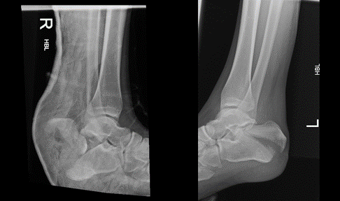
Figure 1. Preoperative X rays (lateral view) showing the bilateral tongue-type calcaneal fractures. A below knee backslap is applied to the right sided open fracture.
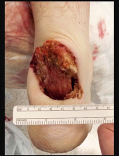
Figure 2. Intra-operative medical photograph of the right open calcaneal fracture. Photograph is taken after washout.
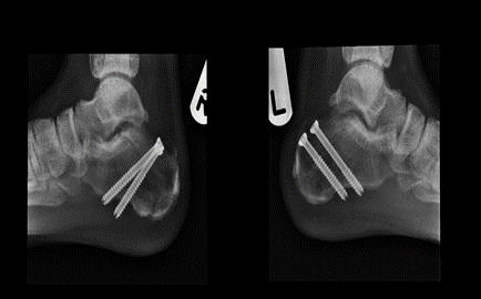
Figure 3. Twelve weeks postoperative follow up X rays (lateral view).
Despite that calcaneal fractures represent 50-60% of all fractured tarsal bones [6], the rate of bilateral involvement and open fractures is considerably low, estimated at 8-11.4% and 2.4%, respectively [1,7]. Both factors contribute to significantly poorer outcomes [3,8].
Essex-laprosty categorized calcaneal fractures into 2 major types,the tongue-type and the depression-type fractures [9]. Tongue-type calcaneal fractures are longitudinal fractures that passes through the calcaneal tuberosity posteriorly involving a portion of the articular surface. They are usually pulled superiorly by the Achilles tendon and the gastroc-soleus complex [10]. Due to the thin layer of soft tissue and the superficial nature of the fracture, compromise of the soft tissue envelope overlying the posterior calcaneal tuberosity can occur in up to 21% of this type of calcaneal fractures [11]. This soft tissue compromise requires immediate reduction to preserve viability.
In this case report, the patient had bilateral tongue type calcaneal fractures; an open fracture on the right side and a closed one on the left. In addition to the open fracture component, the closed left sided fracture was showing skin tenting. These two factors necessitated urgent surgical intervention.
Both fractures were managed by open reduction and internal fixation using two 6.5 mm cannulated screws. This open approach has been historically compared to the percutaneous closed approach. More wound problems and deep infections were observed with open reduction techniques, while revision and hardware removal predominated in closed reduction techniques [12]. However, a recent systematic literature review of reports on surgical management of tongue- type showed good outcomes and less complications without definite advantage for either techniques, open or closed [12].
Overall, the patient has shown a good recovery with early restoration of function. The right sided open fracture took the full expected recovery time on partial weight bearing (6 weeks) before total weight bearing was allowed (12 weeks after the injury). The left sided closed fracture showed a remarkable good healing and the patient was allowed to fully weight bear after 6 weeks of non-weight bearing and 3 weeks of partial weight bearing (9 weeks after the injury). This shows an earlier regaining of mobility compared to another article reporting 6 weeks of non-weight bearing and additional 6 weeks of partial weight bearing prior to giving the green light for total weight bearing [13].
This case report shows successful management of bilateral tongue type calcaneal fractures, with a Gustilo Type 2 open fracture on the right side and a closed fracture with skin tenting on the left side. Open reduction and internal fixation by cannulated screws yielded satisfactory results without complications and with early restoration of function.
Funding: Not applicable.
Conflicts of interest/Competing interests: the authors confirm that they don’t have any competing interests.
Ethics approval: the patient gave an informed consent for the case report.
Consent to participate: the patient gave an informed consent for the case report.
Consent for publication: the authors and the patient consent for publication.
Availability of data and material: Not applicable
Code availability: Not applicable
Almigdad Ali (AA): Conception and design, acquisition, and interpretation of data, drafting and revising the article.
Noman Niazi (NN): Treating surgeon, acquisition, and interpretation of data, drafting, and revising the article.
Anand Pillai (AP): Treating surgeon, acquisition, and interpretation of data, drafting, and revising the article.
- Vosoughi AR, Borazjani R, Ghasemi N, Fathi S, Mashhadiagha A, et al. (2022) Different types and epidemiological patterns of calcaneal fractures based on reviewing CT images of 957 fractures. Foot Ankle Surg 28: 88-92. [Crossref]
- Mitchell MJ, McKinley JC, Robinson CM (2009) The epidemiology of calcaneal fractures. Foot (Edinb) 19: 197-200. [Crossref]
- Leite CBG, Macedo RS, Saito GH, Sakaki MH, Kojima KE, et al. (2018) Epidemiological study on calcaneus fractures in a tertiary hospital. Rev Bras Ortop 53: 472-476. [Crossref]
- Bohl DD, Ondeck NT, Samuel AM, Diaz-Collado PJ, Nelson SJ, et al. (2017) Demographics, Mechanisms of Injury, and Concurrent Injuries Associated With Calcaneus Fractures: A Study of 14 516 Patients in the American College of Surgeons National Trauma Data Bank. Foot Ankle Spec 10: 402-410. [Crossref]
- Dooley P, Buckley R, Tough S, McCormack B, Pate G, et al. (2004) Bilateral calcaneal fractures: operative versus nonoperative treatment. Foot Ankle Int 25: 47-52. [Crossref]
- Davis D, Seaman TJ, Newton EJ. Calcaneus Fractures. In: StatPearls [Internet]. Treasure Island (FL): StatPearls Publishing; 2021. [Crossref]
- De Boer AS, Van Lieshout EMM, Van 't Land F, Misselyn D, Schepers T, et al. (2018) Soft tissue complications and timing of surgery in patients with a tongue-type displaced intra-articular calcaneal fracture: An international retrospective cohort study. Injury 49: 425-429. [Crossref]
- Tennent TD, Calder PR, Salisbury RD, Allen PW, Eastwood DM (2001) The operative management of displaced intra-articular fractures of the calcaneum: a two-centre study using a defined protocol. Injury 32: 491-496. [Crossref]
- Essex-Lopresti P (1952) The mechanism, reduction technique, and results in fractures of the OS calcis. Br J Surg 39: 395–419. [Crossref]
- Chhabra N, Sherman SC, Szatkowski JP (2013) Tongue-type calcaneus fractures: a threat to skin. Am J Emerg Med 31: 1151.e3-e4. [Crossref]
- Gardner MJ, Nork SE, Barei DP, Kramer PA, Sangeorzan BJ, et al. (2008) Secondary soft tissue compromise in tongue-type calcaneus fractures. J Orthop Trauma 22: 439-445. [Crossref]
- van der Vliet QMJ, Potter JM, Esselink TA, Houwert RM, Hietbrink F, et al. (2020) Open Versus Closed Operative Treatment for Tongue-Type Calcaneal Fractures: Case Series and Literature Review. J Foot Ankle Surg 59: 264-268. [Crossref]
- Hegde A, Mathias LJ, Ballal A, Shetty V, Shetty A (2016) A Prospective Study on Radiological and Functional Outcome of Displaced Tongue Type Intra-Articular Calcaneal Fractures Treated by Percutaneous Screw Fixation. J Clin Diagn Res 10: RC01-RC04. [Crossref]