We present the case of a 55-year-old woman who consulted for chronic constipation and asthenia. The patient had no relevant medical history except for long-term treatment with anthraquinone laxatives. Routine laboratory blood revealed anemia. A colonoscopy was performed which showed a blackish colonic pigmentation of the entire colon mucosa (melanosis coli) and a 10-mm sessile polyp located 50 cm from the anal verge (Figure 1A&B). Endoscopic resection of the polyp was performed (Figure 1C). The pathology report described an enteric adenocarcinoma arising from a tubular adenoma with positive resection margins. Elective surgery was decided given the pathological results. Therefore, the patient underwent a preoperative tattoo with India ink and laparoscopic left colectomy was performed (Figure 2A&B). The postoperative course was uneventful, and she was discharged on the 6th postoperative day. After one year of follow-up (Figure 2C), the patient remains asymptomatic and there is no clinical or radiological sign of recurrent or metastatic disease.
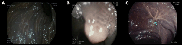
Figure 1. Colonoscopy showing melanosis coli (A) and a 10-mm sessile polyp (B). (C) Preoperative colonoscopy showing colonic scar post-polypectomy (arrow).
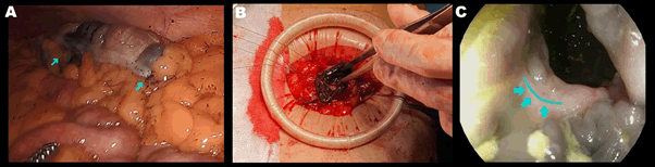
Figure 2. (A) Intraoperative laparoscopic view of a non-palpable colon lesion tattooed with India ink (arrows). (B) Intraoperative view while performing a hand-sewn end-to-end anastomosis. Be aware that dark colonic mucosa (melanosis coli) could be misdiagnosed as ischemic colitis. (C) Follow-up endoscopy showing the anastomosis (arrows).
Melanosis coli is a benign reversible condition characterized by a blackish coloration of the colonic mucosa. It is associated with the chronic use of anthraquinone laxatives [1]. There is no clear association with colorectal cancer, and it is usually reported as an incidental finding during colonoscopy. Although the diagnosis of melanosis coli can be suspected from endoscopic imaging, the definitive diagnosis is based on the pathology report.
There is an association between melanosis coli and a higher polyp detection rate due to the greater visibility of the polyps, given their contrast with a dark background [2]. However, this dark coloration of the colonic mucosa can be misdiagnosed with an ischemic mucosa [3,4]. Moreover, performing a successful intestinal anastomosis can be a dilemma because surgeons may think that the anastomosis is not well vascularized and perform unnecessary colon resections. Pathology studies can be helpful in diagnosis, although they may not always be available. Therefore, Intraoperative diagnosis remains a challenge for the surgeon.
In conclusion, the contrast between dark colonic mucosa and intestinal polyps makes their detection and endoscopic resection easier. But melanosis coli can also be easily confused with intestinal ischemia or anastomosis failure, which can lead to unnecessary colon resections. Therefore, it is important to be aware of the existence of this entity due to its possible clinical implications.
We have no conflicts of interest to disclose and there has been no significant financial support for this work that could have influenced its outcome.
We attest to the fact that all Authors listed on the title page have contributed significantly to the work, have read the manuscript, attest to the validity and legitimacy of the data and its interpretation, and agree to its submission to the Journal of Global Surgery.
The manuscript is approved by all Authors and in case of acceptance of the manuscript the copyright is transferred to Global Surgery.
- Yang N, Ruan M, Jin S (2020) Melanosis coli: A comprehensive review. Gastroenterol Hepatol 43: 266–72. [Crossref]
- Abu Baker F, Mari A, Feldman D, Suki M, Gal O, et al. (2018) Melanosis Coli: A Helpful Contrast Effect or a Harmful Pigmentation? Clin Med Insights Gastroenterol 11:1179552218817321. [Crossref]
- Moeller J, Solomon R, Kiffin C, Ditchek JJ, Davare DL (2019) Melanosis Coli: A Case of Mistaken Identity-A Case Report. Perm J 23: 18–063. [Crossref]
- Chaudhary BN, Sharma H, Nadeem M, Niayesh MH (2007) Ischemic colitis or melanosis coli: A case report. World J Emerg Surg 2: 25.