Introduction
Angiomatosis is a nonneoplastic proliferative vascular lesion, which occurs mostly in soft tissues. They grow up vertically between body compartments. We present an angiomatosis case that originated from mediasten, grew up into the thoracic spinal canal and removed by microsurgical laminotomy and costotransversectomy surgical technique [1,2].
Case
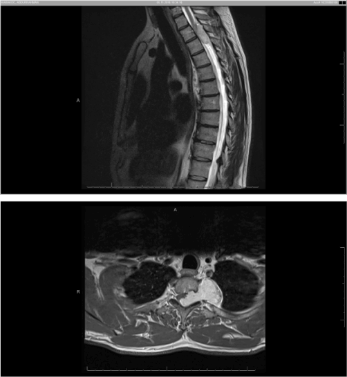
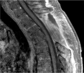
A 49 years old male patient admitted to our hospital with pain and numbness in both legs had been increasing progressively for 3 weeks after a horseshoe trauma. He had paraparesis and hypoesthesia on his neurological examination.. Magnetic Resonance Imaging (MRI) revealed a mass in 43x23 mm size with heterogeneous contrast enhancement. The lesion was in upper mediastinum, at T2 vertebra level, in left paravertebral area, extending to the spinal canal by expanding neural foramen at level T2- T3 (Figure 1). The patient was operated by neurosurgeons and thoracic surgeons together. T2 and T3 total microsurgical laminectomy, left 3rd costostransversectomy and total mass excision were applied to the patient at prone position. The postoperative neurological examination was normal. Histopathological diagnosis was “Angiomatosis”, a rare benign entitiy characterised by vascular proliferation consisting of mostly ectatic vascular structures showing multiple lobular patterns, separated by relatively good boundary from surrounding soft tissues (Figure 2).

Figure 1. In MRI; The lesion was in upper mediastinum, in left paravertebral area, extending to the spinal canal by expanding neural foramen.

Figure 2: Post operative MRI showing in operation area.
Discussion
Mediastinal angiomatosis can spread into soft tissues and cause spinal compression symptoms, especially at the epidural space at the thoracic region. During the resection of such large tumors, it is not possible to access the tumor boundaries by only laminectomy with the posterior approach. At the same time, posterolateral approach has begun to be abandoned in the world of neurosurgery due to thoracotomy, chest tube need, additional pleural complications, prolongation of operation time and increased risk of infection. In addition to microsurgical laminectomy with posterior approach, facetectomy and postotransversectomy appear to be more minimally invasive [1-5].
Conclusion
We report that this p2021 Copyright OAT. All rights reservd in upper mediastinum, enwrapping the spinal cord and causin neural compression was treated successfully via microsurgical laminectomy and costatransversectomy techniques.
References
- Tang EK, Chu PT, Goan YG, Hsieh PP, Lin JC (2016) Dumbbell-Mimicked Mediastinal Angiomatosis. Ann Thorac Surg 102: e555-e556. [Crossref]
- Zairi F, Nzokou A, Sunna T, Obaid S, Weil A, et al. (2017) Minimally invasive costotransversectomy for the resection of large thoracic dumbbell tumors. Br J Neurosurg 31: 179-183. [Crossref]
- Mazur M, Mumert M, Schmidt M (2015) Treatment of costal osteochondroma causing spinal cord compression by costotransversectomy: case report and review of the literatüre. Clin Pract 5: 734. [Crossref]
- Yun T, Suzuki H, Tagawa T, Iwata T, Mizobuchi T, et al. (2016) Cavernous hemangioma of the posterior mediastinum with bony invasion. Gen Thorac Cardiovasc Surg 64: 43-46. [Crossref]
- Pham M, Cohen J, Tuchman A, Commins D, Acosta F (2016) Large solitary osteochondroma of the thoracic spine: Case report and review of the literature. Surg Neurol Int 7: S323-S327. [Crossref]


