Differences in the cancers (treated and untreated) individuals and uninfected controls were identified with a NMR metabolomics approach. The current study was limited in sample size but provided original insights for modelling and simulation of 13C, 15N, 17O NMR chemical shifts, 17O and 14N electric field gradients and measurement of 13C and 15N chemical shifts in DNA/RNA of human gum cancer cells, tissues and tumors using NMR biospectroscopic profiling of gum cancer cells, tissues and tumors for novel systems diagnostics. This work has demonstrated the reliability, simplicity, and predictive ability of NMR–based metabolomics in discriminating between the experimental groups studied in our sample.
NMR biopectroscopy, gum cancer, cancer cells, cancer tissues, cancer tumors, modelling, simulation, chemical shifts, electric field gradients
Modelling and simulation of 13C, 15N, 17O NMR chemical shifts, 17O and 14N electric field gradients and measurement of 13C and 15N chemical shifts in DNA/RNA of human gum cancer cells, tissues and tumors using NMR biospectroscopic profiling of gum cancer cells, tissues and tumors for novel systems diagnostics was applied to determine the effect of human gum cancer infection on blood and plasma components. NMR biospectroscopic profile of the infected samples showed specific biomolecular information including reduction in body components especially DNA and RNA as compared to the healthy samples. Therefore, modelling and simulation of 13C, 15N, 17O NMR chemical shifts, 17O and 14N electric field gradients and measurement of 13C and 15N chemical shifts in DNA/RNA of human gum cancer cells, tissues and tumors using NMR biospectroscopic profiling of gum cancer cells, tissues and tumors for novel systems diagnostics may be a suitable candidate for evaluating human gum cancer infection related changes in blood and plasma samples thus providing useful information that can really help in diagnosis especially at individual level and potentially early screening of the cancer [1-10].
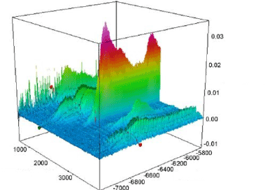
NMR biospectroscopy and its fingerprinting capabilities or rapid, high–throughput, and non–destructive analysis of a wide range of sample types producing a characteristic chemical “fingerprint” with a unique signature profile for modelling and simulation of 13C, 15N, 17O NMR chemical shifts, 17O and 14N electric field gradients and measurement of 13C and 15N chemical shifts in DNA/RNA of human gum cancer cells, tissues and tumors using NMR biospectroscopic profiling of gum cancer cells, tissues and tumors for novel systems diagnostics. Nuclear magnetic resonance (NMR) biospectroscopy and an array of mass spectrometry (MS) techniques provide selectivity and specificity for modelling and simulation of 13C, 15N, 17O NMR chemical shifts, 17O and 14N electric field gradients and measurement of 13C and 15N chemical shifts in DNA/RNA of human gum cancer cells, tissues and tumors using NMR biospectroscopic profiling of gum cancer cells, tissues and tumors for novel systems diagnostics, but demand costly instrumentation, complex sample pretreatment, are labor–intensive, require well–trained technicians to operate the instrumentation, and are less amenable for implementation in clinics. The potential for NMR biospectroscopy techniques to be brought to the bedside gives hope for modelling and simulation of 13C, 15N, 17O NMR chemical shifts, 17O and 14N electric field gradients and measurement of 13C and 15N chemical shifts in DNA/RNA of human gum cancer cells, tissues and tumors using NMR biospectroscopic profiling of gum cancer cells, tissues and tumors for novel systems diagnostics in the clinic. We discuss the utilization of current NMR biospectroscopy methodologies on biologic samples as an avenue towards rapid cost saving diagnostics for modelling and simulation of 13C, 15N, 17O NMR chemical shifts, 17O and 14N electric field gradients and measurement of 13C and 15N chemical shifts in DNA/RNA of human gum cancer cells, tissues and tumors using NMR biospectroscopic profiling of gum cancer cells, tissues and tumors for novel systems diagnostics (Figure 1).

Figure 1. Simulation of 13C, 15N, 17O NMR chemical shifts, 17O and 14N electric field gradients and measurement of 13C and 15N chemical shifts in DNA/RNA of human gum cancer cells, tissues and tumors using NMR biospectroscopic profiling of gum cancer cells, tissues and tumors for novel systems diagnostics
NMR biospectroscopic profile of the infected samples showed specific biomolecular information including reduction in body components especially DNA and RNA as compared to the healthy samples. Therefore, modelling and simulation of 13C, 15N, 17O NMR chemical shifts, 17O and 14N electric field gradients and measurement of 13C and 15N chemical shifts in DNA/RNA of human gum cancer cells, tissues and tumors using NMR biospectroscopic profiling of gum cancer cells, tissues and tumors for novel systems diagnostics may be a suitable candidate for evaluating human gum cancer infection related changes in blood and plasma samples thus providing useful information that can really help in diagnosis especially at individual level and potentially early screening of the cancer. Future studies with larger subject numbers are warranted to expand upon the present findings.
This study was supported by the Cancer Research Institute (CRI) Project of Scientific Instrument and Equipment Development, the National Natural Science Foundation of the United Sates, the International Joint BioSpectroscopy Core Research Laboratory Program supported by the California South University (CSU), and the Key project supported by the American International Standards Institute (AISI), Irvine, California, USA.
- Heidari, C. Brown, “Study of Composition and Morphology of Cadmium Oxide (CdO) Nanoparticles for Eliminating Cancer Cells”, J Nanomed Res., Volume 2, Issue 5, 20 Pages, 2015.
- A. Heidari, C. Brown, “Study of Surface Morphological, Phytochemical and Structural Characteristics of Rhodium (III) Oxide (Rh2O3) Nanoparticles”, International Journal of Pharmacology, Phytochemistry and Ethnomedicine, Volume 1, Issue 1, Pages 15–19, 2015.
- A. Heidari, “An Experimental Biospectroscopic Study on Seminal Plasma in Determination of Semen Quality for Evaluation of Male Infertility”, Int J Adv Technol 7: e007, 2016.
- A. Heidari, “Extraction and Preconcentration of N–Tolyl–Sulfonyl–Phosphoramid–Saeure–Dichlorid as an Anti–Cancer Drug from Plants: A Pharmacognosy Study”, J Pharmacogn Nat Prod 2: e103, 2016.
- A. Heidari, “A Thermodynamic Study on Hydration and Dehydration of DNA and RNA−Amphiphile Complexes”, J Bioeng Biomed Sci S: 006, 2016.
- A. Heidari, “Computational Studies on Molecular Structures and Carbonyl and Ketene Groups’ Effects of Singlet and Triplet Energies of Azidoketene O=C=CH–NNN and Isocyanatoketene O=C=CH–N=C=O”, J Appl Computat Math 5: e142, 2016.
- A. Heidari, “Study of Irradiations to Enhance the Induces the Dissociation of Hydrogen Bonds between Peptide Chains and Transition from Helix Structure to Random Coil Structure Using ATR–FTIR, Raman and 1HNMR Spectroscopies”, J Biomol Res Ther 5: e146, 2016.
- A. Heidari, “Future Prospects of Point Fluorescence Spectroscopy, Fluorescence Imaging and Fluorescence Endoscopy in Photodynamic Therapy (PDT) for Cancer Cells”, J Bioanal Biomed 8: e135, 2016.
- A. Heidari, “A Bio–Spectroscopic Study of DNA Density and Color Role as Determining Factor for Absorbed Irradiation in Cancer Cells”, Adv Cancer Prev 1: e102, 2016.
- A. Heidari, “Manufacturing Process of Solar Cells Using Cadmium Oxide (CdO) and Rhodium (III) Oxide (Rh2O3) Nanoparticles”, J Biotechnol Biomater 6: e125, 2016.

