A 40-year-old female formal dancer revealed non-symptomatic diagnosed inflammation on the base of between the middle and the lateral cuneiforms following magnetic resonance (MR) scans. It remains unclear if there are any differences in gait function between a person who have sustained non-symptomatic diagnosed inflammation on the base of cuneiforms and those who have not. To see its relationship to the gait patterns, dynamic plantar pressure was obtained during running and it was compared against T2* values as well as plantar pressure measuring from twenty healthy subjects. In this case of a dancer’s foot, the gait was modified possibly due to the non-symptomatic diagnosed inflammation on the base of cuneiforms, and hence the plantar pressure on the heel was increased compared with healthy non-dancer controls.
T2* map, MRI, Foot, Metatarsal, Plantar pressure, Dancer
T2*-weighted mapping has been recently used to evaluate ultrastructural morphological alteration including extracellular fluid content, hence detecting early-stage degeneration within cartilage [1], which is not detectable on the conventional magnetic resonance (MR) images. However, it remains unclear how this subtle detection on T2* maps relate to the human gait.
Recently we rarely recruited a former female dancer who had a pre-existing non-symptomatic inflammation on the base of between the middle and the lateral cuneiforms on T2* maps, which is not easy to evaluate its relationship to the gait patterns mainly because of the ethical issues. This case report presented subtle inflammation on the distal cuneiforms detecting by T2* mapping and its relationship to the plantar pressure during gait.
We examined a 40-year-old female former dancer (63kg, 167cm, 22.59 kg/m2) who at the time was involved in recreational sports activities such as yoga and Pilates. She used to be dance professionally (contemporary dance), with over 10 years of experience, but it had been five years since she quit dancing at this level. At the time of enrolment in this study, she reported no pain and no lower limb injuries. We obtained her written informed consent form approved by the University of Auckland Human Participants Ethics Committee (reference number: 016488).
Magnetic resonance data acquisition and analysis
MR scans of the right foot and ankle (dominant foot) were acquired using a 3-Tesla MR scanner (Siemens Skyra 3T, Erlangen, Germany) with a sixteen-channel foot coil while she was lying down in a natural position. Following sequence were used for MRI: (1) Sagittal T1 turbo spine echo, repetition time 600 ms, echo time 14 ms, flip angle 140 ̊, slice thickness 2.5 mm, field of view 250 mm, Bandwidth 257 Hz/Pixel, time of acquisition 2.57 minutes, (2) sagittal proton density turbo spine echo spair, repetition time 2530 ms, echo time 44 ms, flip angle 151 ̊ , slice thickness 2.5 mm, field of view 250 mm, Bandwidth 250 Hz/Pixel, time of acquisition 4.45, (3) sagittal T2 de3d water excitation, repetition time 12.9 ms, echo time 5 ms, flip angle 28 ̊ , slice thickness 0.6 mm, field of view 250 mm, Bandwidth 310 Hz/Pixel, time of acquisition 4.51, and (4) sagittal and coronal T2 star mapping, repetition time 980 ms, echo time 4.36 ms, flip angle 180 ̊ , slice thickness 3 mm, field of view 250 mm, Bandwidth 230 Hz/Pixel, time of acquisition 4.16 minutes. All the MR scans were analysed by Osirix software Lite v.9.0.1 (Pixmeo, Geneva, Switzerland).
Based on the MR scans, subtle inflammation on the base of between the middle and the lateral cuneiforms was identified (Figure 1). However, despite this pre-existing inflammation, she had no pain or discomfort during walking or running.
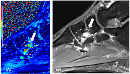
Figure 1. Sagittal view. (A) T2* mapping and (B) a sagittal PD (proton density) TSE (turbo spin-echo) spair sequence showed the pre-existing inflammation at the base of between the middle and the lateral cuneiforms
Plantar pressure comparison against healthy subjects
To see how this non-symptomatic inflammation affects gait patterns, the plantar pressure was measured from the right foot using the Novel emed® pressure platform during running unshod with natural pace. Pressure map was subdivided into eleven region of interests (ROIs), including hallux, toe regions (T2-T5), metatarsals 1-5 (M1, M2, M3, M4, M5), medial/lateral mid foot (MM, LM), medial/lateral heel (HM, HL). Peak pressure (kPa), peak force (N), contact area (cm2), and impulse (Ns) were obtained from each ROI, and these parameters were compared against twenty (10 female and 10 male) healthy controls (age: 30.1 ± 8 years, weight: 66.12 ± 9.7 kg, height: 1.71 ± 0.08 m).
SPSS (IBM Corporation, Version 20) was employed for statistical tests. The differences in plantar pressure parameters between the case subject and the 20 healthy controls were evaluated with a one-way T-test (two-tailed) with a significance set at p=0.05.
The plantar pressure distribution (Figure 2) seemed to be very different between the case subject and the 20 healthy controls. In the case subjects, the load underneath MH and LH revealed the greatest values among the eleven ROIs during unshod running, whereas the 20 healthy controls showed the highest values under M1. Mean peak force and impulse also demonstrated a similar disparity, with the greatest values beneath the heel regions in the case subject while the 20 healthy controls presented the greatest values beneath M1. More specifically, mean peak pressure underneath MH and LH were significantly greater by 40.26% and 43.36%, respectively, in the case subject when compared with the 20 healthy controls. The MM, LM, and T1 also showed similar trends of significantly higher values (48.86%, 16.12%, and 16.73%, respectively) in the case subject when compared with 20 healthy controls. Mean peak force and impulse underneath T1 (21.61%, 17.82%, respectively), MM (42.23%, 42.25%, respectively), LM (31.47%, 34.47%, respectively), and LH (23.98%, 48.68%, respectively) also presented greater values in the case subject compared with 20 healthy controls. In contrast, all of the metatarsal regions showed lower loading in the case subject by comparison to the healthy cohort– mean peak plantar pressure underneath M2 and M3 were significantly lower in the case subject (Figure 2). Mean peak force and impulse underneath M1 and M2 also showed significantly lower values in the case subject. The case subject generally showed lower values in the contact area than the 20 healthy individuals, except for LM.
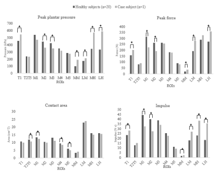
Figure 2. Peak mean plantar pressure, force, contact area, and impulse during running unshod. * indicates a significant (p=0.05) difference between two groups
This case study shows that non-symptomatic inflammation, which was detected by T2* maps, does modify the plantar pressure distribution during natural gait. Based on the pedobarography analysis, the contribution of the heel was greatly increased in the case subject, while the healthy controls showed greater contribution of the metatarsal regions. The results from the healthy controls were in line with other unshod analysis studies [2,3], reporting that unshod walking and/or running lead to greater loading under the forefoot regions, although it must be noted that they both had different study design, gait types, measuring devices, and shod condition. It has been well documented that unshod runners (even first-time unshod runners) likely adopt a forefoot strike technique [4,5], which results in a load increasing underneath the lateral forefoot [3], while shod runners generally run with heel strike techniques. Heel strike landing with bare feet may cause greater deformation of the fat-pad under the calcaneus compared with a heel strike in shoes [6].
Contrary to our expectations, however, the case subject showed significantly increased loading under the heel regions during unshod running. This unexpected gait pattern was possibly due to the pre-existing inflammation at the base of her cuneiforms. Alteration of T2 and/or T2* has been associated with early-stage degeneration in cartilage [1] and a higher T2 signal was observed in severely osteoarthritic knee cartilage when compared with normal cartilage [7]. Recently, an acute alteration of T2 and/or T2* value was observed in the knee and the ankle [8] cartilage after long-distance running. It has also been reported that female ballet dancers showed greater T2 values in the ankle cartilage when compared with age-matched healthy controls [9]. Although there is no study looking at the relationship between the T2* signal on the foot and gait pattern, the T2* values indicate changes to the cartilage that affects gait patterns to off-load the forefoot, and hence increase pressure on the heel.
This case study presents the evaluation of gait biomechanics in a former dancer who had pre-existing, non-symptomatic inflammation at the base of between the middle and the lateral cuneiforms. The results may suggest that the subtle inflammation, detected by T2* mapping, does modify gait and may lead to decreased involvement of metatarsal regions, while increasing involvement of heel regions when compared with healthy subjects. In the present case, the gait was altered to off-load the forefoot and metatarsals, and hence increase pressure on the heel.
The authors do not have any conflict of interest which could have influenced the results of this case report.
There were no sources of funding in relation to this case report.
- Crema MD, Roemer FW, Marra MD, Burstein D, Gold GE, et al. (2011) Articular cartilage in the knee: Current MR imaging techniques and applications in clinical practice and research. Radiographics 31: 37-61. [Crossref]
- Bergstra S, Kluitenberg B, Dekker R, Bredeweg S, Postema K, et al. (2015) Running with a minimalist shoe increases plantar pressure in the forefoot region of healthy female runners. J Sci Med Sport 18: 463-468. [Crossref]
- D’AoÛt K, Pataky TC, De Clercq D, Aerts P (2009) The effects of habitual footwear use: Foot shape and function in native barefoot walkers. Footwear Sci 1: 81-94.
- Hatala KG, Dingwall HL, Wunderlich RE, Richmond BG (2013) Variation in foot strike patterns during running among habitually barefoot populations. PLoS One 8: e52548. [Crossref]
- Lieberman DE, Venkadesan M, Werbel WA, Daoud AI, D’Andrea S, et al. (2010) Foot strike patterns and collision forces in habitually barefoot versus shod runners. Nature 463: 531-535. [Crossref]
- De Clercq D, Aerts P, Kunnen M (1994) The mechanical characteristics of the human heel pad during foot strike in running: An in vivo cineradiographic study. J Biomech 27: 1213-1222. [Crossref]
- Dunn TC, Lu Y, Jin, H, Ries MD, Majumdar S, et al. (2004) T2 relaxation time of cartilage at MR imaging: Comparison with severity of knee osteoarthritis. Radiology 232: 592-598. [Crossref]
- Schutz UHW, Ellermann J, Schoss D, Wiedelbach H, Beer M, et al. (2014) Biochemical cartilage alteration and unexpected signal recovery in T2* mapping observed in ankle joints with mobile MRI during a transcontinental multistage footrace over 4486 km. Osteoarthritis Cartilage 22: 1840-1850. [Crossref]
- Cha JG, Yi JS, Han JK, Lee YK (2015) Comparison of quantitative cartilage T2 measurements and qualitative MR imaging between professional ballet dancers and healthy volunteers. Radiology 276: 199-206. [Crossref]