Background: Individuals who have sustained concussion frequently develop impairments in oculomotor function. Some providers have attempted to improve these impairments and aid the overall recovery from concussion by prescribing vision therapy.
Objective: To review and assess the validity of common clinical oculomotor findings and subsequent vision therapy for concussion.
Methods: A literature review was conducted via OVID MEDLINE and ePub databases. Three hundred and sixty-three records were obtained. Results were pared down to include only those with adequate power and relevance to the study questions. Eleven studies on examination and 2 studies on treatments met inclusion criteria.
Results: There is reasonable evidence to support the use of near-point convergence, pursuits, and saccades (when computerized) aiding the diagnosis of concussion. Adequately powered randomized controlled trials do not exist to support of the use of vision therapy for the treatment of concussion.
Conclusions: Oculomotor examination findings are useful in clinical practice to aid the diagnosis of concussion, but further research using adequately powered randomized controlled trials are required to clarify the efficacy of vision therapy in the treatment of concussion.
concussion, oculomotor, mild traumatic brain injury, saccadic dysfunction, vergence, pursuits, smooth pursuit eye movement, vision therapy
Concussion is a common sports and occupational health problem [1] with numerous clinical trajectories [2]. The visual trajectory is associated with prolonged recovery [3] and academic difficulties in adolescents [4]. Brain structures involved in visual function appear to have a vulnerability to shear stress and are thus commonly impacted [5-7], with oculomotor symptoms in over 60% of concussed children and adolescents [8] and over 90% of concussed adults [9].
Examination techniques assessing oculomotor function are based on eye movements [10], including vergence, saccades, and pursuits [11], and are not commonly assessed in primary care.
Vergence movements align the fovea with targets at different distances from the eye. They involve disconjugate adjustments (convergence and divergence) as opposed to conjugate adjustments (e.g., eyes moving the same direction to track a moving object) and are part of the near reflex triad which includes accommodation and pupillary constriction. Near point convergence is tested by having the patient focus on the examiner's finger from a distance and gradually bringing the finger closer to the patient (Figure 1). The point at which the image of the finger becomes double is measured from the eye.
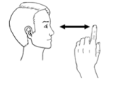
Figure 1. Examination of near point convergence
Saccades are rapid, ballistic movements of the eyes, that abruptly change the focal point. This includes smaller movements made while reading and larger movements scanning a crowd. They are tested by the examiner holding fingers at far ends of the visual fields while the patient looks back and forth between fingers (Figure 2). With abnormal saccades, the patient is unable to correctly fix on the object after ballistic movements.
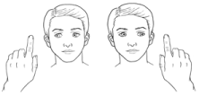
Figure 2. Examination of saccadic eye movements
Smooth pursuit eye movements are slow, voluntary tracking movements designed to keep a moving object on the fovea. They are examined by having the patient fix their gaze on the examiner's finger while it is slowly moved in the horizontal and vertical planes (Figure 3). An abnormal finding is difficulty with performing this task.
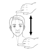
Figure 3. Examination of (vertical) smooth pursuit eye movements
This review attempts to clarify the utility of (1) oculomotor examination findings as they relate to the diagnosis of concussion and (2) proposed oculomotor treatments for concussion.
An OVID MEDLINE and ePub database search was conducted for articles published in the last 10 years in English with keywords relating to the above-noted oculomotor tests and treatments for concussion. A total of 363 records were obtained. A total of 11 studies of oculomotor examination findings and 2 studies of oculomotor treatments met our inclusion criteria. Only articles relevant to the study purpose were used. Sports-related, incidental, and blast-related were all considered if the patient met the definition of concussion. Case reports, studies with small sample size (approximately 20 participants or fewer), and uncontrolled studies were given less or no consideration. Random automatized naming tools (King-Devick and others) and vestibulo-oculomotor tests were excluded as they incorporate multiple oculomotor systems at once. Treatment with optic devices (lenses, etc.) was not reviewed.
Oculomotor examination findings for concussion
Results are summarized in Table 1.
Table 1. Articles on oculomotor examination findings that met inclusion criteria.
Authors |
Topic |
Study design |
Key findings |
Near point convergence dysfunction |
Thiagarajan et al. [12] |
Vergence dysfunction in concussion |
Review |
Mild traumatic brain injury is associated with a range of vergence dysfunctions in adults with increased distance of break and recovery in cases vs controls studies reviewed |
Szymanowicz et al. [14] |
Vergence dysfunction in concussion |
Case-control
(N= 31) |
Significant difference (increased distance) of NPC break and recovery between case and control groups |
DuPrey et al. [15] |
Vergence dysfunction in concussed athletes |
Retrospective cohort (N=270) |
Concussed athletes with NPC had significantly longer recovery periods (51.6 days) than those without convergence insufficiency (14.7 days) |
Kawata et al. [16] |
Vergence dysfunction with sub-concussive head injury |
Prospective cohort study (N= 29) |
Higher impact sub-concussive head trauma in university football players is associated with prolongation of NPC from baseline with return to baseline after 3 weeks. |
Saccadic Dysfunction |
Hunfalvay et al. (2019) [24] |
Saccades in concussion |
Case-control (N=287) |
Horizontal saccadic dysfunction has sensitivity of 0.77 and specificity of 0.78 in predicting concussion. |
Smooth Pursuit Eye Movement Dysfunction |
Murray et al. [26] |
SPEM after concussion |
Case-control (N=36) |
Abnormalities in SPEM amplitude and velocity were associated with concussion vs control groups. |
Wetzel et al. [27] |
SPEM after concussion |
Case-control (N=146) |
Patients with post-concussion syndrome had abnormalities in tracking movements used in SPEM vs controls. |
Multiple Abnormalities |
Snegireva et al. [21] |
Eye tracking technology in concussion |
Systematic review and meta-analysis |
Saccadic and pursuit dysfunction is seen (by computerized assessment) in the context of concussion and remains affected in the acute (<30 days) phase. |
Capo-Aponte et al. [13] |
Visual dysfunction after blast-induced concussion |
Case-control
(N=40) |
Significant difference (increased distance) of NPC after blast-induced concussion vs controls. Significant differences in errors in smooth pursuit and saccadic eye movements. |
Hunt et al. [17] |
Oculomotor abnormalities in concussion |
Systematic review |
Exploratory studies find that concussion appears to be associated with saccadic abnormalities (greater amplitudes smaller peak accelerations slower velocities and less accurate target prediction) as well as abnormalities of smooth pursuit eye movements. |
Cifu et al. [23] |
Oculomotor abnormalities post-concussion |
Case-control (N=86) |
Military veterans with concussion had significantly poorer smooth pursuit tracking ability as well as altered saccadic amplitudes accelerations and velocities. |
Abnormalities of near point convergence
In adults, abnormalities of near point convergence after concussion have been reported in numerous studies [12-14]. The normal finding in adults is a break in unified visual image at 5 cm from the eye and recovery at 7 cm. Convergence abnormalities appear to be a reliable marker for prolonged concussion recovery; those with this abnormality have taken more than 50 days (as opposed to approximately 15 days) to recover from concussion [15].
Sub-concussive head injury in athletes (as measured by accelerometers) has also been associated with mildly impaired NPC [16], which may confound the assessment of the acutely or previously injured patient.
Saccadic dysfunction
Dysfunctional saccadic eye movements can be measured by computer and are those with greater amplitude, smaller peak acceleration, slower velocity, and lower accuracy with target prediction [17]. Several systematic reviews have demonstrated that saccadic eye movements are affected by concussion. Previously, only class 3-4 level evidence indicated significant reduction in saccade velocity vs. controls [17-23]; however, newer, adequately powered case-controlled research suggests a sensitivity of 0.77 and specificity of 0.78 for predicting concussion [24].
Smooth pursuit eye movements (SPEM)
Impairments in SPEM in concussion has been previously studied [25] and discussed in a 2016 systematic review [17]. More recently, two case-control studies (one with a study population of 146 and the other of 36) have supported the finding of abnormalities of smooth pursuit eye movements in concussion [26,27].
Treatment for oculomotor manifestations of concussion
A number of therapies have been proposed for the treatment of visual/oculomotor dysfunctions associated with concussion [28-31]. These include laboratory-based versional, vergence, and accommodative eye movement training, simulated reading exercises, balance training and visual awareness exercises, driving simulators [32], light filtering and photochromic lenses, and use of glasses with various tints and prism combinations [33]. These treatments have been utilized in non-concussed patients [34]; however, they have only recently been applied to concussion.
This review presents the best available information with regards to proposed treatments, summarized in Table 2.
Table 2. Articles on effects of oculomotor treatments for concussion meeting inclusion criteria.
Authors |
Topic |
Study design |
Key findings |
Peters et al. [36] |
Vision therapy in concussion |
Cohort study (N=137) |
Concussed hockey players who participated in therapy had symptom resolution in 5.8 weeks versus 12.3 weeks in those who did not. |
Gallaway et al. [34] |
Vision therapy in concussion |
Retrospective case series (no control group) (N=218) |
85% of patients who completed vision therapy protocol had improvements in NPC base-out demand and/or concussion symptoms. |
Vision therapy
There is no single consensus method of vision therapy; however, many propose to improve concussion symptoms through various oculomotor exercises targeting components of the oculomotor system. An example of a proposed therapy is the Computer Oculomotor Rehabilitation (COR) Program for concussion [35], which includes exercises in gaze fixation (central, horizontal, vertical) for set periods of time, predictable saccades and non-predictable saccades (horizontal and vertical), smooth pursuits (horizontal and vertical), vestibulo-ocular reflex in different positions of gaze (central, horizontal, vertical, oblique), and simulated reading. One can modify the base-in, -out, -up, or -down of spectacle prisms to alter horizontal or vertical vergence on demand. The program allows variation in parameters for each eye movement trained, including speed of movement, amplitude of movement, and other modifications to alter the degree of difficulty. Large, controlled trials have not yet assessed these programs.
A retrospective chart review of concussed professional hockey players (N=137) compared those who received a form of vision therapy and those who did not [36]. Vision therapy consisted of a review of their visual prescription, anti-glare and 40% blue tint glasses for those with photophobia, avoiding prism spectacles, challenge the oculomotor system by playing catch with a bean bag while adding difficulty by talking to the patient, and playing catch with glasses with base-in and -out prisms (15 prism diopters), then wearing yoked prisms (yoked right then left), and using a balance board while wearing the above noted glasses. Those who participated had symptom resolution in 5.8 weeks versus 12.3 weeks in those who did not.
An uncontrolled retrospective case series [34] assessed a vision therapy program that consisted of once or twice weekly 45-minute office sessions and home-based sessions using therapy described in the Convergence Insufficiency Treatment Trials (CITT) [37] (a trial assessing the interventions of pencil push-ups vs office-based vision therapy in non-concussed individuals), with the addition of exercises for saccadic and pursuits using Hart Charts, thumb rotations, an use of the Sanet Vision Integrator software. Specific protocols were not further described. Eighty-five percent of participants who completed vision therapy had significant improvements in NPC, base-out demand, and/or symptoms.
Many of the studies reviewed were not considered due to low sample size or other methodological limitations [30,38-46].
Abnormalities in vergence appear to be the most reliably affected by concussion. However, history of sub-concussive impacts may affect interpretation. Saccadic and pursuit abnormalities do appear to be associated with concussion but are less easily detected without computerized detection methods and are thus of less utility in a primary care clinical setting.
There is insufficient evidence at this time to establish the medical requirement of most oculomotor treatments for concussion patients. The literature surrounding therapies for concussion is unfortunately limited by small sample size, lack of control groups, and other methodological limitations. Only one controlled cohort study met our inclusion criteria, and demonstrated possible benefit from vision therapy, though it was limited by lack of randomization (participants chose whether to opt-in or out for therapy). At this time, it is therefore not possible to draw definitive conclusions regarding the efficacy of these interventions. Large, randomized controlled trials or Bayesian-style meta-analysis of the existing smaller studies would better establish any potential benefit of vision therapies for oculomotor impairments in concussion.
There appears to be validity to the oculomotor examination findings in relation to concussion, with vergence chief among them in the primary care setting. At the time of this review, further research is required to be able to clarify the potential efficacy of vision therapy for concussion-related oculomotor disorders.
The authors wish to acknowledge Melissa McIvor who provided illustrations and Nicole Askin for performing literature searches.
- Langer L, Levy C, Bayley M (2020) Increasing Incidence of Concussion: True Epidemic or Better Recognition? J Head Trauma Rehabil 35: E60–E66.
- Craton N, Ali H, Lenoski S (2017) COACH CV: the seven clinical phenotypes of concussion. Brain sci 7: 119. [Crossref]
- Master CL, Master SR, Wiebe DJ, Storey EP, Lockyer JE, et al. (2018) Vision and vestibular system dysfunction predicts prolonged concussion recovery in children. Clin J Sport Med 28: 139-145. [Crossref]
- Swanson MW, Weise KK, Dreer LE, Johnston J, Davis RD, et al. (2017) Academic difficulty and vision symptoms children with concussion. Optom Vis Sci 94: 60-67. [Crossref]
- Nevin NC (1967) Neuropathological changes in the white matter following head injury. J Neuropathol Exp Neurol 26: 77-84. [Crossref]
- Lipton ML, Kim N, Park YK, Hulkower MB, Gardin TM, et al. (2012) Robust detection of traumatic axonal injury in individual mild traumatic brain injury patients: intersubject variation, change over time and bidirectional changes in anisotropy. Brain Imaging Behav 6: 329-342. [Crossref]
- Maruta J, Lee SW, Jacobs EF, Ghajar J (2010) A unified science of concussion. Ann N Y Acad Sci 1208: 58-66. [Crossref]
- Master CL, Scheiman M, Gallaway M, Goodman A, Robinson RL, et al. (2016) Vision diagnoses are common after concussion in adolescents. Clin Pediatr (Phila) 55: 260-267. [Crossref]
- Ciuffreda KJ, Kapoor N, Rutner D, Suchoff IB, Han ME, et al. (2007) Occurrence of oculomotor dysfunctions in acquired brain injury: a retrospective analysis. Optometry 78: 155-161. [Crossref]
- Hunt AW, Mah K, Reed N, Engel L, Keightley M (2016) Oculomotor-based vision assessment in mild traumatic brain injury: a systematic review. J Head Trauma Rehabil 31: 252-261. [Crossref]
- Purves D, Augustine GJ, Fitzpatrick D, et al. editors (2001) Neuroscience. 2nd edition. Sunderland (MA): Sinauer Associates; Types of Eye Movements and Their Functions. [Crossref]
- Thiagarajan P, Ciuffreda KJ, Ludlam DP (2011) Vergence dysfunction in mild traumatic brain injury (mTBI): a review. Ophthalmic Physiol Opt 31: 456-468. [Crossref]
- Capo-Aponte JE, Urosevich TG, Temme LA, Tarbett AK and Sanghera NK (2012) Visual dysfunctions and symptoms during the subacute stage of blast-induced mild traumatic brain injury. Mil Med 177: 804-813. [Crossref]
- Szymanowicz D, Ciuffreda KJ, Thiagarajan P, Ludlam DP, Green W, et al. (2012) Vergence in mild traumatic brain injury: a pilot study. J Rehabil Res Dev 49: 1083-1100. [Crossref]
- DuPrey KM, Webner D, Lyons A, Kucuk CH, Ellis JT, et al. (2017) Convergence insufficiency identifies athletes at risk of prolonged recovery from sport-related concussion. Am J Sports Med 45: 2388-2393. [Crossref]
- Kawata K, Rubin LH, Lee JH, Sim T, Takahagi M, et al. (2016) Association of football subconcussive head impacts with ocular near point of convergence. JAMA Ophthalmol 134: 763-769. [Crossref]
- Hunt AW, Mah K, Reed N, Engel L, Keightley M, et al. (2016) Oculomotor-Based Vision Assessment in Mild Traumatic Brain Injury: A Systematic Review. J Head Trauma Rehabil 31: 252-261. [Crossref]
- Heitger MH, Anderson TJ, Jones RD, Dalrymple‐Alford JC, Frampton CM, et al. (2004) Eye movement and visuomotor arm movement deficits following mild closed head injury. Brain 127: 575-590. [Crossref]
- Heitger MH, Jones RD, Dalrymple-Alford JC, Frampton CM, Ardagh MW, et al. (2006) Motor deficits and recovery during the first year following mild closed head injury. Brain Inj 20: 807-824. [Crossref]
- Kraus MF, Little DM, Donnell AJ, Reilly JL, Simonian N, et al. (2007) Oculomotor function in chronic traumatic brain injury. Cogn Behav Neurol 20: 170-178.
- Snegireva N, Derman W, Patricios J, Welman KE (2018) Eye tracking technology in sports-related concussion: a systematic review and meta-analysis. Physiol Meas 39: 12TR01. [Crossref]
- Cochrane GD, Christy JB, Almutairi A, Busettini C, Swanson MW, et al. (2019) Visuo-oculomotor function and reaction times in athletes with and without concussion. Optom Vis Sci 96: 256-265. [Crossref]
- Cifu DX, Wares JR, Hoke KW, Wetzel PA, Gitchel G, et al. (2015) Differential Eye Movements in Mild Traumatic Brain Injury Versus Normal Controls. J Head Trauma Rehabil 30: 21-28. [Crossref]
- Hunfalvay M, Roberts CM, Murray N, Tyagi A, Kelly H, et al. (2019) Horizontal and vertical self-paced saccades as a diagnostic marker of traumatic brain injury. Concussion 4: CNC60. [Crossref]
- Suh M, Basu S, Kolster R, Sarkar R, McCandliss B, et al. (2006) Increased oculomotor deficits during target blanking as an indicator of mild traumatic brain injury. Neurosci Lett 410: 203–207. [Crossref]
- Murray NG, Szekely B, Islas A, Munkasy B, Gore R, et al. (2020) Smooth Pursuit and Saccades after Sport-Related Concussion. J Neurotrauma 37: 340-346.
- Wetzel PA, Lindblad AS, Raizada H, James N, Mulatya C, et al. (2018) Eye Tracking Results in Postconcussive Syndrome Versus Normative Participants. Invest Ophthalmol Vis Sci 59: 4011-4019. [Crossref]
- Armstrong RA (2018) Visual problems associated with traumatic brain injury. Clin Exp Optom 101: 716-726. [Crossref]
- Ciuffreda KJ, Han Y, Kapoor N, Ficarra AP (2006) Oculomotor rehabilitation for reading in acquired brain injury. NeuroRehabilitation 21: 9–21. [Crossref]
- Thiagarajan P, Ciuffreda KJ, Capo‐Aponte JE, Ludlam DP, Kapoor N (2014) Oculomotor rehabilitation for reading in mild traumatic brain injury (mTBI): An integrative approach. NeuroRehabilitation 34: 129–146. [Crossref]
- Ciuffreda KJ, Rutner D, Kapoor N, Suchoff IB, Craig S, et al. (2008) Vision therapy for oculomotor dysfunctions in acquired brain injury: a retrospective analysis. Optometry 79: 18-22. [Crossref]
- Akinwuntan AE, Wachtel J, Rosen PN (2012) Driving simulation for evaluation and rehabilitation of driving after stroke. J Stroke Cerebro Dis 21: 478–486. [Crossref]
- Jackowski MM, Sturr JF, Taub HA, Turk MA (1996) Photophobia in patients with traumatic brain injury: uses of light‐filtering lenses to enhance contrast sensitivity and reading rate. Neurorehabilitation 6: 193–201. [Crossref]
- Gallaway M, Scheiman, M, Mitchell GL (2017) Vision therapy for post-concussion vision disorders. Optom Vis Sci 94: 68-73. [Crossref]
- Ciuffreda KJ, Yadav NK, Thiagarajan P, Ludlam DP (2017) A novel computer oculomotor rehabilitation (COR) program for mild traumatic brain injury (MTBI). Brain Sci 7: 99. [Crossref]
- Peters M (2015) The Peters/Price (see to play) vision concussion protocol: diagnosis and treatment. Optom Vis Perform 3: 126-138.
- Scheiman M, Mitchell GL, Cotter S, Kulp MT, Cooper J, et al. (2005) A randomized clinical trial of vision therapy/orthoptics versus pencil pushups for the treatment of convergence insufficiency in young adults. Optom Vis Sci 82: E583-E595. [Crossref]
- Thiagarajan P, Ciuffreda KJ (2015) Short-term persistence of oculomotor rehabilitative changes in mild traumatic brain injury (mTBI): A pilot study of clinical effects. Brain Inj 29: 1475-1479.
- Kapoor N, Ciuffreda KJ, Han Y (2004) Oculomotor rehabilitation in acquired brain injury: a case series. Arch Phys Med Rehabil 85: 1667-1678. [Crossref]
- Scheiman M, Talasan H, Mitchell GL, Alvarez TL (2017) Objective assessment of vergence after treatment of concussion-related CI: a pilot study. Optom Vis Sci 94: 74. [Crossref]
- Berryman A, Rasavage K, Politzer T, Gerber D (2020) Oculomotor Treatment in Traumatic Brain Injury Rehabilitation: A Randomized Controlled Pilot Trial. Am J Occup Ther 74: 7401185050p1-7401185050p7. [Crossref]
- Thiagarajan P, Ciuffreda KJ (2014) Versional eye tracking in mild traumatic brain injury (mTBI): effects of oculomotor training (OMT). Brain Inj 28: 930-943. [Crossref]
- Yadav NK, Thiagarajan P, Ciuffreda KJ (2014) Effect of oculomotor vision rehabilitation on the visual-evoked potential and visual attention in mild traumatic brain injury. Brain Inj 28: 922-929. [Crossref]
- Thiagarajan P (2013) Oculomotor rehabilitation for reading dysfunction in mild traumatic brain injury. (Doctoral dissertation, State University of New York).
- Thiagarajan P, Ciuffreda KJ (2014) Effect of oculomotor rehabilitation on accommodative responsivity in mild traumatic brain injury. J Rehabil Res Dev 51: 175–191. [Crossref]
- Thiagarajan P, Ciuffreda KJ (2013) Effect of oculomotor rehabilitation on vergence responsivity in mild traumatic brain injury. J Rehabil Res Dev 50: 1223–1240. [Crossref]
Editorial Information
Editor-in-Chief
Shangming Zhang
Penn State Hershey Medical Center, USA
Article Type
Review Article
Publication history
Received: January 11, 2021
Accepted: January 25, 2021
Published: February 01, 2021
Copyright
©2021 Morrow C. This is an open-access article distributed under the terms of the Creative Commons Attribution License, which permits unrestricted use, distribution, and reproduction in any medium, provided the original author and source are credited.
Citation
Morrow C, Craton N (2021) Oculomotor Examination and Treatment for Concussion. Neuro Neurosurg 5: DOI: 10.15761/NNS.1000140.