A lady in her 60s with metastatic non-small cell lung cancer presented with a 3-month history of involuntary eye movements associated with a progressive decline in her mobility and functioning due to dizziness and imbalance. She was noted to have an oculopalatal tremor and investigations revealed a metastatic lesion in the posterior medulla with bilateral hypertrophic olivary degeneration. She was commenced on sodium valproate with good effect. Oculopalatal tremor is a rare presentation of brainstem or cerebellar damage which results in myoclonus of the soft palate associated with a synchronous or asynchronous pendular vertical nystagmus. It is often due to disruption of the dentato-rubro-thalamic tract, also known as the Guillain-Mollaret triangle, resulting in inferior olivary nucleus hypertrophy. It is difficult to treat with no established consensus or guidelines on its management.
Oculopalatal tremor (OPT) is a rare condition that occurs secondary to damage in the brainstem or cerebellum disrupting the dentato-rubro-thalamic tract, or Guillain-Mollaret triangle, and causing hypertrophy of the inferior olivary nucleus [1]. It commonly occurs secondary to a vascular event, such as haemorrhage or ischaemia, but can also be associated with demyelination and structural lesions [2]. Patients often report oscillopsia secondary to a pendular vertical nystagmus that is associated with palatal myoclonus [3]. OPT is often difficult to treat with limited evidence-based data. Some of the available treatment options include gabapentin, memantine, clonazepam, valproate and botulinum toxin. This report covers the case of a 61-year-old lady who presented with OPT secondary to metastatic non-small cell lung cancer, followed by a review of the literature.
Case report
A 61-year-old lady was referred for admission following review by her palliative care physician in the community. She was noted to have visual hallucinations and gradual decline in her physical and cognitive function due to troubling involuntary eye movements for the past three months. This included difficulty with self-feeding and showering and significant ataxia leaving her bedbound and dependent on family for assistance. Prior to this, she was living independently with her husband and daughter and previously worked as an oncology nurse. She is a lifelong non-smoker and does not consume regular alcohol.
There was a background of non-small cell lung cancer (NSCLC) with cerebral and bone metastases diagnosed almost two years prior to the current presentation, which was initially diagnosed after a hospital presentation with persistent vertigo and ataxia. The patient underwent whole brain radiotherapy one year later, followed by maintenance therapy with gefitinib, an endothelial growth factor receptor inhibitor. She also had a history of osteoarthritis with bilateral total knee replacements and hypertension managed with candesartan.
During admission, Neurology was consulted to assess and manage the troubling eye movements with a goal to improve the patient’s function and quality of life. On examination, she was noted to have a symmetric convergent and vertical pendular nystagmus of high amplitude which persisted despite fixation or gaze in any direction and loss of horizontal eye movements that were not overcome by the vestibulo-ocular reflex (Supplementary video 1). There was synchronous rhythmic oscillation of the soft palate with an estimated frequency of 2 to 3 Hz. Visual acuity was 6/21 bilaterally and she also had a left seventh cranial nerve palsy and dysarthria. Examination of the upper and lower limbs revealed dysmetria bilaterally but was otherwise unremarkable and there was mild axial ataxia. She was unable to mobilise due to her troubling ocular symptoms. She did not report a clicking sound in her ears.
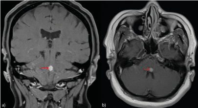
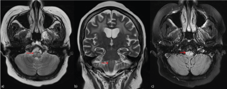
Magnetic resonance imaging (MRI) of the brain, at the time of diagnosis of NSCLC, showed multiple small enhancing lesions consistent with metastases in the left parietal lobe, right cerebellar hemisphere, right frontal lobe and in the brainstem at the pontomedullary junction, abutting the floor of the fourth ventricle (Figure 1). A subsequent MRI twelve months later (prior to the current presentation) showed interval development of radiation-induced leukoencephalopathy and bilateral hypertrophic olivary degeneration (HOD) demonstrated by an increased signal within the inferior olives on T2-weighted and fluid-attenuated inversion recovery (FLAIR) imaging as depicted in (Figure 2). Cerebrospinal fluid (CSF) did not show evidence of an inflammatory cause (WCC 1 x 10⁶/L, protein 0.25 g/L) or leptomeningeal disease and an electroencephalogram (EEG) showed non-specific generalised slowing. Antineuronal antibodies were negative on serum and CSF.
The patient was diagnosed with OPT in the setting of HOD from her metastatic NSCLC and commenced on sodium valproate at a dose of 500mg twice daily, with therapeutic serum levels achieved on this dose. The patient was also commenced on dexamethasone 4mg daily by her palliative care physician, and underwent physical and occupational therapy throughout her admission.
Video 1: Eye movements
The video shows the patient’s large vertical and convergent pendular nystagmus with loss of horizontal eye movements. The latter part of the video shows a palatal myoclonus.
Figure 1: MRI brain on diagnosis
T1-weighted MRI post-contrast a) coronal and b) axial sequence at the time of diagnosis of NSCLC showing a metastatic deposit in the posterior midbrain in the floor of the fourth ventricle (red arrows)
One week post the commencement of sodium valproate, the patient was noted to have an improvement in the amplitude of her eye movements which translated to an improvement in her symptoms and function. She was able to feed herself and drink from a cup or bottle with minimal difficulty. On review, in the outpatient clinic, one month following discharge, her eye movements had remained stable and she continued to have sustained benefits on sodium valproate with improved quality of life.
Discussion
OPT, previously also known as oculopalatal myoclonus, is an acquired syndrome of rhythmic soft palate oscillation together with a convergent or pendular vertical nystagmus which may be synchronous or asynchronous [1]. It was first described by Herbert Spencer in 1886 in the case of a twelve-year-old girl with a cerebral tumour and termed ‘pharyngeal and laryngeal nystagmus’ [4]. OPT is a delayed complication of an injury to the brainstem or cerebellum that can occur weeks to years following the insult but typically within 6 to 12 months [1-3].
Pathophysiology
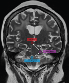
OPT was historically attributed to disruption of the Guillain-Mollaret triangle (Figure 3), also known as the dentato-rubro-thalamic tract, which connects the inferior olivary nucleus (ION) in the ventral medulla to the contralateral cerebellar dentate nucleus (via the inferior cerebellar peduncle), the dentate nucleus to the contralateral red nucleus in the midbrain (via the superior cerebellar peduncle) and the red nucleus to the ipsilateral ION (via the central tegmental tract) [1-3]. Disruption of the central tegmental tract and dentato-rubral tract results in denervation and secondary ION hypertrophy with new soma-to-soma electronic coupling via gap junctions that synchronises the ION neurons and leads to sustained aberrant signalling resulting in OPT [5,6]. Olivo-dentate fibres in the inferior cerebellar peduncle are not involved in the development of HOD as they are deafferent fibres. In the case presented, a metastatic deposit at the pontomedullary junction in the midline (Figure 2) likely disrupted bilateral central tegmental tract with the development of bilateral olivary hypertrophy and oculomotor symptoms twelve months following diagnosis of NSCLC.
More modern models have implicated the role of the cerebellum as a modulator of the rhythmic excitation by inhibiting the inferior olivary neurons through gamma-aminobutyric acid (GABA) synaptic transmission [6,7]. The signal produced by the ION is too small to cause ocular oscillations, even with synchronisation of the neurons. Disinhibition from cerebellar damage causes maladaptive learning and amplification of the low-amplitude signal produced by the ION [2].
Functional imaging supports the role of the dentato-rubro-thalamic dysfunction in the pathogenesis of OPT with functional MRI demonstrating an increase blood flow in the ION in patients studied [8]. Positron emission tomography studies have been inconsistent with one study showing increased glucose metabolism in the medulla of seven patients with OPT [9] and no hypermetabolism in another study of nine patients [10].
On a cellular level, the neurons become vacuolated with severe fibrillary gliosis, demyelination and astrocytic proliferation resulting in pseudohypertrophy [1]. On MRI this often appears as a lesion of high signal intensity in the ION on T2-weighted and fluid-attenuated inversion recovery (FLAIR) sequences, reflecting increased water content and gliosis [11]. Hypertrophy of the inferior olives occurs, lasting a few years before the nucleus disintegrates and permanent atrophy ensues as a result of neuronal and astrocytic death [5,12].
Figure 2: MRI prior to admission (twelve months post initial diagnosis)
T2-weighted MRI a) axial, b) coronal and c) FLAIR sequences showing the development of hypertrophic olivary degeneration as demonstrated by increased T2 signal (red arrows)
Figure 3: Guillain-Mollarettriangle
The Guillain-Mollaret triangle is formed by connections between the red nucleus in the midbrain, inferior olivary nucleus in the medulla and contralateral cerebellar dentate nucleus.
Aetiology
The most common aetiology is a vascular insult, such as an ischaemic or haemorrhagic stroke of the posterior circulation, often from vascular malformations of the brainstem [1,2]. OPT can also occur due to demyelination (multiple sclerosis), inflammation (neurosarcoidosis), infection (abscess, toxoplasmosis), other space occupying lesions (tumours), trauma or may be iatrogenic, secondary to neurosurgery [1-3]. There have even been cases reported secondary to GAD antibody-associated encephalitis [13,14].
Clinical presentation
The palatal tremor in OPT is a segmental myoclonus caused by rhythmic contraction of the levator veli palatini muscle, which is innervated by the vagus nerve from the nucleus ambiguous [3]. The tremor often persists in sleep, lacks voluntary modulation, and may also spread to involve adjacent structures that derive from the branchial arches, such as the larynx, pharynx or diaphragm [15]. In contrast, ‘essential’ palatal tremor involves the tensor veli palatini muscle which is innervated by the trigeminal nerve and tends to dissipate during sleep. An audible clicking sound may ensue due to the synchronous collapse of the eustachian tube, which is at the insertion site of the tensor veli palatini muscle. It is not associated with abnormal ocular movements and no structural lesion can be found [3]. The palatal tremor in OPT is almost always asymptomatic.
The nystagmus in OPT has a typical frequency of 1-3 Hz and is often synchronous with the palatal tremor [2]. It is often of larger amplitude and faster velocity than that of acquired pendular nystagmus from other causes, such as multiple sclerosis (MS), and usually has a vertical, torsional or mixed vertical-torsional trajectory [5,16]. In cases of unilateral or dissociated nystagmus, hypertrophy of the ION occurs on the contralateral side. In patients with bilateral conjugate or dissociated nystagmus, the ION hypertrophy may be unilateral or bilateral [17]. As in the patient presented, this is typically a very visually disabling condition due to oscillopsia, an illusion of the environment moving, and decreased visual acuity causing significant limitations in mobility and function [5,18]. In some cases, symptoms associated with OPT may settle a few years after onset. The patient presented also had concomitant bilateral horizontal gaze palsies and a left-sided facial nerve palsy secondary to the pontomedullary metastatic lesion which has also been seen other reports of OPT due to the close proximity of the facial and abducens nucleus to the central tegmental tract. Involvement of the medial longitudinal fasciculus may also give rise to a one-and-a-half syndrome [12,19].
Investigation
The diagnosis of OPT is clinical but investigations may be helpful in the determining the aetiology. OPT is characterised by unilateral or bilateral HOD with T2/FLAIR hyperintensity on MRI and may be accompanied by a preceding lesion in the Guillain-Mollaret triangle [20] as demonstrated in the case presented (figure 2). HOD can be distinguished from other medullary masses such as those caused by tumours and infection by the absence of contrast enhancement and from infarction and multiple sclerosis by ION enlargement [21]. There have been three distinct MRI stages described in literature that correspond to the pathological changes in the inferior olives [13]. In the first stage there is increased T2 signal in the ION without hypertrophy that occurs as early as one month after the initial insult and corresponds to ION gliosis, increased water content, and demyelination. Hypertrophy of the ION develops four to six months later and in the final stage, after three to four years, the hypertrophy resolves and the olives become atrophic due to neuronal cell death, although the increased T2 signal may persist. MR angiogram may help diagnose a thrombus or vascular lesion such as an aneurysm or arteriovenous malformation [3].
If the aetiology is unclear on imaging, blood tests including angiotensin-converting enzyme (ACE), anti-glutamic acid decarboxylase (GAD) antibodies, and anti-thyroid peroxidase antibodies should be considered in the right clinical setting [3]. Cerebrospinal fluid can aid in the diagnosis of multiple sclerosis (oligoclonal bands), neurosarcoidosis (ACE) and lymphoma (flow cytometry and cytology) [3]. Computed tomography (CT) or positron emission tomography (PET) imaging may be required to exclude a metastatic malignancy.
Treatment
OPT occasionally resolves spontaneously [2,22] but tends to persist for years and although the palatal tremor does not usually bother the patient, the associated oscillopsia or ataxia from the ocular manifestations may be disabling. Given the rarity of this condition, there is insufficient evidence-based treatment and hence there are no established guidelines. The definitive treatment of OPT involves the medical or surgical treatment of the underlying aetiology, which may include surgical resection, however, in most cases the approach to treatment is symptomatic relief.
Certain medications have been proven to be efficacious in the treatment of acquired pendular nystagmus and from other causes, such as MS and essential palatal tremor [5]. Unlike MS, OPT tends to be treatment resistant. The most evidence comes from treatment with GABA agonists, such as gabapentin and baclofen, which is purported to enhance GABA transmission in the Guillain-Mollaret triangle, or N-methyl-D-aspartate (NMDA) receptor antagonists, such as memantine, which reduces output from the ION and cerebellum [1]. Studies have shown that these medications may be effective in improving visual acuity, reducing oscillopsia and improving oscillation amplitude and frequency in those with acquired pendular nystagmus [23-26]. These studies often involve small numbers of patients and there is limited evidence in the treatment of OPT. A crossover trial conducted by Thurtell et al. [25] in 10 patients with acquired nystagmus, four of whom had OPT, compared gabapentin with memantine. Both were found to reduce the eye speed and improve visual acuity, with the best improvement from gabapentin. Gabapentin is recommended at a dose of 300 to 600mg four times a day and is generally well tolerated but may worsen ataxia and increase falls. A 6-month open label study of memantine in six patients with OPT only showed a modest and transient improvement in nystagmus and visual acuity in 2 of 6 patients and without improvement in quality of life [27].
If therapy with a single agent is limited, combination therapy targeting both the inferior olive and the cerebellum may increase effectiveness. Anticholinergics, such as trihexyphenidyl, have shown moderate effectiveness in reducing eye and palate movements but can cause drowsiness and other unwanted anticholinergic effects [28]. Clonazepam and other anti-seizure medications, such as sodium valproate have also been used with various success in some patients [29,30] as proved successful in the present case.
Retrobulbar botulinum toxin administration or injection into the recti in cases of acquired nystagmus and oscillopsia may offer temporary reprieve but carries a risk of retrobulbar haemorrhage and varying degrees of external ophthalmoplegia and ptosis [31]. There has also been limited success with rectus muscle tenotomy or disinsertion in reducing the nystagmus amplitude [18,32]. Bilateral deep brain stimulation of the red nucleus was trialled in one patient with no benefit [33].
OPT remains a difficult condition to treat and a combination of both medical and botulinum toxin or surgical management may be required. Quantitative oculomotor testing is occasionally employed to measure the effects of treatment. In the present case, the use of sodium valproate proved efficacious in managing both the patient’s symptoms and providing improved perception of quality of life, presumably due to its psychotropic effects complementing its role in the movement disorder.
Conclusion
Oculopalatal tremor is a rare condition causing myoclonus of the soft palate associated with a pendular vertical nystagmus. It is often caused by ischaemic or haemorrhagic stroke of the brainstem but can be caused by tumours as in the case presented. OPT is often difficult to treat but agents such as valproate, clonazepam, gabapentin and memantine can be used. Sodium valproate may provide dual intervention, as per the current case, addressing both the movement disorder and enhancing quality of life, presumably based on its additional psychotropic properties. Further studies are required into the optimal management of this condition, though given its rarity, prospective randomised studies would likely be difficult.
Authorship
T.B.T.N. was responsible for the conceptualisation and drafting of the manuscript; R.G.B was responsible for review and editing of the manuscript
Acknowledgements
Nil
Funding
This research received no external funding
Competing interest
The authors declare no conflict of interest
References
Tilikete C, Desestret V (2017) Hypertrophic Olivary Degeneration and Palatal or Oculopalatal Tremor. Front Neurol 8: 302. [Crossref]
Jang J, Borruat FX (2014) Oculopalatal tremor: variations on a theme by Guillain and Mollaret. Eur Neurol 72: 144-149. [Crossref]
Bhattacharjee S (2020) Palatal tremor – pathophysiology, clinical features, investigations, management and future challenges. Tremor and Other Hyperkinetic movements 10: 1-12. [Crossref]
Borruat FX (2013) Oculopalatal tremor: current concepts and new observations. Curr Opin Neurol 26: 67-73. [Crossref]
Kang S, Shaikh AG (2017) Acquired pendular nystagmus. J Neurol Sci 375: 8-17. [Crossref]
Shakih AG, Hong S, Liao K, Tian J, Solomon D, et al. (2010) Oculopalatal tremor explained by a model of inferior olivary hypertrophy and cerebellar plasticity. Brain 133: 923-940. [Crossref]
Nitschke MF, Kruger G, Bruhn H, Klein C, Gehrking E, et al. (2001) Voluntary palatal tremor is associated with hyperactivation of the inferior olive: a functional magnetic resonance imaging study. Mov Disord 16: 1193-1195. [Crossref]
Dubinsky RM, Hallett M, Di Chiro G, Fulham M, Schwankhaus J (1991) Increased glucose metabolism in the medulla of patients with palatal myoclonus. Neurology 41: 557-562. [Crossref]
Moon SY, Cho SS, Kim YK, Kim SE, Kim JH, et al. (2008) Cerebral glucose metabolism in oculopalatal tremor. Eur J Neurol 15: 42-49. [Crossref]
Aladdin Y, Scozzafava J, Muayqil T, Saqqur M (2008) Oculopalatal tremor with one-and-a-half syndrome after pontine haemorrhage. Neurology 71: e39-e41. [Crossref]
Goyal M, Versnick E, Tuite P, Cyr JS, Kucharczyk W, et al. (2000) Hypertrophic olivary degeneration: Metaanalysis of the temporal evolution of MR findings. AJNR AM J Neuroradiol 21: 1073-1077. [Crossref]
Ip MF, Li SH, Wai TY, et al. (2019) Oculopalatal tremor (OPT) in a patient with anti-GAD brainstem encephalitis: a case report. Mov Disord 34.
Turgunkhujaev O, Seliverstov YU, Fominykh V (2022) Unilateral hypertrophic olivary degeneration in anti-GAD-associated encephalomyelitis. Mov Disord 37. [Crossref]
Deuschl G, Wilms H (2002) Clinical spectrum and physiology of palatal tremor. Mov Disord 17: S63-S66. [Crossref]
Tilikete C, Jasse L, Pelisson D, Vukusic S, Durand-Dubief F, et al. (2011) Acquired pendular nystagmus in multiple sclerosis and oculopalatal tremor. Neurology 76: 1650-1657. [Crossref]
Kim JS, Moon SY, Choi KD, Kim JH, Sharpe JA (2007) Patterns of ocular oscillation in oculopalatal tremor: Imaging correlations. Neurology 68: 1128-1135. [Crossref]
Talks SJ, Elston JS (1997) Oculopalatal myoclonus: eye movement studies, MRI findings and the difficulty of treatment. Eye (Lond) 11: 19-24. [Crossref]
Wolin MJ, Trent RG, Lavin PJ, Cornblath WT (1996) Oculopalatal myoclonus after the one-and-a-half syndrome with facial nerve palsy. Ophthalmology 103: 177-180. [Crossref]
Kitajima M, Korogi Y, Shimomura O, Sakamoto Y, Hirai T, et al. (1994) Hypertrophic olivary degeneration: MR imaging and pathologic findings. Radiology 192: 539-543. [Crossref]
Gao Q, Li Z, Guo C, Wang S, Liu X, et al. (2022) Hypertrophic olivary degeneration: a description of four cases of and a literature analysis. Quant Imaging Med Surg 12: 3480-3488. [Crossref]
Shery T, Proudlock FA, Sarvanathan N, McLean RJ, Gottlob I (2006) The effect of gabapentin and memantine in acquired and congenital nystagmus: a retrospective study. Br J Ophthalmol 90: 839-843. [Crossref]
Nerrsnt E, Abouaf L, Pollet-Villard F, Vie AL, Vukusic S, et al. (2020) Gabapentin and memantine for treatment of acquired pendular nystagmus: effects on visual outcome. J Neuroophthalmol 40: 198-206. [Crossref]
Thurtell MJ, Joshi AC, Leone AC, Tomsak RL, Kosmorsky GS, et al. (2010) Crossover trial of gabapentin and memantine as treatment for acquired nystagmus. Ann Neurol 67: 676-680. [Crossref]
Averbuch-Heller L, Tusa RJ, Fuhry L, Rottach KG, Ganser GL, et al. (1997) A double-blind controlled study of gabapentin and baclofen as treatment for acquired nystagmus. Ann Neurol 41: 818-825. [Crossref]
Leigh RJ, Burnstine RH, Ruff RL, Kasmer RJ (1991) Effect of anticholinergic agents upon acquired nystagmus: a double-blind study of trihexyphenidyl and tridihexethyl chloride. Neurology 41: 1737-1741. [Crossref]
Martins AI, Soares-dos-Reis R, Jorge A, Duque C, Pereira DJ, et al. (2022) A 6-month trial of memantine for nystagmus and associated phenomena in oculopalatal tremor. Front Neurol 13: 921341. [Crossref]
Borggreve F, Hageman G (1991) A case of idiopathic palatal myoclonus: treatment with sodium valproate. Eur Neurol 31: 403-404. [Crossref]
Lefkowitz D, Harpold G (1985) Treatment of ocular myoclonus with valproic acid. Ann Neurol 17: 103-104. [Crossref]
Ruben ST, Lee JP, O’Neil D, Dunlop I, Elston JS (1994) The use of botulinum toxin for treatment of acquired nystagmus and oscillopsia. Ophthalmology 101: 783-787. [Crossref]
Wang D, Sanchez J, Foote KD, Sudhyadhom A, Bhatti MT, et al. (2009) Failed DBS for palliation of visual problems in a case of oculopalatal tremor. Parkinsonism Relat Disord 15: 71-73. [Crossref]
Editorial Information
Editor-in-Chief
Shangming Zhang
Penn State Hershey Medical Center, USA
Article Type
Case Report
Publication history
Received: August 10, 2023
Accepted: September 22, 2023
Published: September 26, 2023
Nguyen T (2023) Oculopalatal tremor secondary to metastatic non-small cell lung cancer: A case report and literature review. Neuro Neurosurg 6: DOI: 10.15761/NNS.1000144.
Corresponding author
Shangming Zhang
Penn State Hershey Medical Center, USA
Video 1: Eye movements
The video shows the patient’s large vertical and convergent pendular nystagmus with loss of horizontal eye movements. The latter part of the video shows a palatal myoclonus.
Figure 1: MRI brain on diagnosis
T1-weighted MRI post-contrast a) coronal and b) axial sequence at the time of diagnosis of NSCLC showing a metastatic deposit in the posterior midbrain in the floor of the fourth ventricle (red arrows)
Figure 2: MRI prior to admission (twelve months post initial diagnosis)
T2-weighted MRI a) axial, b) coronal and c) FLAIR sequences showing the development of hypertrophic olivary degeneration as demonstrated by increased T2 signal (red arrows)
Figure 3: Guillain-Mollarettriangle
The Guillain-Mollaret triangle is formed by connections between the red nucleus in the midbrain, inferior olivary nucleus in the medulla and contralateral cerebellar dentate nucleus.