Pediatricians are the first-line doctors that assess a child or an adolescent and provide treatment or referral for further evaluation. Most of the pediatric surgical conditions are usually seen in pediatric office but the identification of the surgically unwell child is of great importance, as urgent surgery might be needed and a delay in diagnosis can have dire consequences. As a result, Pediatricians need to be familiar with these conditions.
The aim of this article is to describe the most frequent surgical conditions, to provide a framework of each of them so that an adequate differential diagnosis can be made. We will also emphasize on some key points in the presentation of each disease that will lead the Pediatrician to the right therapeutic decision.
The most frequently described surgical conditions is life threatening and they present with abdominal pain; there are also conditions presenting with inguinal, scrotal or labial mass, acute scrotum, empty scrotum and male genital abnormalities.
The role of a Pediatrician is well-known worldwide and it is primarily to assess for the first time the health status of a child and coordinate care, alone or with other physicians if necessary. The aim is to lead to effective and appropriate provision of care, taking an advocacy position for the patient or family when needed. The role of Pediatricians in cases of pediatric surgery is mainly to assess the condition of the child or adolescent and distinguish if there is a surgical pediatric condition and if there is the need for urgent surgical treatment or not. Afterwards, proper directions and guidance are given, or the patient is referred to a pediatric surgeon. This article aims to present some frequent conditions that Pediatricians have to diagnose, focus on their main characteristics and provide some important information which will ease the diagnosis of these conditions compared to other diseases.
The first basic and more important stage in the assessment of a sick child is the medical history. The General Practitioner must acquire a precise and detailed medical history from both the child and the parents, if possible, as in 60% of the cases the physician can set the diagnosis only from history taking and the clinical examination [1]. If the child is too young or unable to communicate, parents’ testimony is necessary. The next important stage is the clinical examination, which is much different than the adults’. Vital signs should always be obtained, search for external signs of trauma must be performed and general status, appearance and hydration status must be assessed. Furthermore, the area of the body that most often presents with pathology, the abdomen, should be examined. Careful abdominal examination is crucial; one should inspect the genitalia as well as the hernia orifices. When it comes to examining a male child, the scrotum must be examined for possible testicular pathology. Key points in the examination of the genitourinary system are shown in (Table 1). Next, a head-to-toe examination should be performed, and along with the medical history, a first diagnosis is made and the child is referred to a specialist if necessary. It is advised by the authors that children up to the age of four to five (preschool children) should be initially examined by a pediatrician, as the clinical presentation of diseases is more diffuse and can confuse the Pediatrician. When a Pediatrician examines preschool children, he or she should be accustomed to the variety in the normal values concerning the vital signs, which are dependent on the age of the child. One should perform a very thorough clinical examination, with the child as relaxed as possible. The clinical presentation of the diseases differs so gravely in children that the cause of abdominal pain in children tends to be one outside of the abdomen in approximately 50% of the cases [2]. This percentage is higher in younger children, which is why a careful head to toe examination is essential.
Table 1. Key components in the physical examination of the genitourinary system
Component |
What to check for |
General examination |
Blood pressure, height, weight |
Abdomen |
Abdominal masses, tenderness, palpable stool in colon, umbilical assessment (drainage, mass) |
Flank |
Costovertebral angle tenderness, masses |
Back |
Spinal bony abnormalities, soft tissue masses, sinus tracts, sacral anomalies, hairy patch, cutaneous pigmentation, abnormal gluteal cleft |
Perineum |
Position/patency of anus, hemorrhoids, fissures, bruises, tears/lacerations, position/appearance of urogenital orifices, wetness |
Note: One important stage of the assessment of a sick child is clinical examination Careful abdominal examination is essential, including the genitalia and hernial orifices. Some of the key points of the examination of the male genitourinary system are mentioned in the table above.
A child or adolescent presenting with abdominal pain is common for the Pediatrician, and the cause often needs surgical treatment. The assessment must be careful because it is of great importance as the underlying pathology may be life threatening.
As mentioned above, careful history acquisition and clinical evaluation must be performed. Special notice must be given to worrying features and signs as they may declare the urgency of the pathology. Such features are localized tenderness and the presence of guarding. Localized tenderness could suggest a possible internal trauma, if it coincides with history and clinical signs, or it might suggest bowel perforation or plaster formation. The presence of guarding usually suggests acute abdomen. It is due to inflammation of the peritoneal membrane e.g. peritonitis rising from a ruptured appendicitis. On the other hand, the absence of guarding is not as important and may be very confusing when examining young children. If a surgical condition is suspected, referral to a pediatrician, if possible, or re-examination and follow-up are necessary. Below we describe some of the causes seen in children and adolescents with abdominal pain.
Appendicitis
The commonest surgical cause of abdominal pain in children is appendiceal inflammation and this accounts for approximately 30% of childhood admissions for abdominal pain [3]. Appendicitis is caused by obstruction in the opening of the appendix to the caecum, commonly caused by a fecalith or due to lymphoid tissue hyperplasia. The child presents complaining about a sharp or stabbing periumbilical pain that migrates to the right lower abdominal quadrant. Typically, anorexia, fever, guarding, vomiting and/or diarrhea are present and occur in all age groups but rarely in infants. During the examination, the patient tries not to move because of the pain. Typical signs as the Mc Burney’s sign, psoas sign and Rovsing’s sign may be positive, rectal tenderness might exist in some cases and in the case of an abscess formation, it may be palpable at the right lower abdominal quadrant [4]. However, diagnosis can be challenging, as a great proportion of children might not present with these classic symptoms.
Usual laboratory tests include full blood cell count which may reveal elevated or normal titers of white blood cells (WBC) and absolute neutrophil counts (ANC) [5]. A 20% may present without leukocytosis, so careful evaluation of the likelihood for appendicitis is needed. C-reactive protein can be helpful identifying patients with perforated appendicitis or abscess formation, but predicted value is limited. Urinalysis and urine pregnancy tests are usually performed additionally to exclude urinary causes and pregnancy in girls of appropriate age. Abdominal ultrasound is the first imaging test of choice to be performed and may reveal a dilated appendix, free fluid in the abdominal cavity and an appendicolith [6]. If abdominal ultrasound results in inconclusive findings, computed tomography of the abdomen, either with or without contrast, is the next imaging technique of choice which sets the diagnosis [7]. Computed tomography has better sensitivity and specificity than ultrasound and is capable of visualizing a dilated appendix, free fluid in the abdominal cavity, mesenteric standing, appendicolith as well as abscess or flegmon that are consistent with perforated appendix. Keep in mind that dose reduction protocols should be implemented. Over the last years, the use of MRI has proven reliable for the diagnosis of appendicitis as it does not expose the child to ionizing radiation. However, it has a high cost, it is not as easily available and is not considered appropriate for younger children [8].
Clinical pathways have been developed and have the potential to improve the diagnostic process reducing radiation exposure. Such pathways include the use of Pediatric Appendicitis Score (PAS) where patients are deemed as low (PAS score 1-3), moderate (PAS score 4-7) and high risk (PAS score 8-10). Low risk patients are discharged with recommendations or alternative diagnoses are considered. High risk patients should receive surgery consult. Moderate risk patients undergo ultrasound and based on its findings along with white blood cell count, a risk for appendicitis is estimated in order to plan for further management (Figure 1) [9].
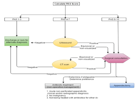
Figure 1. Clinical pathway in the evaluation of a child with suspected appendicitis
PAS Score: cough/percussion/hopping tenderness in the right lower quadrant (2 points), anorexia (1point), pyrexia (1point), nausea/emesis (1point), right lower quadrant tenderness (2 points), migration of pain (1point), leukocytosis (WBC ≥ 10,000) (1 point), polymorphonuclear neutrophilia (1 point).
Note: BBD = Bladder Bowel Dysfunction; DMSA = dimercaptosuccinic acid; IV = intravenous; VCUG = voiding cystourethrography; VUR = vesicoureteral reflux.
Appendectomy is considered the standard treatment for children with acute appendicitis. However non-operative management with antibiotics is an appropriate treatment option in selected patients, especially those with absence of an appendicolith on imaging [10].
Abdominal trauma (blunt or penetrating)
Children who have sustained trauma and present with abdominal pain must be thoroughly evaluated for intra-abdominal injuries e.g. solid organ ruptures. The main cause is accidents, mainly road accidents, but cases of child abuse are becoming more often as the years pass, so a thorough medical history and examination are vital.
The children usually present with localized abdominal pain that started after the accident. There are also cases where the pain is delayed, or it may even present as an extra abdominal pain at first e.g. backache or shoulder ache at occasions of spleen ruptures or hematoma [11]. Other times, the pain might be non-specific and the child may have multiple complaints. During the examination, abdominal tenderness is detected and skin lesions are recognized, which may reflect the mechanism of injury. If the patient complains of pain at the left shoulder, splenic injury must be suspected [12]. Presence of blood at the urethra or in the urine, indicate trauma of the urinary tract or the kidneys and a urinalysis should be performed when abdominal injury is suspected. As stated above, always look for cigarette burns, subdural hemorrhages in an infant/young toddler as well as other signs of abuse. Tachycardia is a reliable sign of hypovolemia in children.
Initially full blood cell count performed in search for the presence of low hematocrit and hemoglobin. In the suspicion of solid organ laceration, abdominal ultrasound and/ or computed tomography scan of the upper and lower abdomen with IV contrast are the imaging tests of choice when searching for blood or fluid in the abdominal cavity. When the patient is unstable and there is a need for an emergency laparotomy, IVP might provide important information about the urinary tract. Other tests are a chest plain radiography that may show pneumothorax or air under the diaphragm, a sign of perforation, and full skeletal plain radiographs, in case of multiple complaints.
Constipation
Constipation is a very common situation in children. During history taking the main causes usually become apparent. Often a poor diet in fibers and little fluid intake are present and less often there is a history of cerebral palsy, developmental delay, spinal cord problems or psychological problems [13]. The child would be often weaning to its parents who may have suggested toilet training and stress e.g stress to go to school in the morning, or other types of stress may have already appeared.
The child mentions vague abdominal pain with colicky characteristics, painful defecations and fecal incontinence [14]. When it comes to infants, they may squeeze anal and buttocks muscles and extend their legs to prevent stooling. Medication with known constipating agent (e.g. iron) may be present. During clinical examination, findings may be minimal (mild abdominal tenderness, some stool in rectum). In severe cases or in young children, abdominal distension may be seen but there is absence of guarding or rebound tenderness (signs that indicate peritonitis). A fecal mass may be palpable on abdominal or rectal examination. Other conditions that could arise are anal fissure or hemorrhoids, a spinal cord abnormality, imperforate anus or anal stenosis and psychological distortion [14].
The test of choice is simple abdominal X- ray, usually without contrast and at standing stance. If there is suspicion of an underlying condition or if the constipation insists after the initial treatment, an abdominal X- ray with contrast or CT scan should be performed in order for the stool to be visualized in the colon and to exclude other conditions.
Gastroenteritis
The child with gastroenteritis presents with vague abdominal pain, usually colicky or cramping. The severity of pain may mimic appendicitis but almost always no rebound is present. Nausea and vomiting are often present along with diarrhea with or without the presence of mucus in stool [15]. The child has traveled recently or has been in contact with sick individuals or ingested suspicious food and drink. The presence of diarrhea more than 10 days suggests parasitic or non-infectious cause. Other symptoms might be fever, chills, myalgia, rhinorrhea and other upper respiratory tract symptoms.
During the examination, the Pediatrician discovers diffuse abdominal pain without evidence of peritonitis, hyperactive bowel sounds and probably signs of volume depletion (tachycardia, hypotension, dry mucous membranes, poor capillary refill or sunken fontanelle in infants). The presence of mucus in the stool suggests bacterial or parasitic cause. Other findings may be low grade fever, lethargy and/ or irritability, reduced response to noxious stimuli and abnormally low or high body temperature. The Pediatrician must be very careful even when the diagnosis of gastroenteritis is most likely as many other diseases can mimic gastroenteritis, especially in young children. Some of these conditions are displayed in (Table 2).
Table 2. Causes of nausea and vomiting in childhood
Acute vomiting, usually in the context of diarrhea |
Gastrointestinal infections |
– viral (e.g. rotavirus, adenovirus, calicevirus) |
– bacterial (e.g. campylobacter, shigella, salmonella) |
– protozoal (e.g. Giardia, cryptosporidium) |
Food poisoning |
– staphylococcus toxin |
Non-gastrointestinal infection |
– urinary tract infection |
– meningitis |
– septicemia |
Surgical |
– appendicitis |
– intussusception |
– malrotation with or without volvulus |
Food allergy (following recent introduction of new food in the first 2 years of life) |
– cow’s milk protein allergy |
– Coeliac disease |
Acute vomiting that presents as vomiting alone |
• Pyloric stenosis (in infants) |
• Appendicitis |
• Raised intracranial pressure |
• Meningitis |
• Surgical obstruction |
• Metabolic disease |
Note: General Practitioner must be very careful even when the diagnosis of gastroenteritis is the more probable and apparent as many other diseases can mimic gastroenteritis, especially in young children. The table shows the most usual conditions in which the typical symptom of gastroenteritis, the symptom of vomiting can be also found.
The first test of choice is full blood cell count which may show elevated white blood cell count probably of granulosa type, if the infection is bacterial. Serum electrolytes are important to consider and in this disease, we may find normal or low sodium and/ or potassium. Stool microscopy and culture are advised if the diarrhea lasts more than 7-10 days. Usual findings include fecal leukocytes, ova or parasites. If the culture should be positive, it describes the responsible agent in bacterial gastroenteritis. Other tests include blood urea and creatinine levels in case of renal failure with hemolytic uremic syndrome, blood culture in case of sepsis and endoscopy with biopsy.
Urinary Tract Infection
Urinary tract infection is an important cause of severe bacterial infection in infants and children. The most common pathogen is E. coli, followed by Proteus, Klebsiella and Staphylococcus. Thorough history taking and physical examination are as important as the laboratory tests. UTI in newborns presents with fever, vomiting, sepsis, lethargy, irritability, icterus and avoidance of supine position. UTI in infants presents with fever, vomiting, diarrhea, lethargy, poor feeding, flank pain, incontinence and smelly urine. Older children present with symptoms similar to the ones adults present with; namely dysuria, increased urinary frequency and urgency and backache in case of pyelonephritis. In (Table 3) the clinical features of UTIs are presented [16]. During the clinical examination, fever >39.0˚C could be present [17]. On palpation, suprapubic and/or costovertebral angle tenderness may be present, irritability, foul smelling urine and/or gross hematuria. Cloudy urine with fishy smell refers to E. coli infection.
Table 3. Clinical features of urinary tract infection in children
Age group |
Most common |
Less common |
Least common |
<3 months |
Fever |
Poor feeding |
Abdominal pain Prolonged |
| |
Vomiting |
Failure to thrive |
jaundice |
| |
Lethargy |
|
Hematuria |
| |
Irritability |
|
Offensive urine |
>3 months (preverbal) |
Fever |
Abdominal pain |
Lethargy |
| |
|
Loin tenderness |
Irritability |
| |
|
Vomiting |
Hematuria |
| |
|
Poor feeding |
Offensive urine |
| |
|
|
Failure to thrive |
Verbal |
Frequency |
Dysfunctional voiding Changes to continence |
Malaise |
| |
Dysuria |
Abdominal pain |
Vomiting |
| |
Nausea |
Loin tenderness |
Hematuria |
| |
|
Fever |
Offensive urine |
| |
|
|
Cloudy urine |
| |
|
|
Suspected sexual abuse |
| |
|
|
Hypertension |
At first, urine dipstick should be performed which usually reveals positive leukocyte esterase and/ or positive nitrite. Absence of significant leukocyturia can rule out UTI with high certainty [18]. Afterwards, samples must be taken for urine microscopy and urine culture. In microscopy, more than 4 WBC per high power field or presence of bacteria, suggest infection. The culture is considered positive if there are more than 1.000 CFU/ml after suprapubic aspiration, more than 10.000 CFU/ ml after catheter sampling and more than 100.000 CFU/ ml from clean-catch midstream.
Other tests include renal ultrasound when suspecting pyelonephritis, which may reveal dilatation of the renal pelvis or ureters, distension of thick walled bladder, renal abscess and voiding cystourethrogram (VCUG) in case of vesicoureteral reflux. The use of dimercaptosuccinic acid scan (DMSA) is the gold standard in case of renal scars [19].
The use of antibiotics must be based on resistance patterns and prior use of antibiotics by the child. In case of pyelonephritis, it is recommended to start with intravenous cefotaxime for 3 days and then switch to 11 days of oral cefixime. In case of cystitis, oral trimethoprim or nitrofurantoin for 5 days is first line therapy. Children with recurrent urinary tract infections, especially those with risk factors for pyelonephritic scars will benefit from low-dose prophylactic treatment with trimethoprim or nitrofurantoin for a period of 6 months [20].
An algorithm for the assessment of UTI is presented in Figure 2.
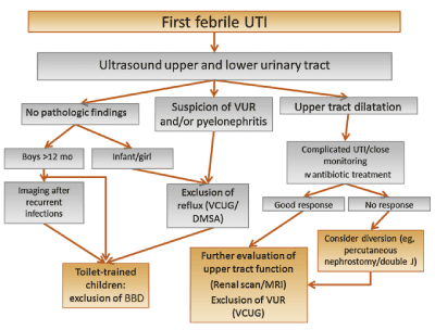
Figure 2. Algorithm for diagnosis and treatment of febrile UTI
Respiratory tract infection and pneumonia
At young children, abdominal pain could be the symptom of respiratory tract infection or pneumonia. This is more frequent in younger children (20). The child usually presents with cough, sputum production (purulent in case pneumonia), rhinorrhea, sore throat, nasal congestion, shortness of breath, fever, chills, vomiting, diarrhea, anorexia. During the examination, clinical signs as tachypnea, decreased breath sounds, crackles/rales on auscultation of the lungs, dullness on percussion, abdominal tenderness and distension without guarding or rebound are often found.
Full blood count and chest X- ray are the examinations of choice and in case of pneumonia, consolidation and pleural effusion may be found. Sputum is taken for culture to identify the infecting organism and chest ultrasonography is sometimes performed as a guiding tool for fluid collection. Computed tomography with contrast may be performed in order to assess the consolidation of the lung parenchyma and the pleuritic fluid, if the consolidation is loculated, empyema is suspected.
Primary dysmenorrhea
In case of primary dysmenorrhea, there is a history of recurrent crampy abdominal pain associated with menstruation. Lower abdominal tenderness with normal pelvic examination are the most characteristic findings. No further examinations are advised in the beginning. If the pain is persistent or if its origin is uncertain, then an ultrasound of the lower abdomen and the pelvis should be performed to rule out any other cause. If the results are not clear, then a computed tomography of the lower abdomen and the pelvis is the next step.
Other uncommon causes that can cause or mimic abdominal pain in children and adolescents are included in (Table 4).
Table 4. Uncommon causes which present as abdominal pain in children and adolescents
Malrotation with midgut volvulus |
Incarcerated inguinal hernia |
Adhesions with intestinal obstruction |
Necrotizing enterocolitis |
Peptic ulcer disease |
Ectopic pregnancy |
Diabetic ketoacidosis |
Hirschsprung associated enterocolitis |
Hemolytic uremic syndrome |
Primary bacterial peritonitis |
Myocarditis |
Mesenteric lymphadenitis |
Ruptured ovarian cyst |
Foreign body ingestion |
Colic |
Inflammatory bowel disease |
Pancreatitis |
Cholecystitis |
Intraabdominal abscess |
Dietary protein allergy |
Malabsorption |
Meckel’s diverticulum |
Abdominal migraine |
Wandering spleen |
Henoch-Schonlein |
Hepatitis |
Sickle cell anemia crisis |
Malignant solid tumor |
Urolithiasis |
Testicular torsion |
Ovarian torsion |
Toxins |
Acute porphyria |
Familial Mediterranean fever |
|
|
Note: Abdominal pain is the commonest cause that will bring a child to the GP. In several cases of abdominal pain, the cause is not the same.
As abdominal pain is the commonest cause that will bring a child to the Pediatrician, it is not the only one. There are other pediatric surgical conditions that will be initially diagnosed by a Pediatrician and will then be referred to a pediatric surgeon. We describe below the most common of these causes and we analyze the key features of each one, to provide a guide for the consideration of Pediatricians.
Inguinal, scrotal or labial mass (groin mass)
Several conditions can present with an abnormal inguinal, scrotal or labial mass in a child. A complete list of such causes is included in (Table 5). The most common are inguinal hernia and hydrocele and are analyzed downwards.
Table 5. Causes that present as an inguinal, scrotal or labial mass in children and adolescents
Trauma causes |
Neoplastic disorders |
Iliopsoas bursitis |
Lipoma |
Hip fracture |
Melanoma |
Iliopectineal hip bursitis |
Lymphoma, Hodgkin’s/non- Hodgkin’s |
| |
Metastasis, lymph node |
| |
Carcinoma or sarcoma of the testis |
Infections |
Infected organ |
Arthritis (pyogenic/ septic) |
Adenitis, lymph node |
Lyme disease |
Epididymitis, acute |
Cat- scratch |
Epididymorchitis |
Herpes simplex, genital |
Orchitis |
Impetigo |
Cellulitis |
Syphilis |
Balanoposthitis |
Tuberculosis |
Testicular abscess |
Tularemia |
|
Anatomic, foreign body, structural disorders |
Congenital |
Hernia, inguinal indirect/inguinal direct/femoral |
Cryptorchidism/ undescended testis |
Spermatocele |
Hydrocele of the cord testis |
Varicocele |
Hydrocele, tunica vaginalis |
Torsion of testis/ spermatic cord |
|
Epididymis cyst |
|
Vegetative, autonomic, endocrine disorders |
Arteriosclerotic, vascular, venous disorders |
Hydradenitis suppurativa |
Femoral artery pseudo aneurysm |
Note: A complete list of conditions can present as an abnormal inguinal, scrotal or labial mass in a child.
Inguinal hernia
A hernia is the protrusion of an organ or tissue through an abnormal opening of the abdominal wall that normally contains it. The inguinal canal is an oblique channel passing through the abdominal wall, inside of which the spermatic cord passes, starting from the abdomen and ending into the scrotum in boys while in girls the round ligament ends into the labia majora. The type of hernia produced due to the dysfunction and loosening of the canal is indirect. When the canal is widely patent intestine passes through towards the scrotum or the processus vaginalis creating the hernia. Primary inguinal hernia occurs in 1 to 5% of newborns and 9- 11% of prematures. The incidence is higher at the first year of life. The repair of an inguinal hernia is the most commonly performed surgical procedure in children [21]. The incidence is three or four times higher in boys than girls and more frequent at the right side.
Children with hernia present with intermittent protrusion of a mass, often reducible, or incarceration. The mass appears when the intra-abdominal pressure rises, mainly when the infant cries or struggles (Figure 3). Infants are usually irritable and crying and many vomit or have abdominal distension. When incarcerated, the hernia presents as a soft inguinal or labial mass, tender and often surrounded by edema. General Practitioners must be very careful of incarceration which occurs in 14-31% of infants younger than a year and need immediate surgery as strangulation of the arteries and tissue necrosis occurs within 2 hours [22]. In case of incarceration pain is the main symptom, but with development of mechanical obstruction, symptoms like abdominal distension, vomiting, no gas discharge and loss of peristalsis can present.
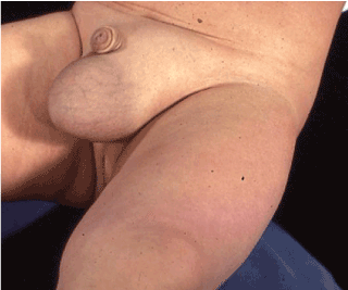
Figure 3. Inguinal-scrotal mass on the left side
Diseases that are similar to inguinal hernia and may confuse the GP are: hydrocele, varicocele, testicular torsion, appendix testis torsion, retractile testis, femoral hernia, inguinal lymphadenitis, testicular cancer (Figure 4). Ultrasound scanning of the area is helpful and has 93% accuracy whereas radiographs and lab tests are not [23]. The presence of peristalsis is diagnostic for hernia and hyperemia of scrotal tissue is a sign of strangulation [24].
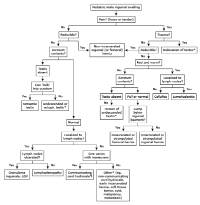
Figure 4. Differential diagnosis of pediatric male inguinal swelling
Initial management should be manual reduction if the patient has no systemic symptoms, but failure to do so suggests urgent operation [25].
Hydrocele
A hydrocele is a collection of peritoneal fluid between the parietal and visceral layers of the tunica vaginalis. Hydroceles may be communicating or non-communicating. A narrowly patent processus vaginalis that only permits passage of peritoneal fluid results in a communicating hydrocele. Non-communicating hydroceles have no connection to the peritoneum; the fluid is produced by the mesothelial lining of the tunica vaginalis. Hydroceles are common in newborns and the majority resolves spontaneously, usually by the first year. In older children and adolescents, non-communicating hydroceles may be idiopathic or may emerge secondary to epididymitis, orchitis, testicular torsion, torsion of the appendix testis or appendix epididymis, trauma or tumor.
Hydrocele usually presents with a cystic scrotal mass that is painless. During examination, the communicating hydrocele enlarges with increase of intra-abdominal pressure e.g. with the valsava maneuver; this is useful to rule out other conditions. The diagnosis is made by the clinical presentation along with the transillumination test of the scrotum, in which the cystic fluid collection becomes apparent. Doppler ultrasonography is the preferred imaging technique to rule out other conditions [26]. Ultrasound shows unechogenic content.
Persistent communicating or non-communicating permanently large and tight hydroceles are best treated by scrotal surgery. Most hydroceles can resolve spontaneously and surgical management is not mandatory before 18 months of age [25].
Varicocele
A varicocele is a collection of dilated and tortuous veins in the pampiniform plexus surrounding the spermatic cord in the scrotum. It is caused because of reno-testicular venous reflux that leads to venous stasis, reflux of renal and adrenal metabolites and higher scrotal temperature. Compression of the left renal vein between the superior mesenteric artery and the aorta is the commonest mechanism. Wilms tumor and congenital vena cava anomalies can cause secondary varicocele. They occur in 85-95% of the cases at the left side because the left spermatic vein enters the left renal vein at 90˚ angle while the right spermatic vein drains at a more obtuse angle, directly into the inferior vena cava. Approximately 10-25% of adolescent males have varicocele and 10-15% of them have fertility problems [27].
Presentation is usually asymptomatic, sometimes a dull ache exists or fullness of the scrotum is felt upon standing and they can present with a smaller testicle. During the examination in standing position the varicocele resembles a “bag of worms” (Figure 5). When the patient is examined again supine, the varicoceles may disappear or become smaller and fewer. Idiopathic varicocele disappears when supine whereas secondary varicocele does not. The exam of choice is Doppler ultrasonography and is most frequently suggested if a varicocele insists when supine, if it has acute onset or if it is right sided. The main criterion is continuous reflux pattern during quiet inspiration. The causes that must be excluded are thrombus in the right renal vein, thrombosis with clot propagation down the Inferior Vena Cava and abdominal mass. Renal ultrasonography must be performed to exclude renal pathology, vascular anomaly and Wilms tumor [28].
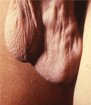
Figure 5. Varicocele on the left side
The presence of small testis, bilateral palpable varicocele and a large varicocele are indications for surgery, while others varicoceles should be followed-up [29].
Acute scrotum
The most common causes of acute scrotal pain in children and adolescents are testicular torsion, torsion of the appendix testis and epididymitis. Other causes include torsion of the appendix epididymis, trauma, Henoch-Schonlein purpura and orchitis (Table 6).
Table 6. Differential diagnosis in acute scrotum
| |
Torsion of spermatic cord |
Torsion of the appendix testis/epididymis |
Epididymitis |
Orchitis |
Onset of symptoms |
rapid |
slow |
slow |
slow |
Pain |
severe |
mild |
mild/severe |
severe |
Systemic signs |
vegetative symptoms |
absent |
absent |
absent |
Scrotal swelling |
present |
mild |
present |
present |
Pain on palpation |
predominantly testis |
predominantly at cranial pole of testis |
predominantly epididymis |
severe |
Fever |
absent |
absent |
Usually present |
severe |
Cremasteric reflex |
absent |
present |
present |
present |
Testicular blood flow on Colour Doppler US |
absent, rarely present |
present |
accentuated |
accentuated |
| |
Hydrocele |
Scrotal incarcerated hernia |
Idiopathic scrotal oedema |
Henoch- Schőnlein purpura |
Onset of symptoms |
slow |
rapid |
slow |
slow |
Pain |
absent |
mild/severe |
absent |
mild |
Systemic signs |
absent |
initially absent |
absent |
rash, abdominal and articular pain |
Scrotal swelling |
present |
present |
present |
mild |
Pain on palpation |
absent |
severe |
absent |
mild, diffuse |
Fever |
absent |
absent |
absent |
absent |
Cremasteric reflex |
present |
present |
present |
present |
Testicular blood flow on Colour Doppler US |
present |
present |
present |
present |
Testicular torsion and torsion of the appendix testis
Both testicular torsion and torsion of the appendix testis present with the abrupt onset of severe pain with the pain being more severe in the case of testicular torsion. In testicular torsion, the testicle may lie transversely in the scrotum and may be retracted and/or swollen; the cremaster reflex is absent in most occasions. The testis is tender and it is not relieved with elevation of the scrotum or testis. In torsion of the appendix testis, the pain is initially localized to the region of the appendix testis, although with progression and the development of a reactive hydrocele, diffuse swelling and tenderness may occur. A pathognomonic “blue dot” sign may be apparent. A normal cremaster reflex may be present, and the testis is not tender on palpation.
Diagnosis is made with the use of color Doppler ultrasonography or nuclear scan of the scrotum. Presence of blood flow does not exclude torsion. A snail shell shaped mass sign (spiral twisting of the vessels, sudden enlargement of the cord and interruption of the linear course) is currently the most reliable sign indicative of testicular torsion [30].
The management of testicular torsion must be as immediate as possible and an urgent surgical exploration is needed within the first 24 hours, if left untreated, the testis starts to necrotize (Figure 6). The management of torsion of the appendix testis is supportive [31]. At the time of ischemia, it is recommended to try the manual detorsion by pulling the testis down, rotating it at the opposite direction of the torsion laterally from the midline.
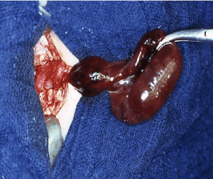
Figure 6. Intraoperative findings after torsion of testis
Epididymitis
Inflammation of the epididymis is known as epididymitis. It occurs more frequently among late adolescents with sexual activities, but also occurs in younger boys who deny sexual activity. Factors are: sexual activity, heavy physical exertion, direct trauma. Bacterial epididymitis in pre-pubertal boys is linked to structural anomalies of the urinary tract. Among sexually active males, chlamydia, N. gonorrhea, E. coli and viruses are most common pathogens.
Presentation is with acute or sub-acute onset of pain and swelling isolated to epididymis. History of urination frequency, dysuria, urethral discharge and/or fever may be present. On physical examination, the testis is normal, the scrotum may be inflamed and scrotal edema exists in at least 50% [33]. Sometimes an inflammatory nodule may be felt. Cremaster reflex is usually normal and pain is relieved when the testis is elevated (Prehn’s sign). Laboratory exams may reveal leukocytosis and pyuria. Although urinalysis and urine culture must be performed to all patients with epididymitis, only 15% of patients have positive urinalysis [33]. Scrotal ultrasound may show elevated blood flow to epididymis and can exclude torsion of the spermatic cord. Renal and bladder ultrasound can be added to exclude other urogenital pathology [34].
If urinalysis is negative, antibiotics are not indicated. Local cold compress, anti-inflammatory drugs and rest are recommended. In case of pyuria, intravenous antibiotics should immediately be started.
Empty Scrotum
Empty scrotum is the condition where the testis is absent from the scrotum. During clinical examination, undescended testes are often located between the external inguinal ring and the scrotum but can be also found inside the peritoneal cavity or extra-peritoneal space. Almost 90% of the testes are felt in the inguinal region or can be massaged into the inguinal area by pressing with one hand near the antero-superior iliac spine and massaging downwards and medially down the scrotum. If after this maneuver the testis remains in the scrotum, the diagnosis is retractile testis. If the testis is not palpable, the diagnosis is ectopic testis. Truly impalpable testes are discovered in 10-20% of cases with empty scrotum [35].
Complications of undescended testis are infertility, malignancy (abdominal testis) and testicular torsion. Preferred imaging studies are ultrasonography of the abdomen, inguinal area and scrotum. If the results are inconclusive, computed tomography, magnetic resonance and laparoscopy are the next examinations of choice. Management is mostly surgical with removal of the ectopic testis or orchiopexy of the retractile testis into the scrotum.
Phimosis
Phimosis refers to the inability of the distal foreskin to retract over the glans penis (Figure 7). Physiologic phimosis occurs naturally in newborn males. Pathologic phimosis defines the inability to retract the foreskin after it was previously retractable or after puberty. Natural phimosis results from adhesions between the epithelial layers of the inner prepuce and glans. These adhesions spontaneously dissolve with intermittent foreskin retraction and erections, so that as males grow, phimosis resolves. Secondary phimosis occurs after many episodes of balanitis or balanoposthitis, which result to scarring of the preputial orifices of the distal foreskin and consequently, to pathologic phimosis. A differential diagnosis for the causes of balanoposthitis is presented in (Figure 8). Patients with phimosis are at risk to develop paraphimosis when the foreskin is forcibly retracted past the glans or when the patient or caretaker forgets to replace the foreskin. Ten percent of males have natural phimosis at 3 years of age and 1-5% at 16 years old [36].
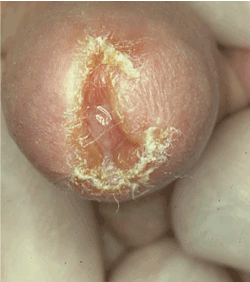
Figure 7. Phimosis is inability to retract the distal foreskin over the glans penis
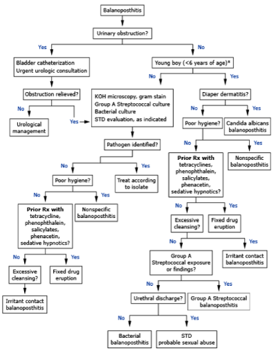
Figure 8. Differential diagnosis of the possible causes of balanoposthitis based on signs and symptoms
From the medical history, the parent usually notices inability to retract the foreskin or ballooning of the foreskin when the child urinates. Other findings are referred painful erections, hematuria, recurrent urinary tract infections, preputial pain or weak urinary stream. On examination, the foreskin cannot be retracted. In natural phimosis, the preputial orifice is unscarred and healthy. In pathologic phimosis, a contracted white fibrous ring may be visible around the preputial orifice. Phimosis rarely needs immediate treatment but should be referred to a pediatric urologist for further examination and follow up. Treatment is initially medical consisting of special maneuvers with the foreskin and surgical as a choice of last resort.
Paraphimosis
Paraphimosis is the entrapment of a retracted foreskin behind the coronal sulcus. It only occurs to uncircumcised or partially circumcised males, usually if the foreskin is not replaced after its retraction in males with phimosis. Penile piercings increase the risk of developing paraphimosis if pain and swelling prevent reduction of a retracted foreskin. With time, impairment of venous and lymphatic flow to the glans leads to venous engorgement and worsening of the swelling. As the swelling progresses, arterial supply is compromised, leading to penile infarction, necrosis, gangrene, and eventually, auto-amputation.
The child usually presents with a painful, swollen glans penis. A preverbal infant may present only with irritability. Occasionally, paraphimosis may be an incidental finding noted by a caretaker of a debilitated patient. Most usual causes are, forgotten retracted foreskin after bathing or voiding, adolescents in the setting of vigorous sexual activity and males with chronic balanoposthitis.
During clinical examination, retracted foreskin behind the glans penis is discovered. The foreskin forms a tight, constricting ring around the glans. Flaccidity of the penile shaft proximal to the area of paraphimosis is seen, the glans becomes extremely erythematous and edematous and areas with necrosis or stiffness may exist. Paraphimosis needs emergency care by pediatric urologist as necrosis is possible [37].
- Paley L, Zornitzki T, Cohen J, Friedman J, Kozak N, et al. (2011) Utility of Clinical Examination in the Diagnosis of Emergency Department Patients Admitted to the Department of Medicine of an Academic Hospital. Arch Intern Med 171: 1393-1400. [Crossref]
- Scholer SJ, Pituch K, Orr DP, Dittus RS (1996) Clinical outcomes of children with acute abdominal pain. Pediatrics 98: 680-685. [Crossref]
- Bundy DG, Byerley JS, Liles EA, Perrin EM, Katznelson J, et al. (2007) Does this child have appendicitis? JAMA 298: 438-451. [Crossref]
- Bachoo P, Mahomed AA, Niman GK, Youngson GG (2001) Acute appendicitis: the continuing role for active observation. Pediat Surg Int 17: 125-128. [Crossref]
- Wang LT, Prentiss KA, Simon JZ, Doody DP, Ryan DP (2007) The use of white blood cell count and left shift in the diagnosis of appendicitis in children. Pediatr Emerg Care 23: 69-76. [Crossref]
- Dilley A, Wesson D, Munden M, Hicks J, Brandt M, et al (2001) The impact of ultrasound examinations on the management of children with suspected appendicitis: a 3-year analysis. J Pediatr Surg 36: 303-308. [Crossref]
- Doria AS, Moineddin R, Kellenberger CJ, Epelman M, Beyene J, et al. (2006) US or CT for Diagnosis of Appendicitis in Children and Adults? A Meta-Analysis. Radiology 241: 83-94. [Crossref]
- Kulaylat AN, Moore MM, Engbrecht BW, Brian JM, Khaku A, et al. (2015) An implemented MRI program to eliminate radiation from the evaluation of pediatric appendicitis. J Pediatr Surg 50: 1359-1363. [Crossref]
- Saucier A, Huang EY, Emeremni CA, Pershad J (2014) Prospective evaluation of a clinical pathway for suspected appendicitis. Pediatrics 133: e88-e95. [Crossref]
- Gonzalez DO, Deans KJ, Minneci PC (2016) Role of non-operative management in pediatric appendicitis. Semin Pediatr Surg 25: 204-207. [Crossref]
- Miller D, Garza J, Tuggle D, Mantor C, Puffinbarger N (2006) Physical examination as a reliable tool to predict intra-abdominal injuries in brain-injured children. Am J Surg 192: 738-742. [Crossref]
- Young KD, Seidel JS (1998) Delayed diagnosis of splenic injury after falls from less than 10 feet. Pediatr Emerg Care 14: 413-415. [Crossref]
- Rasquin A, Di Lorenzo C, Forbes D, Guiraldes E, Hyams JS, et al. (2006) Childhood functional gastrointestinal disorders: child/adolescent. Gastroenterology 130: 1527-1537. [Crossref]
- Fishman L, Rappaport L, Schonwald A, Nurko S (2003) Trends in referral to a single encopresis clinic over 20 years. Pediatrics 111: e604-e607. [Crossref]
- Staat MA, Azimi PH, Berke T, Roberts N, Bernstein DI, et al. (2002) Clinical presentations of rotavirus infection among hospitalized children. Pediatr Infect Dis J 21: 221-227. [Crossref]
- National Institute for Health and Clinical Excellence. Urinary tract infection in children: diagnosis, treatment and long-term management. Royal College of Obstetricians and Gynaecologists; National Collaborating Centre for Women’s and Children’s Health. 2007.
- Shaikh N, Morone NE, Lopez J, Chianese J, Sangvai S, et al. (2007) Does this child have a urinary tract infection? JAMA 298: 2895-2904. [Crossref]
- Aspevall O, Hallander H, Gant V, Kouri T (2001) European guidelines for urinalysis: a collaborative document produced by European clinical microbiologists and clinical chemists under ECLM in collaboration with ESCMID. Clin Microbiol Infect 7: 173-178. [Crossref]
- Hoberman A, Wald E, Hickey R, Baskin M, Charron M, et al. (1999) Oral versus initial intravenous therapy for unirary tract infections in young febrile children. Pediatrics 104: 79-86. [Crossref]
- Rosenberg AR, Rossleigh MA, Brydon MP, Bass SJ, Leighton DM, et al. Evaluation of acute urinary tract infection in children by dimercaptosuccinic acid scintigraphy: a prospective study. J Urol 148: 1746-1749. [Crossref]
- Kanegaye JT, Harley JR (1995) Pneumonia in unexpected locations: an occult cause of pediatric abdominal pain. J Emerg Med 13: 773. [Crossref]
- Kapur P, Caty MG, Glick PL (1998) Pediatric hernias and hydroceles. Pediatr Clin North Am 45: 773. [Crossref]
- Zamakhshary M, To T, Guan J, Langer JC (2008) Risk of incarceration of inguinal hernia among infants and young children awaiting elective surgery. CMAJ 179: 1001-1005. [Crossref]
- Erez I, Schneider N, Glaser E, Kovalivker M (1992) Prompt diagnosis of 'acute groin' conditions in infants. Eur J Radiol 15: 185-189. [Crossref]
- Dogra WS, Gottlieb RH, Oka M, Rubens DJ (2003) Sonography of the scrotum. Radiology 227: 18-36. [Crossref]
- Lau ST, Lee Z, Caty MG (2007) Current management of hernias and hydroceles. Semin Pediatr Surg 16: 50-57. [Crossref]
- Spencer Barthold J, Kass EJ. Abnormalities of the penis and scrotum. In: Clinical Pediatric Urology, 4th, Belman AB, King LR, Kramer SA. (Eds), Martin Dunitz Ltd, London 2002. 1093.
- Skoog SJ, Roberts KP, Goldstein M, Pryor JL (1997) The adolescent varicocele: what's new with an old problem in young patients? Pediatrics 100: 112. [Crossref]
- Zampieri N, Cervellione RM (2008) Varicocele in adolescents: a 6-year longitudinal followup observational study. J Urol 180: 1653-1656. [Crossref]
- Barroso U, Andrade DM, Novaes H, Netto JMB, Andrade J (2009) Surgical treatment of varicocele in children with open and laparoscopic Palomo technique: a systematic review of the literature. J Urol 181: 2724-2728. [Crossref]
- Waldert M, Klatte T, Schmidbauer J, Remzi M, Lackner J, et al. (2010) Color Doppler sonography reliably identifies testicular torsion in boys. Urology 75: 1170-1174. [Crossref]
- Pillai SB, Besner GE (1998) Pediatric testicular problems. Pediatr Clin North Am 45: 813-830. [Crossref]
- Kadish HA, Bolte RG (1998) A retrospective review of pediatric patients with epididymitis, testicular torsion, and torsion of testicular appendages. Pediatrics 102: 73-76. [Crossref]
- Siegel A, Snyder H, Duckett JW (1987) Epididymitis in infants and boys: underlying urogenital anomalies and efficacy of imaging modalities. J Urol 138: 1100-1103. [Crossref]
- Cisek LJ, Peters CA, Atala A, Bauer SB, Diamond DA, et al. (1998) Current findings in diagnostic laparoscopic evaluation of the nonpalpable testis. J Urol 160: 1145-1149. [Crossref]
- McGregor TB, Pike JG, Leonard MP (2007) Pathologic and physiologic phimosis: approach to the phimotic foreskin. Can Fam Physician 53: 445-448. [Crossref]
- DeVries CR, Miller AK, Pacher MG (1996) Reduction of paraphimosis with hyaluronidase. Urology 48: 464-465. [Crossref]
Editorial Information
Editor-in-Chief
Article Type
Case Report
Publication history
Received date: March 03, 2021
Accepted date: March 11, 2021
Published date: March 15, 2021
Copyright
©2021 Sakellaris G. This is an open-access article distributed under the terms of the Creative Commons Attribution License, which permits unrestricted use, distribution, and reproduction in any medium, provided the original author and source are credited.
Citation
Sakellaris G, Kastritsi O, Kontakis M, Stelios S and Petra G. (2021) Office paediatric surgery and urology: An update – Clinical evaluation and recommendations for diagnosis and treatment. Case Rep Imag Surg 4: doi: 10.15761/CRIS.1000155

Figure 1. Clinical pathway in the evaluation of a child with suspected appendicitis
PAS Score: cough/percussion/hopping tenderness in the right lower quadrant (2 points), anorexia (1point), pyrexia (1point), nausea/emesis (1point), right lower quadrant tenderness (2 points), migration of pain (1point), leukocytosis (WBC ≥ 10,000) (1 point), polymorphonuclear neutrophilia (1 point).
Note: BBD = Bladder Bowel Dysfunction; DMSA = dimercaptosuccinic acid; IV = intravenous; VCUG = voiding cystourethrography; VUR = vesicoureteral reflux.

Figure 2. Algorithm for diagnosis and treatment of febrile UTI

Figure 3. Inguinal-scrotal mass on the left side

Figure 4. Differential diagnosis of pediatric male inguinal swelling

Figure 5. Varicocele on the left side

Figure 6. Intraoperative findings after torsion of testis

Figure 7. Phimosis is inability to retract the distal foreskin over the glans penis

Figure 8. Differential diagnosis of the possible causes of balanoposthitis based on signs and symptoms
Table 1. Key components in the physical examination of the genitourinary system
Component |
What to check for |
General examination |
Blood pressure, height, weight |
Abdomen |
Abdominal masses, tenderness, palpable stool in colon, umbilical assessment (drainage, mass) |
Flank |
Costovertebral angle tenderness, masses |
Back |
Spinal bony abnormalities, soft tissue masses, sinus tracts, sacral anomalies, hairy patch, cutaneous pigmentation, abnormal gluteal cleft |
Perineum |
Position/patency of anus, hemorrhoids, fissures, bruises, tears/lacerations, position/appearance of urogenital orifices, wetness |
Note: One important stage of the assessment of a sick child is clinical examination Careful abdominal examination is essential, including the genitalia and hernial orifices. Some of the key points of the examination of the male genitourinary system are mentioned in the table above.
Table 2. Causes of nausea and vomiting in childhood
Acute vomiting, usually in the context of diarrhea |
Gastrointestinal infections |
– viral (e.g. rotavirus, adenovirus, calicevirus) |
– bacterial (e.g. campylobacter, shigella, salmonella) |
– protozoal (e.g. Giardia, cryptosporidium) |
Food poisoning |
– staphylococcus toxin |
Non-gastrointestinal infection |
– urinary tract infection |
– meningitis |
– septicemia |
Surgical |
– appendicitis |
– intussusception |
– malrotation with or without volvulus |
Food allergy (following recent introduction of new food in the first 2 years of life) |
– cow’s milk protein allergy |
– Coeliac disease |
Acute vomiting that presents as vomiting alone |
• Pyloric stenosis (in infants) |
• Appendicitis |
• Raised intracranial pressure |
• Meningitis |
• Surgical obstruction |
• Metabolic disease |
Note: General Practitioner must be very careful even when the diagnosis of gastroenteritis is the more probable and apparent as many other diseases can mimic gastroenteritis, especially in young children. The table shows the most usual conditions in which the typical symptom of gastroenteritis, the symptom of vomiting can be also found.
Table 3. Clinical features of urinary tract infection in children
Age group |
Most common |
Less common |
Least common |
<3 months |
Fever |
Poor feeding |
Abdominal pain Prolonged |
| |
Vomiting |
Failure to thrive |
jaundice |
| |
Lethargy |
|
Hematuria |
| |
Irritability |
|
Offensive urine |
>3 months (preverbal) |
Fever |
Abdominal pain |
Lethargy |
| |
|
Loin tenderness |
Irritability |
| |
|
Vomiting |
Hematuria |
| |
|
Poor feeding |
Offensive urine |
| |
|
|
Failure to thrive |
Verbal |
Frequency |
Dysfunctional voiding Changes to continence |
Malaise |
| |
Dysuria |
Abdominal pain |
Vomiting |
| |
Nausea |
Loin tenderness |
Hematuria |
| |
|
Fever |
Offensive urine |
| |
|
|
Cloudy urine |
| |
|
|
Suspected sexual abuse |
| |
|
|
Hypertension |
Table 4. Uncommon causes which present as abdominal pain in children and adolescents
Malrotation with midgut volvulus |
Incarcerated inguinal hernia |
Adhesions with intestinal obstruction |
Necrotizing enterocolitis |
Peptic ulcer disease |
Ectopic pregnancy |
Diabetic ketoacidosis |
Hirschsprung associated enterocolitis |
Hemolytic uremic syndrome |
Primary bacterial peritonitis |
Myocarditis |
Mesenteric lymphadenitis |
Ruptured ovarian cyst |
Foreign body ingestion |
Colic |
Inflammatory bowel disease |
Pancreatitis |
Cholecystitis |
Intraabdominal abscess |
Dietary protein allergy |
Malabsorption |
Meckel’s diverticulum |
Abdominal migraine |
Wandering spleen |
Henoch-Schonlein |
Hepatitis |
Sickle cell anemia crisis |
Malignant solid tumor |
Urolithiasis |
Testicular torsion |
Ovarian torsion |
Toxins |
Acute porphyria |
Familial Mediterranean fever |
|
|
Note: Abdominal pain is the commonest cause that will bring a child to the GP. In several cases of abdominal pain, the cause is not the same.
Table 5. Causes that present as an inguinal, scrotal or labial mass in children and adolescents
Trauma causes |
Neoplastic disorders |
Iliopsoas bursitis |
Lipoma |
Hip fracture |
Melanoma |
Iliopectineal hip bursitis |
Lymphoma, Hodgkin’s/non- Hodgkin’s |
| |
Metastasis, lymph node |
| |
Carcinoma or sarcoma of the testis |
Infections |
Infected organ |
Arthritis (pyogenic/ septic) |
Adenitis, lymph node |
Lyme disease |
Epididymitis, acute |
Cat- scratch |
Epididymorchitis |
Herpes simplex, genital |
Orchitis |
Impetigo |
Cellulitis |
Syphilis |
Balanoposthitis |
Tuberculosis |
Testicular abscess |
Tularemia |
|
Anatomic, foreign body, structural disorders |
Congenital |
Hernia, inguinal indirect/inguinal direct/femoral |
Cryptorchidism/ undescended testis |
Spermatocele |
Hydrocele of the cord testis |
Varicocele |
Hydrocele, tunica vaginalis |
Torsion of testis/ spermatic cord |
|
Epididymis cyst |
|
Vegetative, autonomic, endocrine disorders |
Arteriosclerotic, vascular, venous disorders |
Hydradenitis suppurativa |
Femoral artery pseudo aneurysm |
Note: A complete list of conditions can present as an abnormal inguinal, scrotal or labial mass in a child.
Table 6. Differential diagnosis in acute scrotum
| |
Torsion of spermatic cord |
Torsion of the appendix testis/epididymis |
Epididymitis |
Orchitis |
Onset of symptoms |
rapid |
slow |
slow |
slow |
Pain |
severe |
mild |
mild/severe |
severe |
Systemic signs |
vegetative symptoms |
absent |
absent |
absent |
Scrotal swelling |
present |
mild |
present |
present |
Pain on palpation |
predominantly testis |
predominantly at cranial pole of testis |
predominantly epididymis |
severe |
Fever |
absent |
absent |
Usually present |
severe |
Cremasteric reflex |
absent |
present |
present |
present |
Testicular blood flow on Colour Doppler US |
absent, rarely present |
present |
accentuated |
accentuated |
| |
Hydrocele |
Scrotal incarcerated hernia |
Idiopathic scrotal oedema |
Henoch- Schőnlein purpura |
Onset of symptoms |
slow |
rapid |
slow |
slow |
Pain |
absent |
mild/severe |
absent |
mild |
Systemic signs |
absent |
initially absent |
absent |
rash, abdominal and articular pain |
Scrotal swelling |
present |
present |
present |
mild |
Pain on palpation |
absent |
severe |
absent |
mild, diffuse |
Fever |
absent |
absent |
absent |
absent |
Cremasteric reflex |
present |
present |
present |
present |
Testicular blood flow on Colour Doppler US |
present |
present |
present |
present |


