A 62-year-old female patient was referred for exploring right hand tumefaction. This tumefaction goes back more than 6 months. There was no pain or trauma history nor fever or weight loss.
The physical examination found a mass on the right hand occupying the entire length of the third metacarpal. It was hard, painless on palpation, fixed to the deep plane, without inflammatory signs.
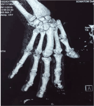
The standard X-ray showed osteo-condensation of the whole third metacarpal (Figure 1). The computed tomography (CT) showed a third metacarpal hypertrophy with cortical thickening, without periosteal reaction or bone lysis, making strongly evoke fibrous dysplasia (Figure 2). No surgical treatment was indicated, only monitoring every 6 months was recommended.
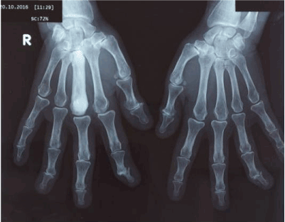
Figure 1. Radiograph of the face of both hands showing the appearance of osteo-condensation occupying the whole 3rd right metacarpal.
Fibrous dysplasia is a sporadic bone disease, representing 0.8% of all primary bone tumors. It is a benign lesion, often asymptomatic. It can affect one bone (monostotic) or several bones (polyostotic). The monostotic form mainly affects the ribs, but hardly ever touches the hand.
During skeletal formation and growth, lesions of fibrous dysplasia are developed and they have a variable natural evolution. The etiology has been linked to an activating mutation in the gene that encodes the α subunit of stimulatory G protein (Gsα) located at 20q13.2-13.3 This malignity transformation is rare. Clinical observation and patient education if asymptomatic lesion is the unique treatment required.
2021 Copyright OAT. All rights reserv
Figure 2. CT and 3D reconstruction of the right hand showing bone hypertrophy and cortical thickening of the third metacarpal.