Osteoarthrosis (OA) is a locomotor system disease which often leads to disability. Overall OA affects 13.9% of adults aged 25 and older and 33.6% (12.4 million) of those 65+; an estimated 26.9 million US adults in 2005 up from 21 million in 1990 [1]. Issues concerning relationship between osteoarthrosis and osteoporosis were studied from the end of 1960 and continue to this day [2,3]. According to the American Association of Rheumatologists (New York), the diagnostic criteria for OA include 10 indicators: joints pain at night, marginal osteophytes and nodules, subchondral sclerosis, cystic changes in epiphyses and others [4]. However, this is only a qualitative characteristic. But for detail characteristics of the process and understanding of OA and OP relationship, these subjective factors are not sufficient. Objective criteria are necessary to evaluate bone quality.
The purpose is to study the density of bones forming hip and knee joints using multislice computed tomography (MSCT), within the age norm and in patients with idiopathic coxarthrosis and gonarthrosis of II-III grade.
Subjects
Roentgenography and MSCT examinations are performed in 135 patients with OA of hip (59) and knee joints (76) at three age groups: first - of 25 to 35 y.o., second – 36-45 y.o, third – 46-60 y.o. and older. 60 patients were studied with no clinical and radiological manifestations OA (control group, patients with pelvic pathology).
Procedures
The studies were performed using computer tomographs GE Lihgt Speed VCT (GE Medical Systems, Wisconsin, USA), Toshiba Aquilion-64 (Toshiba Medical Systems corporation, Toshigi 324-8550, Japan). Technical conditions of spiral scanning: software LowerExtremity. Processing of axial slices was done in the mode of multiplanar reconstruction (MPR) in frontal and sagittal planes. The bone density was measured in Hounsfield units (HU). At the third stage we performed analysis of topographic-anatomic changes in mode 3D-reconstruction(VRT) with bone and soft-tissue filters of working stations. The bone density was measured at standard points (Figure 1).
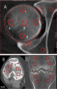
Figure 1. Hip joints MSCT, axial view(A). Knee-joint MSCT, axial view(B), MPR (C). Scheme of bone density measurement in different parts of femoral head, acetabulum, different parts of knee-joint, HU.
Statistical analyses
Descriptive statistics (mean ± SD) were computed to describe the subject population. For statistical analysis we used statistic software Attestat. Student t-test was used to calculate the value of differences in density in three groups of patients. Mean value and standard deviation were calculated for indices of bone density. For all analyses, statistical significance was established at p < 0.05.
Density of different femoral head parts was decreased with age in patients without clinical and radiological OA implications. Minimal density is evident in lateral, medial and anterior head poles (Figure 2).
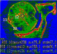
Figure 2. MSCT hip joint. Axial plane. Colormap. Measurement of density in different parts of the head.
Maximal dense is a central head part (point 5, Table 1) (Table 1).
Table 1. Density of different femoral head points in patients with OA varied with age, N=59 (HU)
Age |
Points for measuring the density in the head of the femur (Figure 1), HU |
1 |
2 |
3 |
5 |
5 |
25-35 |
348,2 ± 18,6 |
254,5 ± 10,9 |
275,6 ± 23,1 |
252,8 ± 16,3 |
460,4 ± 31,2* |
36-45 |
303,1 ± 17,9 |
135,6 ± 14,1 |
277,4 ± 19,9 |
267,6 ± 18,2 |
513,7 ± 38,4† |
46-60 |
280,6 ± 11,8 |
104,2 ± 9,7 |
275,5 ± 15,9 |
215,6 ± 19,2 |
538,3 ± 26,7‡ |
* †‡ p<0,05
Density of pubic and ischium bones had no significant variations at different age groups. According to our data, in patients at the age of 25-35 y. o., at norm, subchondral layer density of pubic and ischium bones was 750-800HU, was increasing with age, reaching in the roof area 950 ± 36 HU at the older age group.
Subchondral layer density of pubic and ischial bones in patients with coxarthrosis varied depended on age. Density of subchondral acetabular layer was decreasing. With age, in patients without clinical implications of OA, bone density of femoral condyles was decreasing; density of lateral condyle was higher. In patients without clinical implications of OA, bone density of tibial condyles was lower than femoral condyles density and was decreasing with age. With age, in patients without clinical implications of OA, subchondral bone density of femoral condyles was slightly decreasing, but was not less than 600 HU. Medial condyle index of tibial bone was 800 HU. In patients at the age of 25-35 y. o., with OA of knee joint condyles density was lower, especially for medial condyle. In the next age group density index increases for subchondral zone medial tibial condyle, at the age of 56-60 y. o., all condyles density decreases. Bone density was lower at subchondral layer in comparison with age norm, except patients with significant Varus knee joint deformity. The older patients are, the lower is the bone density. Sometimes, density subchondral zone lateral condyle was decreased up to 300-400 HU (point 24, Figure 3).
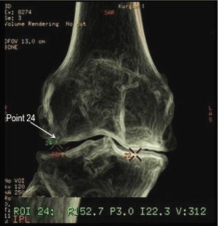
Figure 3. MSCT knee-joint.VRT. The arrow indicates the location of point 24.
Typical feature of bony density quantitative change in patients with OA is a general density decreasing of femoral and tibial condyles and significant bone induration at subchondral zone, which cannot be treated as sclerotic because bone density at this area is no more than 400-600 HU, while at sclerotic zones the bone density is 1600-2000 HU and more. Bone density at subchondral femoral and tibial condyles is lower in comparison with age normal in all patients with OA, except patients with significant Varus or valgus knee joint deformity (point 5, Figure 4).
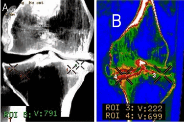
Figure 4. MSCT knee-joint. OA. MIP (A). The density was measured at point 5; MPR, Colormap (B). The density was measured at points 3 and 4.
- In patients with OA at all age groups, density of subchondral pelvic bones parts is significantly lower than density of hip joint bones at normal, it shows relative increase of subchondral layer density, but not true subchondral sclerosis.
- In patients with OA of knee joint at the age of 18-35 y.o., condylar density is lower than at age norm. At the next age group, density of medial tibial condyle increased, and at the age of 46-60 y.o., all condyles density decreases in comparison with age norm.
- Bone density at subchondral femoral and tibial condyles is lower in comparison with age norm in all patients with OA, except patients with significant Varus or valgus knee joint deformity.
In patients with OA “Subchondral sclerosis” is a relative bone induration at subchondral zone, against its condylar density decrease, but not true sclerotic process.
2021 Copyright OAT. All rights reserv
- Lawrence RC, Felson DT, Helmick CG, Arnold LM, Choi H, et al. (2008) Estimates of the prevalence of arthritis and other rheumatic conditions in the United States. Part II. Arthritis Rheum 58: 26-35. [Crossref]
- Zhang J, Chen S, Chen W, Huang Y, Lin R, et al. (2018) Ultrastructural change of the subchondral bone increases the severity of cartilage damage in osteoporotic osteoarthritis of the knee in rabbits. Pathol Res Pract 214: 38-43. [Crossref]
- Linde KN, Puhakka KB, Langdahl BL, Søballe K, Krog-Mikkelsen I, et al. (2017) Bone Mineral Density is Lower in Patients with Severe Knee Osteoarthritis and Attrition. Calcif Tissue Int 101: 593-601. [Crossref]
- [No authors listed] (2002) Recommendations for the medical management of osteoarthritis of the hip and knee: 2000 update. American College of Rheumatology Subcommittee on Osteoarthritis Guidelines. Arthritis Rheum 43: 1905-1915. [Crossref]