Post-exercise T2 prolongation and recovery kinetics of upper arm muscles in non-athletes and athletes
Figure 1 is showing relaxometric maps of the right upper arm of non-athlete and athlete, depicting the time course of exercise induced changes (concentric elbow flexion exercise to failure - weight lifting with 75% of 1RM). T2 maps acquired before exercise, immediately after exercise and 30min and 60min later during recovery are shown.
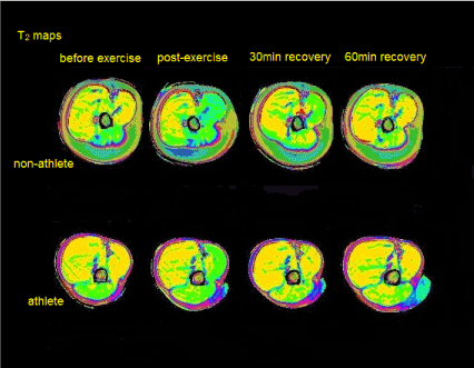
Figure 1 is showing relaxometric maps of the right upper arm of non-athlete and athlete, depicting the time course of exercise induced changes (concentric elbow flexion exercise to failure - weight lifting with 75% of 1RM). T2 maps acquired before exercise, immediately after exercise and 30min and 60min later during recovery are shown.
Protocol: prone position, transverse mid-humerus upper arm images (slice thickness 10mm, FoV 256x512, matrix size 256x128) were obtained by using a multiple spin-echo sequence with TR-2520ms, TE-10, 20, 30, 40,50,60,70, 80,90, 100, 110, 120, 130, 140, 150,160ms on the Magnetom Aera TIM Siemens (Siemens, Erlangen, Germany). T2 time values before, immediately after exercise, as well as after 30min and 60min of recovery were calculated from automatically generated T2 maps in the whole muscle area on the scanner.
Post-processing was obtained by using MIPAV software [1]. Color change in active regions of m.biceps brachii immediately after exercise, as well as 30min and 60min after exercise corresponds to T2 time prolongation and recovery. In non-athlete, T2 recovery with returning to baseline values in m.biceps area was noticed after 60min recovery. In athlete, post-exercise T2 prolongation value in m.biceps area was twice lower than in non-athlete with same relative exercise intensity, and it reestablished baseline values faster, after only 30min of recovery.
Comparing T2 prolongation and recovery kinetics in athletes and non-athletes could provide additional information on metabolic changes in exercising muscle as well as muscle activation patterns in the light of previous exercise history, which could improve treatment strategies for training and rehabilitation exercise protocols [2-4].
Key words
T2 relaxometry, m.biceps brachii, athletes
Acknowledgements
The study was supported by the Ministry of Science and Technological Development, Republic of Serbia, Project: Muscular and neural factors of human locomotion and their adaptation, number: 175037.
We would like to gratefully acknowledge valuable expertise of Dr. sc. hum. Dipl. Phys. Petros Martirosian, from the Section on Experimental Radiology, Department of Diagnostic and Interventional Radiology, University Hospital Tübingen, Tübingen, Germany, who designed protocol MR sequence used in this research.
References
- McAuliffe M, Lalonde F, McGarry D, Gandler W, Csaky K et.al (2001) Medical imaging processing, analysis & visualization in clinical research. IEEE Computer-based Medical Systems 381-386.
- Haddock B, Holm S, Poulsen JM, Enevoldsen LH, Larsson HBW et.al (2016) Assessment of muscle function using hybrid PET/MRI: comparison of 18F-FDG PET and T2-weighted MRI for quantifying muscle activation in human subjects. Eur J Nucl Med Mol Imaging 44: 704-711. [Crossref]
- Hug F, Marqueste T, Le Fur Y, Cozzone PJ, Grelot L et.al (2006) Selective training-induced thigh muscles hypertrophy in professional road cyclists. Eur J Appl Physiol 97: 591-597. [Crossref]
- Varghese J, Scandling D, Joshi R, Aneja A, Craft J et.al (2015) Rapid assessment of quantitative T1, T2 and T2* in lower extremity muscles in response to maximal treadmill exercise. NMR Biomed 28: 998-1008. [Crossref]