We report a case of an adult male with an acute retropharyngeal hematoma secondary to a closed and stable C5-C6 vertebral fracture that caused severe upper airway obstruction resulting in a ‘cannot-intubate-cannot-ventilate’ scenario.
Awareness of this issue and hyper vigilance in suspected cervical vertebral fracture patients can enable adequate preparation for a difficult airway scenario. The need for emergency tracheostomy must be anticipated and suitable help available for the same
acute retropharyngeal hematoma, airway management, emergency airway management cervical spine fracture
Clinical pearls of wisdom
- Retropharyngeal hematoma can develop after apparently minor precipitating injury
- Patient can develop rapid airway obstruction even in seemingly stable injuries
- There may be no warning signs before the airway deteriorates
- A team experienced in tracheostomy is advisable for cervical trauma patients
Acure retropharyngeal hematoma is not an unknown entity to emergency physicians and health care providers. While the potential for a possible emergency airway management is present the rapidity of deterioration is sometimes not accounted for. We present a case of a cervical trauma patient who subsequently developed severe acute airway obstruction and our subsequent management with lessons learnt.
A 51-year-old male pedestrian without prior medical illness, was brought to the ER within 45 minutes of suffering direct trauma and a fall after being hit by a car. Preliminary survey revealed alcohol intoxication, right knee contusion with tibial fracture as a result of direct impact, a few contusions on the scalp with a small cut wound where his head had hit the ground.
He was conscious but incoherent possibly as a result of alcohol intoxication or due to blood loss. He was resuscitated as per the ATLS® protocol and shifted for PAN CT (Trauma protocol). CT revealed a stable fracture of the C5-C6 transverse processes, with a prevertebral hematoma extending from C1 to C5 vertebral level (Figure 1). The patient was monitored throughout with no signs of vital deterioration.
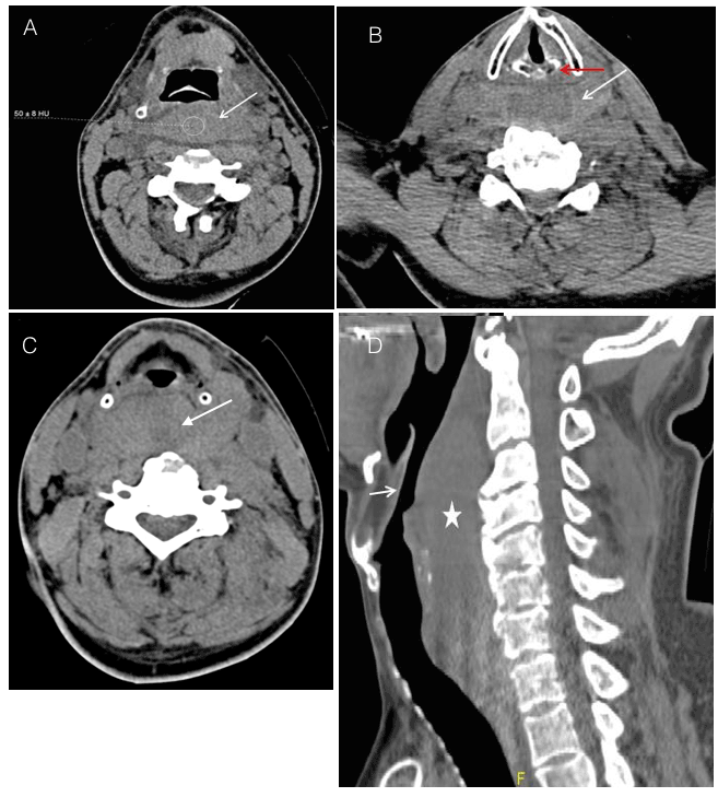
Figure 1. A. Axial unenhanced CT neck at the level of the epiglottis shows prevertebral/retropharyngeal high density (ROI density of 50 +- 8 Hounsfield Unit, white arrow) signifying hematoma.
B. Axial unenhanced CT neck at the level of the vocal cords and arytenoid cartilage (red arrow) shows prevertebral / retropharyngeal high density (white arrow) signifying hematoma.
C. Axial unenhanced CT neck at the level of the hypopharynx showing prevertebral/retropharyngeal high density (white arrow) signifying hematoma displacing the airway anteriorly.
D. Sagittal unenhanced CT neck (right lower corner) shows prevertebral/retropharyngeal high density (white star) signifying hematoma. This is causing significant narrowing of the airway (short white arrow).
Immediately after CT, the patient developed stridor and difficulty breathing which progressed rapidly. An upper airway obstruction was suspected and rapid sequence intubation attempted with Etomidate, Succinylcholine and direct laryngoscopy along with cervical stabilization. The intubation did not succeed and the emergency anesthetist described the Cormak Lehane score as 4 (inability to visualize the epiglottis).
Intubation attempt with a video laryngoscope (Glidescope®) also failed. Against an oxygen saturation of 85%, further attempts at intubation were halted in favor of emergency cricothyroidotomy. The airway was secured and patient shifted to the Operating room for Hemostasis, with possibility of neck exploration.
In the operating room intubation was reattempted with a rigid fiber-optic device (Retro molar) without success. The decision was then taken to re-induce the patient with Fentanyl, Cisatracurium and Propofol with inhalational maintenance. After induction, intubation was attempted again with a Glidescope® which showed a bluish hematoma obscuring the field and causing an anterior displacement of the larynx. A formal tracheostomy was done (Figure 2) and the hematoma was treated conservatively after shifting the patient to the Trauma ICU. He stayed in the TICU for around 12 days where the RPH resolved spontaneously. Post resolution, the tracheostomy tube was removed and the stoma closed gradually as per weaning protocol. His tibial fracture was surgically managed while his cervical fracture was treated conservatively with neck collar. He was discharged home after 1 week in the ward with uneventful outpatient follow-up.
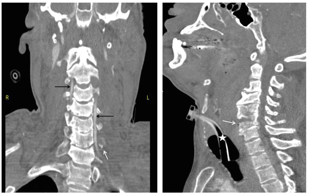
Figure 2. Left image: Coronal reformatted cervical spine post contrast shows the left C6 transverse process fracture (short white arrow). Patent vertebral artery is optimally visualized (black arrows).
Right image: Sagittal reformatted cervical spine demonstrates the left C6 vertebral fracture (short white arrow). Note that a tracheostomy tube (white star) has been inserted.
Retropharyngeal hematoma (RPH) is collection of blood in the retropharyngeal space. The parapharyngeal space, which is continuous with the retropharyngeal space, lies lateral to the pharynx and contains muscles, carotid sheath, the vagus nerve, loose connective tissue, and fat.
Various precipitating factors [1] have been mentioned as causes of RPH such as blunt head or neck trauma, whiplash injury, coagulopathy, central line insertion, stellate ganglion block, sneezing, severe coughing as well as spontaneously. This condition has been scarcely mentioned in literature because of its rarity, but of the few cases that have been described, most have been associated with cervical spine injury [2]. RPH can develop hours or days, even after an apparently minor precipitating injury [3]. Our case has two unique clinical points i.e, RPH causing upper airway obstruction can sometimes develop within a few hours from the history of trauma (within 3 hours in our case). Degree of cervical spine injury has a correlation to RPH. Increased incidence of RPH noted in unstable cervical spine injury than stable injuries. However, RPH causing significant airway obstruction secondary to a stable cervical spine injury within such a short span of 3 hours is a rare finding. The key lesson, as shown in our case, is that a patient may develop rapid airway obstruction after seemingly stable injuries with no prior warning signs. While the hematoma did develop steadily, the airway compromised precipitously with limited time for health care providers to successfully react. A possible reasoning maybe that the patient, to an extent, maintained his airway until the compromise became too severe or till he became fatigued. Either way, sudden tipping of the patient’s airway status may occur in such cases. Additionally, the hematoma, as shown in the CT as well as Glideoscopic image, involved a significant amount of space causing mechanical displacement of the pharynx and larynx. It is to be expected that this would make intubation almost impossible with conventional laryngoscopy or even video assisted techniques.
A high index of clinical suspicion and an early anticipation of possible airway obstruction even in the setting of stable cervical spine injury associated RPH is important. The time taken for RPH to develop and to cause mechanical obstruction can be just enough to allow preparation for a difficult airway as well as availability of fiberoptic technique in Emergency may offer good chance of success [4]. Keeping a team experienced in performing tracheostomy as a backup when the Ambulance service reports a potential neck injury victim can be helpful as well.
The time from initial presentation to complete airway obstruction is very small in cervical trauma patients. Additionally, the initial presentation may be benign and offer a sense of security which can rapidly change and thus health care providers must be ready. In case of acutely expanding treropharyngeal hematoma, even video laryngoscopy in experienced hands is very difficult. A tracheostomy team should be ready as soon as the ambulance team calls and informs of a cervical trauma victim.
2021 Copyright OAT. All rights reserv
None of the above authors have any financial disclosures or conflicts of interest with regards to submission of this case report.
Part of this work has been presented as poster in 1st International Conference in Emergency Medicine and Public Health – Qatar 2016 and poster has been published as conference proceeding in Journal of Emergency Medicine, Trauma & Acute Care, International Conference in Emergency Medicine and Public Health – Qatar 2016:26.
- Myssiorek D, Shalmi C (1989) Traumatic retropharyngeal hematoma. Arch Otolaryngol Head Neck Surg 115: 1130-1132. [Crossref]
- Thomás MD, Torres A, García-Polo J, Gavilán C (1991) Life-threatening cervico-mediastinal haematoma after carotid sinus massage. J Laryngol Otol 105: 381-383. [Crossref]
- Frankfort M, Koeijers JJ, Zwaveling JH, van Mook WNKA (2011) Severe retropharyngeal haematoma after apparent minor injury. Netherlands Journal of Critical Care.
- Lazott LW, Ponzo JA, Puana RB, Artz KS, Ciceri DP, Culp WC (2007) Severe upper airway obstruction due to delayed retropharyngeal hematoma formation following blunt cervical trauma. BMC Anesthesiol 7: 2. [Crossref]