We report a case of primary laryngeal tuberculosis in a 54 year old Indian male, who had hoarseness as his presenting complaint. This case highlights that laryngeal tuberculosis may occur even without pulmonary tuberculosis, and the characteristics of the lesions now appear to be more nonspecific than earlier. Otolaryngologists and airway physicians need to be more alert to the emergence of laryngeal tuberculosis with atypical clinical manifestations and consider it in their differential diagnosis of all laryngeal diseases.
laryngeal tuberculosis, anti tubercular treatment, pulmonary tuberculosis
The incidence of laryngeal tuberculosis has steadily increased over the past two decades due to increased prevalence of HIV infection, immunosuppressive diseases or treatments, the increased survival of aged, immigrants coming from high risk areas, and the emergence of resistant organisms and atypical mycobacteria [1-3].
The clinical pattern of laryngeal tuberculosis has also changed in comparison to the past. Earlier, it affected posterior larynx in most patients, since it directly spreads along the airway [1-6]. Currently, however, it is prevalent in all parts of the larynx and is usually seen in people in their 40s-60s as primary laryngeal tuberculosis. Main complaints are hoarseness and odynophagia, unlike in the past when patients mainly complained of dyspnoea or pulmonary and other constitutional symptoms [1].
Delay in diagnosis of primary laryngeal tuberculosis poses serious threat to the patient due to delayed treatment and complications. We report a 54 year old gentleman who presented with primary laryngeal tuberculosis and was successfully treated with anti-tubercular therapy leading to full clinical recovery.
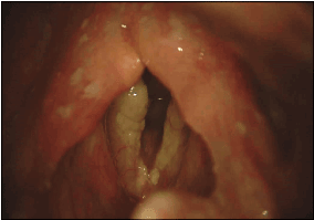
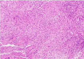
A 54-year-old Indian male presented to the otorhinolaryngology clinic with a 3-months history of dysphonia, dysphagia, and odynophagia. The voice was strained and rough. He had no stridor. The patient had self-treated with oral antibiotics and analgesics without resolution of his symptoms. He had no weight loss, fever, chills, night sweats, cough, haemoptysis, or decrease in appetite. He also denied any allergies and was not on any medications. His past medical history was insignificant. His social history included smoking half pack of cigarettes per day for 30 years and a history of occasional alcohol use. Physical, head and neck, pulmonary examinations and CXR were normal. Flexible fiberoptic examination revealed a grossly abnormal endolarynx with edematous arytenoids, aryepiglottic folds, false and true vocal cords with multiple superficial ulcerations and a fine nodular appearance. There was slough like polypoidal lesion over both the true vocal cords with impairment in adduction (Figure 1). Mantoux test [purified protein derivative test] was positive and ESR was 28 mm/1st hr. Based on the above clinical findings a provisional diagnosis of tuberculosis of larynx was considered first. However, after obtaining informed consent, the patient was taken to the operating room for a direct microlaryngoscopy and biopsy to exclude malignancy. Under direct visualization, the epiglottis, supra-arytenoid tissues, true and false cords were edematous and polypoid in appearance covered with a fine fibrinous exudate with normal subglottis. Multiple biopsies were taken and sent for histopathologic examination, which revealed granulomatous inflammation with necrotising granulomas and psuedoepitheliomatous hyperplasia suggestive of tuberculosis (Figure 2). The patient was discharged on the anti-tubercular medications and asked for a follow- up appointment one month later with the culture report. Cultures of the biopsied tissue returned a diagnosis of pansensitive Mycobacterium tuberculosis, but the sputum cultures were negative. The patient took anti tubercular regimen [ATT] for six months for primary laryngeal tuberculosis and the laryngeal lesions completely healed.

Figure 1. Laryngoscopic image revealing edematous arytenoids, aryepiglottic folds, false and true vocal cords with multiple superficial fibrinous exudate and slough like polypoidal lesion over both the true vocal cords.

Figure 2. Photomicrograph showing confluent epithelioid cell granulomas with giant cells along with lymphocytes in the background. [H&E x 200].
Laryngeal tuberculosis is one of the most frequent diseases causing granulomatous lesions in the larynx, and usually occurs as a complication of pulmonary tuberculosis. Its incidence in the past about 25-40% of general population was thought to be significantly higher than the rate of diagnosis, however, the incidence of this disease has drastically decreased since the introduction of anti-tuberculosis drug therapy, and is reported to be 0.8-1% [1,2,7].
Laryngeal tuberculosis can result from direct bronchogenic spread coming from AFB containing sputum or hematogenous or lymphatic spread [1,2,5]. In the cases of bronchogenic spread, acid fast bacilli in sputum infect the laryngeal mucosa through direct contact and the symptoms are similar to those in the case of pulmonary tuberculosis. The disease would gradually progress after having developed in the posterior portion of the larynx. However, in those cases with hematogenous or lymphatic spread, the spread is quick with predominant laryngeal symptoms such as dysphonia and odynophagia, and the lesions are frequently seen in the epiglottis and anterior larynx and, sometimes, in the pharynx, soft palate and tonsils. Although secondary laryngeal tuberculosis through bronchogenic spread predominated in the past, incidence of primary laryngeal cases without pulmonary involvement is on the rise today.
The incidence of laryngeal tuberculosis was high in people in their 40s-50s, with the age ranging from 25 to 78 years with a male predominance [1,5]. Chronic alcoholism and tobacco abuse were the most common predisposing factors for tuberculosis [8].
Laryngeal tuberculosis is prevalent in those patients whose chief complaint is hoarseness accompanied by dysphagia or odynophagia whereas constitutional symptoms like fever, weight loss, night sweats and fatigue are rare these days. Painful dysphagia attributed as a rare symptom of laryngeal cancer, therefore, would help to differentiate between laryngeal tuberculosis and laryngeal cancer [5,7].
The clinical appearance of types of laryngeal tuberculosis have also changed. Severe ulcerative or granulomatous lesions in the posterior larynx were the most prevalent types in the past, however, it has been reported that more cases of laryngeal tuberculosis affect whole larynx but few with regional infection in the posterior larynx [8-10]. Furthermore, it has been reported that hypertrophic, exophytic, polypoid lesions or non- specific lesions are seen relatively more frequently than ulcerative or granulomatous lesions as in our case [4,5,8,11]. Henceforth it is imperative for the physicians to perform stroboscopic examination and test chest radiograph, sputum study, and laryngeal biopsy when nonspecific infection occurs unilaterally for definitive diagnosis of laryngeal tuberculosis and to differentiate from chronic laryngitis.
In a review by lim et al, nine out of 60 cases [15%] had tuberculosis only involving larynx. Granulomatous lesions were found to be prevalent in those patients with laryngeal tuberculosis accompanying active pulmonary tuberculosis, whereas polypoidal and nonspecific lesions were prevalent in those with inactive pulmonary tuberculosis, and normal lung status [1,8].
Laryngeal tuberculosis can cause posterior glottic stenosis, subglottic stenosis, and vocal cord paralysis due to cricoarytenoid joint or recurrent laryngeal nerve involvement if early treatment is not instituted and may impose tracheostomy depending on the severity of airway obstruction [12]. Clinically, differential diagnosis in many cases is difficult between laryngeal tuberculosis and laryngeal cancer warranting a histopathological examination, as these two diseases may co-exist [8,13,14].
To conclude, with increased incidence of laryngeal tuberculosis, change in its clinical pattern and spread mechanism, increased prevalence of polypoidal or nonspecific changes in larynx, treating physicians should always be aware of atypical clinical features of laryngeal tuberculosis and the possibility of primary laryngeal tuberculosis, for early diagnosis and treatment, thus preventing complications of laryngeal tuberculosis, and the risk of transmission to physicians, hospital personnel and other patients.
2021 Copyright OAT. All rights reserv
Informed written consent was obtained from the patient for publication of the images.
- Lim JY, Kim KM, Choi EC, Kim YH, Kim HS, et al. (2006) Current clinical propensity of laryngeal tuberculosis: review of 60 cases. Eur Arch Otorhinolaryngol 263: 838-842. [Crossref]
- Rizzo PB, Da Mosto MC, Clari M, Scotton PG, Vaglia A, et al. (2003) Laryngeal tuberculosis: an often forgotten diagnosis. Int J Infect Dis 7: 129-131. [Crossref]
- Cleary KR, Batsakis JG (1995) Mycobacterial disease of the head and neck: current perspective. Ann Otol Rhinol Laryngol 104: 830-833. [Crossref]
- Kandiloros DC, Nikolopoulos TP, Ferekidis EA, Tsangaroulakis A, Yiotakis JE, et al. (1997) Laryngeal tuberculosis at the end of the 20th century. J Laryngol Otol 111: 619-621. [Crossref]
- Shin JE, Nam SY, Yoo SJ, Kim SY (2000) Changing trends in clinical manifestations of laryngeal tuberculosis. Laryngoscope 110: 1950-1953. [Crossref]
- Nishiike S, Irifune M, Doi K, Sawada T, Kubo T (2002) Laryngeal tuberculosis: a report of 15 cases. Ann Otol Rhinol Laryngol 111: 916-918. [Crossref]
- Ramadan HH, Tarazi AE, Baroudy FM (1993) Laryngeal tuberculosis: presentation of 16 cases and review of the literature. J Otolaryngol 22: 39-41.
- Ling L, Zhou SH, Wang SQ (2010) Changing trends in the clinical features of laryngeal tuberculosis: a report of 19 cases. Int J Infect Dis 14: e230-235. [Crossref]
- Beg MH, Marfani S (1985) The larynx in pulmonary tuberculosis. J Laryngol Otol 99: 201-203. [Crossref]
- Hunter AM, Millar JW, Wightman AJ, Horne NW (1981) The changing pattern of laryngeal tuberculosis. J Laryngol Otol 95: 393-398. [Crossref]
- Soda A, Rubio H, Salazar M, Ganem J, Berlanga D, et al. (1989) Tuberculosis of the larynx: clinical aspects in 19 patients. Laryngoscope 99: 1147-1150. [Crossref]
- Caldarelli DD, Friedberg SA, Harris AA (1979) Medical and surgical aspects of the granulomatous diseases of the larynx. Otolaryngol Clin North Am 12: 767-781. [Crossref]
- Yarnal JR, Golish JA, van der Kuyp F (1981) Laryngeal tuberculosis presenting as carcinoma. Arch Otolaryngol 107: 503-505. [Crossref]
- Yencha MW, Linfesty R, Blackmon A (2000) Laryngeal tuberculosis. Am J Otolaryngol 21: 122-126. [Crossref]


