Synovial sarcoma is a soft tissue sarcoma that rarely occurs in the oral cavity and the most common site is the tongue. Synovial sarcoma of the oral cavity mostly presents as a slowly growing, painless, and deeply seated mass. Radical surgical excision with negative margins is the treatment of choice and adjuvant radiotherapy is advised. This report presents a 48-year-old male patient with a rapidly growing mandibular gingival mass with underlying alveolar bone destruction. Incisional biopsy was done, and the specimen was stained with Hematoxylin and Eosin. Microscopic sections showed fascicles of proliferated spindle-shaped cells with plump nuclei that invaded the surrounding skeletal muscles. The lesion was positive for Vimentin, CD99, CK and Bcl-2 in immunohistochemistry. The diagnosis of monophasic synovial sarcoma was made, and radical surgical excision was performed. Due to the paucity of synovial sarcoma in the oral region, the information about appropriate treatment for this sarcoma is limited. The knowledge about this rare entity and microscopic characteristics are important and help to have a proper diagnosis and improved treatment plan.
synovial sarcoma, histopathology, oral cavity, cancer, jaw
Sarcomas are a heterogeneous category of cancers originating from the transformed cells of mesenchymal lineage with a wide range of histopathologic types [1]. Synovial sarcoma (SS) is a high-grade soft tissue sarcoma rarely seen in the head and neck area [2]. The most common site is the extremities in young adults without any sex predilection. The posterior of the tongue is the most typical reported site in the oral cavity [3]. The most typical manifestation of oral SS is painless, gradually enlarging mass [1,3]. Microscopically, it shows three major variants: a) monophasic contains only spindle cells or epithelial cells b) biphasic contains epithelial cells arranged into glandular structures with spindle cells arranged into fascicles and c) poorly differentiated subtype with the presence of spindle and/or round blue cells [4,5]. The treatment of choice is radical surgical excision [4,6], and adjuvant radiotherapy with or without chemotherapy is recommended [4,7]. This article aims to report an additional case of primary monophasic synovial sarcoma of the oral cavity.
A 48-year-old male was referred to a private oral and maxillofacial surgery center (Ahvaz, Iran) to evaluate a rapidly growing, ulcerated erythematous mass in the anterior area of mandibular gingiva with dull pain and paresthesia, which had been present for about 4 to 5 months. The mass was soft in consistency, measuring 2.5 × 2 cm (Figure 1). There was no history of previous trauma, and the patient had no other medical problems, but he was a cigarette smoker. The cause of anterior tooth extraction was not unclear. The extra-oral examination was normal. There was no cervical lymphadenopathy, and the laboratory data were unremarkable. The panoramic radiograph and CT scan showed a massive lesion of the anterior mandible with buccal and lingual plate destruction (Figure 2). According to both clinical and radiographic features, malignant lesions such as Non-Hodgkin lymphoma were considered in differential diagnosis, and an incisional biopsy was performed under local anesthesia. The specimen demonstrated a solid, white, and homogenous cut surface, and it was processed for Hematoxylin and Eosin histopathologic study.
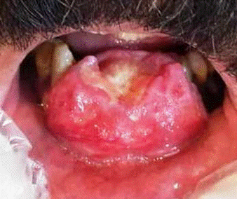
Figure 1. Photograph shows large ulcerated erythematous mass of anterior mandibular gingiva.
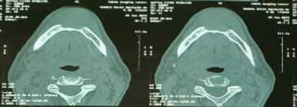
Figure 2. CT scan shows destructive lesion in anterior of mandible (Coronal view).
Microscopic examination showed a malignant mesenchymal neoplasm covered by ulcerated mucosa, composed of fascicles of proliferated spindle-shaped cells with plump nuclei and whorled pattern in some areas that invaded the surrounding skeletal muscles. Areas of necrosis and mitotic figures about 5-6 mitosis per 10HPF were seen (Figure 3). The nature of the spindle cells was determined through immunohistochemistry (IHC). The sections were positive with monoclonal antibodies for Vimentin, CD99, CK (AE1/AE3), Bcl-2 and were negative for S-100, CD34, and EMA. Ki-67 index was about 30% (Figure 4 and Figure 5). The whole-body scan revealed no other organ involvement. The histopathological features combined with those IHC findings were consistent with the primary monophasic synovial sarcoma (SS) diagnosis. The treatment of choice was radical surgery. The microscopic features of the resected tumor confirmed the diagnosis of SS, and the margins were free of tumor. The patient was referred to an oncology center for further treatment, but unfortunately, the patient did not return for follow-up.
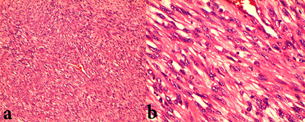
Figure 3. Hematoxylin and Eosin staining demonstrates fascicles of proliferated spindle cells with plump nuclei (a. 100 X and b 400 X).
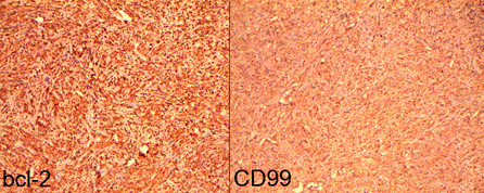
Figure 4. Positive immunoreactivity for bcl-2 and CD99 antibody (immunohistochemistry, 100 X).
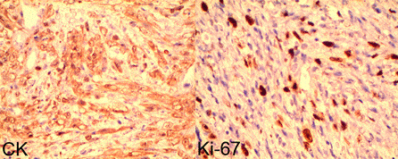
Figure 5. Positive immunoreactivity for CK and Ki-67 index about 30% (immunohistochemistry, 400 X).
About 3% of SSs occur in the head and neck region [6]. SS of the oral cavity is a highly uncommon entity available in scattered case reports [8]. The tumor is supposed to arise from primitive undifferentiated pluripotential mesenchymal cells unrelated to synovial tissue [2]. There is a male predilection [4]. Meer et al. [2] stated that most cases are within the second and third decades of life, and the second largest group is in the fourth and fifth decades of life. Our patient was also in his 5th decade of life. The tongue, especially the base of the tongue, is the most typical location, followed by cheek, soft palate, and retromolar area [2]. It is frequently a slow-growing mass growing in size over 1 to 2 years, which differs from 3 to10 cm [4]. Histopathologically, SS reveals three major variants: a) monophasic contains only spindle cells or epithelial cells b) biphasic contains epithelial cells arranged into glandular structures with spindle cells arranged into fascicles, and c) poorly differentiated subtype with the presence of spindle and/or round blue cells [4]. Mast cells, calcification, PAS-positive material, and mitotic activity had also been reported [1]. The differential diagnosis for monophasic SS comprises leiomyosarcoma, malignant peripheral nerve sheath tumor (MPNST), and solitary fibrous tumor (SFT). Leiomyosarcoma shows spindle cells with abundant eosinophilic cytoplasm; blunt-ended nuclei, cytoplasmic vacuoles; nuclear atypia, and mitotic figures [9]. Immunohistochemical analysis usually shows the presence of desmin, muscle-specific actin, and SMA [10].
In contrast to SS, MPNST shows an intact SMARCB1 and reveals H3 K27 me3 loss. SFT demonstrates hyper- and hypocellularity zones rather than the uniform herringbone pattern of SS and is positive for CD34 and STAT-6 [9]. In addition, the monophasic variant is similar to fibrosarcoma, but it shows no reactivity with epithelial markers [6].
Most SSs demonstrate at least a focal positive immunostaining of spindle cells for cytokeratin or EMA. Bcl-2 and CD99 are frequently expressed in SSs. Calretinin expression is seen in the epithelial and spindle cells of biphasic, monophasic, and poorly differentiated tumors [1]. The transducin-like enhancer of split-1 (TLE-1) is positive in 90% of SSs. It is negative for CD34, actin, myoglobin, and desmin [7]. Over 90% of SSs show t(X;18)(p11.2;q11.2), which results in an SS18-SSX fusion gene [2]. Nevertheless, molecular testing is unnecessary when a definitive diagnosis is made on a clinical, microscopic, and immunohistochemistry basis [6]. Due to the paucity of this tumor in the oral region, the information about appropriate treatment for this sarcoma is limited [6].
Wide surgical excision with negative margins is the treatment of choice [1], and adjuvant radiotherapy with or without chemotherapy is recommended [7]. Metastasis and local recurrence are late events; therefore, long-term follow-up should be performed [3]. The recurrence rate for SS arising from all body regions is about 50% [6]. No significant difference was found in overall survival or disease-specific survival between patients with SS18-SSX1 and SS18-SSX2, though progression-free survival and metastasis-free survival are considered undesirable in patients with SS18-SSX1 [11]. A Ki-67 index higher than 10% may be considerably associated with metastasis. Patients with tumors of a maximum diameter larger than 5 cm have a worse prognosis than smaller neoplasms [6]. Better prognosis is associated with age less than 60 years, extensive calcification, adequate surgery, high degree of tumor differentiation, extensive hemorrhagic necrosis, high mitosis index, and no distant metastasis [4].
SS of the oral cavity is an exceedingly rare lesion and reporting new cases will help our information in this regard. Knowing this rare entity and microscopic characteristics is essential and helpful to have a proper diagnosis and improved treatment plan.
The complete data and materials described in the case report are not publicly available due anonymity purposes but are available from private clinic of Oral and Maxillofacial surgery in Ahvaz, Iran.
- Atarbashi-Moghadam S,Emami Razavi AN,Salehi Zalani S (2019) PrevalenceofHeadandNeckSarcomain aMajorCancerCenterinIran- A10-YearStudy. IranJ Otorhinolaryngol 31: 97-102. [Crossref]
- Meer S,Coleman H,Altini M (2003) Oral synovial sarcoma: a report of 2 cases and a review of the literature. Oral Surg Oral Med Oral Pathol Oral Radiol Endod 96: 306-315. [Crossref]
- Doubi A,Doubi M,Alzaher N,Tulbah A (2019) Synovial sarcoma of the hard palate: The third case in the medical literature. Hematol Oncol Stem Cell Ther 12: 60-63.
- Wang Y, Zhu F, Wang K (2020) Synovialsarcomaof the floor of themouth: a rare case report. BMC Oral Health 20: 5. [Crossref]
- Jayasooriya PR, Madawalagamage LN, Mendis BRRN, Lombardi T (2016) Diagnostic approach to synovial sarcoma of the head and neck illustrated by two cases arising in the face and Oral cavity. Dermatopathology 3: 13-22. [Crossref]
- Wang H, Zhang J, He X, Niu Y (2008) Synovialsarcomain theoralandmaxillofacialregion:reportof4casesandreviewof theliterature. JOralMaxillofac Surg. 66: 161-7. [Crossref]
- Basile LE,Hoch B,Dillon JK (2016) Synovial Sarcoma of the Tongue: Report of a Case. J Oral Maxillofac Surg 74: 95-103. [Crossref]
- Vig T, Thomas M, Pai R, Tirkey AJ, Janakiraman R (2018) PrimarySynovialSarcomaarising from gingivo-buccal sulcus harbouring SS18-SSX2 positive fusion transcript: The 1st reported case in English literature. J Stomatol Oral Maxillofac Surg 119: 220-223. [Crossref]
- Alabdulaaly L, AlDawood Z, Afshar S, Rahbar R, Al-Ibraheemi A, et al. (2020) Calcifyingsynovialsarcomaof the tongue with SS18 rearrangement: a rare variant in a rare location. Oral Surg Oral Med Oral Pathol Oral Radiol 20: S2212-4403(20)31165-2. [Crossref]
- Moghadam SA, Khodayari A, Mokhtari S (2014) Primaryleiomyosarcomaof themandible. J Oral Maxillofac Pathol 18: 308-311. [Crossref]
- Kubo T, Shimose S, Fujimori J, Furuta T, Ochi M (2015) Prognostic value of SS18-SSX fusion type in synovial sarcoma; systematic review and meta-analysis. Springerplus 4 :375. [Crossref]