Abstract
Purpose: Angular deformities of the knee in children are the main complaints in our hospital service and accounts for 13.2%. Several classifications have been proposed in the past. However, there is no known clinical classification that enables to typify the deformities in the entire varus and valgus. The present study aimed to propose a simple classification that can be used even by non-specialists like in African milieu.
Methods: We reviewed the clinical records of 2711 children with deformities of the knee that were followed up in our centre during 10 years. Clinical and radiological angular measurements of the knees were analyzed. There were 1512 boys for 1119 girls, with an average age of 3.5 years.
Results: Out of this, 58.79% presented with genu valgum (Type I) and 30.35% presented with genu varum (type II). A combination of genu valgum and genu varum accounted for 10.84% of the deformities (type III). This was however sub classified based on the affected side, age and severity.
Conclusion: Our results are discussed in the light essentially few African studies published on the subject.
The present classification which has the advantage of being simple and thus easily understandable is also helpful in treatment.
Key words
knee deformities, classification, black African children
Level of evidence
IV, retrospective study.
Introduction
Angular deformities of the childhood reported in previous studies [1,2] are the primary complain and represent 13.2% of all the complains received at the outpatient clinics at the National Centre for the Rehabilitation of the Disabled in Yaoundé, Cameroon. However, all children with angular deformities of the knee consult the physicians for pain, difficulty in walking, and poor cosmetic aspect. These complications are generally corrected by invasive surgical interventions named osteotomies, the specific technique being selected based on the type of knee deformity [3].
Many classifications for these deformities have been proposed previously, but these are too selective and most of them deal with either varus or valgus deformities. The current classifications are too complex for non-specialists, and tend to contradict each other. Clinical assessments of knee angular deformities are based on angular deviations expressed in degrees of varus or degree of valgus [4]. However, the current definitions of “valgus” and “varus” are confusing, leading to poor quality of treatment. The terms “valgus” (“twisted” or “bent” in Latin) “varus” (“crooked” in Latin) are frequently used to describe “knock-kneed” and “bowlegged” limbs, respectively, which are the opposite of their etymology. Therefore, there is an urgent need as expressed rightly by Kamath [4], for a simpler classification of knee angular deformities to ensure proper treatments, even by non-specialists in remote areas.
Hence, the present study aimed to propose a global, simple, and practical classification of these knee deformities that are frequently observed in black African children.
Patients and methods
We reviewed the clinical records of 2711 children who were followed in our orthopedic outpatient clinics from 1998 to 2008. The patients were examined preliminary in 4 positions: nude, lying, standing, and walking. The ultimate diagnosis was based on radiological examination consisted of standard plain X-rays orthoroentgenogram to assess the angular deformities and associated morphological abnormalities.
The laboratory exams, especially, white and red blood count, the dosage of calcium, phosphorus and alkaline phosphates reinforce radiographic exams to determine the etiology of the deformities.
The treatment was conducted according to quality of the deformity (varus or valgus), the quantity (measurement of the angular deformity) and the age. This treatment consisted of serial casts and splints for severe deformities, supplementation of vitamin D, calcium or ion for moderate deformities. The indications for surgery concerned very severe deformities with complications in older children. In these cases, open or closed-wedge osteotomies were undertaken according to the type and severity of knee angulations. In older children with mild deformities stapling to induce early epiphysiodesis was carried out. A great number of children received no treatment at all due to limited resources.
Results
Demographic profiles
From 1998 to 2008, we received a total number of 19100 patients amongst them 2996 children consulted for problems of angular deformities of the knee (20.9%). Incomplete files, 285 cases (9.51%) that could not allow a rational exploitation of the series were excluded. There were 1512 boys and 1199 girls (sex ratio 1:1.2) (Figure 1), and the mean age was 3.5 years (range, 1-19 years). The mean of total follow-up was 148.8 days (from 1 day to 9 years 10 months and 3 days).
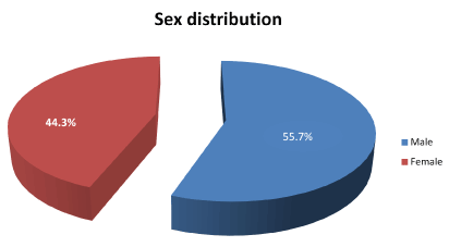
Figure 1. Sex distribution of the total number of reviewed children.
Clinical features of the deformities
We individualized 3 types of deformities (Figure 2). Type I is the valgus deformity (sub-type A for bilateral, sub-type B for unilateral), 1594 children (58.97%) is the most important group. Type II is the varus deformities (sub-type A for bilateral or sub-type B for unilateral), 823 children (30.35%), is the second group. Type III, combination of valgus and varus deformities towards left side, sub-type A or right side, sub-type B (“windswept or strong gale deformities”), counted for 294 children (10.84%). The sub-type C for the 3 above types is determined by age and severity of the deformity. The following Tables 1 and 2 give the detailed distribution of the 3 individualized types.
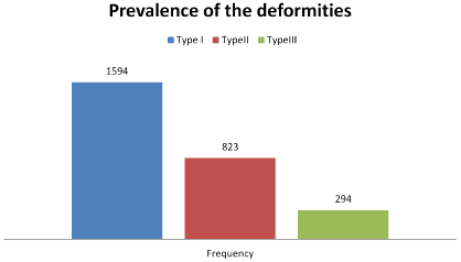
Figure 2: Prevalence of the 3 types of deformities.
Table 1: Distribution of types and sub-types of the deformities according to frequency and angular measurements.
Types |
Sub-types |
Number of children (n) |
Percentage
(%) |
Angular mean values (°) |
Minimum
(°) |
Maximum
(°) |
Type I
Genu valgum
N1=1594
(58.79%) |
Type I A
Bilateral genu valgum |
1447 |
53.37 |
30.4 |
4 |
90 |
Type I B
Unilateral genu valgum |
117 |
4.31 |
13.6 |
3 |
45 |
Type IC
Angulation >30° and age >9 years |
30 |
1.10 |
20.9 |
4 |
57 |
Type II
Genu varum
N2=823
(30.35%) |
Type II A
Bilateral genu varum |
711 |
26.22 |
53.8 |
5 |
110 |
Type II B
Unilateral genu varum |
52 |
1.91 |
30.4 |
5 |
60 |
Type IIC
Angulation> 30° and age ˃ 9 years |
60 |
2.21 |
36.4 |
7 |
110 |
Type III
Combination of genu valgum and genu varum
N3=294
(10.84%) |
Type IIIA
Right genu valgum, left genu varum |
184 |
6.78 |
17.1 |
5 |
55 |
Type IIIB
Left genu valgum, right genu varum |
94 |
3.46 |
17.0 |
5 |
55 |
Type IIIC
Angulation > 30° and
Age˃9 years |
16 |
0.59 |
30.1 |
3 |
87 |
TOTAL |
|
2711 |
99.95 |
27.0 |
3 |
110 |
Table 2: Characteristics of surgical indications.
Types |
Number of children (n) |
Percentage
(%) |
Angular mean values (°) |
Min (°) |
Max
(°) |
Mean age |
IC |
30 |
1.10 |
20.9 |
4 |
57 |
10.1 |
IIC |
60 |
2.21 |
36.4 |
7 |
110 |
8.1 |
IIIC |
16 |
0.59 |
30.1 |
3 |
87 |
8.5 |
TOTAL |
106 |
3.9 |
31.6 |
2 |
110 |
9.0 |
It appears that most children developed Type I or Type III deformities around the age of three, whereas peak prevalence of Type II occurs by the age of 2 (Figure 3).
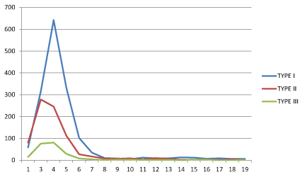
Figure 3. Prevalence of angular deformities of the knee according to age.
Etiology of the deformities
Ricketts diagnosed by clinical examination and confirmed by plain X-rays is the cause of 70.22% of the deformities in 1918 children. Laboratory exams showed evidence of trouble of the metabolism of calcium (80 cases), phosphorus alkaline phosphates (108 cases) and anemia (44 cases) in 232 children. The isolated mineral metabolism disorder was present in 28 children.
The other imputable causes of the deformities were trauma (4 cases), osteogenosa imperfecta (2 cases), juvenile rheumatoid arthritis (1 case), chondroma (1 case) and severe limb length discrepancy (1 case). There were 753 undetermined causes of the deformities.
Treatment
Table 3 summarizes the different methods of treatment applied based on the origin of the deformities in 2708 children. Indications for surgery concerned only 106 cases (3.91%) amongst them 90 osteotomies (3.32%) (Figures 4, 5, 6, 7), 14 stapling (0.51%) and 2 intramedullar nailings (0.07%). These surgical indications concerned children with average age of 9.0 years and average angular deformities of more than 30 degrees (Table 2). Orthopedic treatment concerned 1200 children (44.31%) (Figure 8) while 696 children (25.70) received no treatment (Figure 8). Exclusive medical treatment concerned 706 children (26.07%).
Table 3: The different methods of treatment based on origin of the deformities.
Treatement |
Origine of deformities |
Number
of children (n) |
Percentage (%) |
Ricketts |
Trouble of phosphorus, calcium and alkaline phosphates |
Unknown etiologies |
Other etiologies |
Orthopaedic |
1147 |
9 |
42 |
2 |
1200 |
44.31 |
Medical |
653 |
16 |
36 |
1 |
706 |
26.07 |
No treatment |
22 |
1 |
669 |
4 |
696 |
25.70 |
Surgical |
96 |
2 |
6 |
2 |
106 |
3.91 |
TOTAL |
1918 |
28 |
753 |
9 |
2708 |
100 |
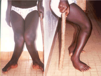
Figure 4. A severe bilateral genu valgum in a 15-year-old girl walking with a stick (Type I C) before surgery [1].
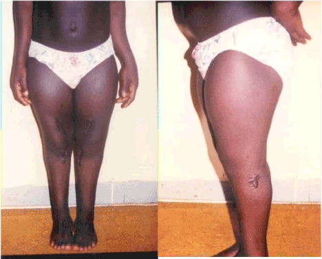
Figure 5. Photograph of the same girl (Type IC). Final aspect after correction [1].
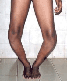
Figure 6. Bilateral recurrent genu varum operated for the first time at childhood (Type IIC) before surgery [2].
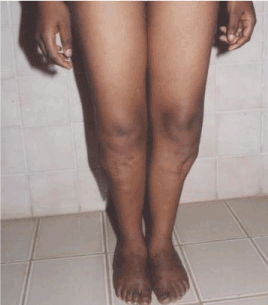
Figure 7. Photograph of the same girl with bilateral genu varum (Type IIC): final aspect after second surgery [2].
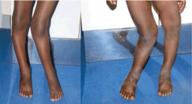
Figure 8. The “windswept or strong gale” deformities towards right side (Type IIIA) and towards left side (Type IIIB).
Discussion
This study and the previous studies carried out in Cameroon [1,2] and others carried out in neighboring countries in Nigeria by Oyemade [5] and Congo by Echarri [6] confirmed the prevalence of angular deformities in Black African Child. This study also confirmed the predominance of male and a peak frequency around 3 years for the majority of children. Rickets was the most imputable origin of these deformities (70.22%). However, there is a lack of reliable and comprehensive classification for the different types of described disorders. Our present study concerning large number of 2711 patients extended to more than 10 years tried to propose a simple and global varus and valgus deformities easily understable even by non-specialists.
The previous classific2021 Copyright OAT. All rights reservge and secondary form), Langenskiöld [8] and Catonné [9], (based on radiological stages focused on skeletal maturation), concern only selective varus deformities. [10] recently proposed a new classification of genu varum based on accompanying torsional deformities of the lower extremities. Most of other classifications are based on radiological exams or sophisticated tests like MRI, Duclou le Pointe et al, 5[11] which are not available every time and everywhere in Africa. The classification that we proposed is based not only on clinical and radiological examination of valgus and varus knee deformities, but also added new criteria of severity of angular measurements and age of the children. These last criteria determine surgical indications and thus were helpful to conduct surgery as we experienced successfully. This surgery concerned 106 patients (3.91%) was indicated to children with a mean angular measurement of 31.6° and an average age of 9.0 years. These data helped to typify the subtype C.
In Cameroon, our study suggests that Type I predominates (58.79%), followed by Type II (30.35 %) and Type III patients (10.84 %). However, it appears in this series that Type II (genu varus) predominate at the age of 2 years. This suggests the conversion of some varus deformities to valgus deformities during growth. Two studies were previously conducted in two neighboring African countries to ours. In Nigeria the valgus and varus deformities are about equally represented with 35.8% and 41.8%, respectively amongst 67 patients [5], while varus deformities dominate the Congolese population with 58.2% amongst 194 rachitic compared to 107 healthy children [6]. Together, these data support region specific differences in the prevalence of specific types of knee angular deformities in Africa. However, these small series could not allow drawing strong and durable conclusions that permits our larger series of 2711 children.
Conclusion
We hope that this simple global proposed classification of angular deformities of the knee in black African children (1) can be used rapidly and easily for epidemiologic studies, (2) can be easily understood as a simple common language and (3) be a helpful therapeutic tool.
Conflicts of interests
none
Acknowledgements
We wish to express our gratitude to Dr Bertrand Zing Awona for his substantial contribution to the statistical analyzes.
References
- Ibrahima F, Pisoh-Tagnyin C, Abolo-Mbenti L, Sosso MA, Eimo Malonga E (2002) Déformations angulaires de genu varum/valgum chez l’enfant et l’adulte jeune. Revue préliminaire 158 cas observés à Yaoundé. Méd Afr Noire 49: 169-175.
- Ibrahima F, Ngo Nonga B, Mouafo Tambo FF, Ngandeu Singwe M, Sosso MA (2010) Aspects diagnostiques et thérapeutiques des déformations d’axe des membres inférieurs de l’enfant africain. Revue de 43 cas Rev Afr Chir Spéc 4: 33-36
- Rossi R, Bonasia DE, Amendola A (2011) The role of high tibial osteotomy in the varus knee. J Am Acad Orthop Surg 19: 590-599. [Crossref]
- Kamath AF, Israelite C, Horneff J, Lotke PA (2010) Editorial: What is varus or valgus knee alignment? a call for a uniform radiographic classification. Clin Orthop Relat Res 468: 1702-1704. [Crossref]
- Oyemade GA (1977) Non-rachitic deformities of the knees in Nigerian children. J Trop Med Hyg 80: 213-218. [Crossref]
- Echarri JJ, Aimé Bazeboso J, Guillén-Grima (2008) Deformaciones raquíticas de miembros inferiores en los niños congoleños. An Sist Sanit Navar 31: 235-240.
- Blount WP (1937) Tibia vara. J Bone Joint Surg 19: 1-29.
- Langenskiold A (1952) Tibia vara; (osteochondrosis deformans tibiae); a survey of 23 cases. Acta Chir Scand 103: 1-22. [Crossref]
- Catonne Y, Dintimille H, Arfi S, Mouchet A (1983) [Blount's disease in the Antilles. Apropos of 26 cases]. Rev Chir Orthop Reparatrice Appar Mot 69: 131-140. [Crossref]
- Joo SY, Park HW, Park KB, Kim BS, Park JS, et al. (2007) A new classification for idiopathic genu vara. Yonsei Med J 48: 833-838. [Crossref]
- Ducou le Pointe H, Mousselard H, Rudelli A, Montagne JP, Filipe G (1995) Blount's disease: magnetic resonance imaging. Pediatr Radiol 25: 12-14. [Crossref]
s