Hodgkin lymphoma (HL) and Non-Hodgkin lymphoma (NHL) are the malignant neoplasm spectrum originating from the lymphoid system and constituting 8% of all malignancies. Although, both are known as primary malignancies of lymph nodes, extranodal involvement can be seen. In this case presentation, in addition to nodal and bone involvement areas in a patient diagnosed as marginal zone lymphoma, rare simultaneous testicular and skin extranodal involvement detected on 18F-FDG PET-CT imaging were presented.
rare skin, testicular extranodal, PET-CT imaging, marginal zone lymphoma, nuclear medicine, Hodgkin lymphoma, Non-Hodgkin lymphoma
Abbreviations:
Hodgkin lymphoma-HL, Non-Hodgkin lymphoma-NHL, Marginal zone lymphomas-MZL
Extranodal involvement areas such as gastrointestinal, head and neck, orbital, central and peripheral nervous system, thorax, bone, skin, breast, testis, thyroid and genitourinary system can be seen in 25-40% of HL and especially NHL patients, although it is known as lymph node malignancy [1,2]. Extranodal involvement is important in terms of prognosis and imaging procedures play a very important role in diagnosis. 18F-FDG PET-CT imaging is a standard method in lymphoma patients and has a special importance in these patients in terms of superiority of CT imaging in detecting extranodal regions. The role of 18F-FDG PET-CT in detection, staging and restaging of patients with extranodal involvement in NHL has also been reported in the literature [3,4]. In this case presentation, in addition to nodal and bone involvement areas in a patient diagnosed as marginal zone lymphoma, rare testicular and skin extranodal simultaneous involvement detected on 18F-FDG PET-CT imaging were presented.
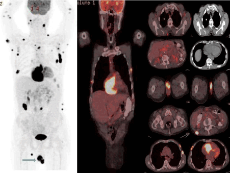
A 72-year-old male patient diagnosed as marginal zone lymphoma following mass excision from the left thoracic cage underwent 18F-FDG PET-CT imaging for initial staging. PET-CT images showed hypermetabolic (SUVmax: 14.66-31.89) conglomereted lymphadenopathies in the bilateral cervical chain, supraclavicular area, bilateral axillary, mediastinal, retroperitoneal, paraaortic, peripancreatic, perigastric, liver hilum, paracoeliac regions, pericardial thickening-effusion and hypermetabolic (SUVmax: 3.32-12.83) bone lesions. Additionally, multiple subcutaneous hypermetabolic (SUVmax: 10.5-24.37) nodules were detected at various levels within the cross-sectional area. In addition; Testis lymphoma FDG uptake (SUVmax: 20.26) was noted in the right hemiscrotum (Figure 1). Testis lymphoma has been proved by excisional biopsy.

Figure 1. 18F-FDG PET-CT Image showing the spread of Lymphoma.
Marginal zone lymphomas (MZL) are defined as a heterogeneous group of lymphomas although, can originate from the spleen, lymph nodes and extra-nodal lymphoid tissue, termed as "marginal zone" and formed of B-lymphocytes. Testicular involvement is a very rare entity. Testicular lymphoma can be primary or secondary to extensive disease. Inguinal and retroperitoneal lymph nodal spread can also be seen in some patients [3]. In our case, there was abnormally increased asymmetric FDG uptake in the enlarged right testis. Also, multiple para-aortic lymph nodes showed abnormal FDG accumulation, as if spreading from involved testis by lymphomatous way. Yin et al. reported a case of a 55-year-old man with NHL of the left testis and of the bilateral adrenals detected by 18F-FDG PET-CT and demonstrated its role in the diagnosis and assessment of therapeutic response [5]. Multiple isolated cases with extranodal involvement of NHL, detected on PET-CT, have been previously reported [5-8]. In this case report, in addition to nodal involvement areas in a patient diagnosed as marginal zone lymphoma, rare areas of testicular and skin extranodal involvement detected by 18F-FDG PET-CT imaging is presented. Patients with abnormal testicular involvement should undergo further examination and confirmation of the finding is necessary.
- Lopez-Guillermo A, Colomo L, Jimenez M, Bosch F, Villamor N, et al. (2005) Diffuse large B-cell lymphoma: clinical and biological characterization and outcome according to the nodal or extranodal primary origin. J Clin Oncol 23: 2797-804. [Crossref]
- Economopoulos T, Papageorgiou S, Rontogianni D, Kaloutsi V, Fountzilas G, et al. (2005) Multifocal Extranodal Non-Hodgkin Lymphoma: a clinicopathologic study of 37 Cases in Greece, a Hellenic Cooperative Oncology Group Study. Oncologist 10: 734-8. [Crossref]
- Paes FM, Kalkanis DG, Sideras PA, Serafini AN. (2010) FDG PET/CT of Extranodal Involvement in Non-Hodgkin Lymphoma and Hodgkin Disease. RadioGraphics 30: 269-291.
- Even-Sapir E, Lievshitz G, Perry C, Herishanu Y, Lerman H, et al. (2007) Fluorine-18 Fluorodeoxyglucose PET/CT Patterns of Extranodal Involvement in Patients with Non-Hodgkin Lymphoma and Hodgkin’s Disease. Radiol Clin N Am 45: 697-709. [Crossref]
- Yin Y, Qing F, Li X, Du B, Li N, et al. (2011) Non-Hodgkin’s lymphoma of the testicle and bilateral adrenals detected by 18F-FDG PET/CT. Exp Theor Med 2: 817-820. [Crossref]
- Julian A, Wagner T, Ysebaert L, Chabbert V, Payoux P. (2011) FDG PET/CT leads to the detection of metastatic involvement of the heart in non-Hodgkin’slymphoma. Eur J Nucl Med Mol Imaging 38: 1174.
- Kaderli AA, Baran I, Aydin O, Bicer M, Akpinar T, et al. (2010) Diffuse involvement of the heart and great vessels in primary cardiac lymphoma. Eur J Echocardiogr 11: 74-76.
- Su HY, Huang HL, Sun CM, Hou SM, Chen ML (2009) Primary cardiac lymphoma evaluated with integration of PET/CT and contrast-enhanced CT. Clin Nucl Med 34: 298-301. [Crossref]

