Objective: To assess differences in renal tract malformations in various degrees of congenital uterine anomalies.
Study Design: A total of 592 women with confirmed uterine anomalies were retrospectively grouped according to the classification of the American Fertility Society (AFS). Of these, 376 (63.5 %) had also characterization of the renal tract status. Uterine anomalies had been diagnosed in endoscopies, imaging techniques and laparotomy. Malformations of the renal tract had mostly been detected by intravenous pyelogram or ultrasound scanning. Presence of renal tract malformation and possible connection with various degrees of Müllerian duct anomaly was the main outcome of the study.
Results: Sixty-five (17.3 %, 95% confidence interval [CI] 13.8 to 21.4) out of 376 women with uterine anomalies had renal tract malformations. Urologic anomaly was found most frequently in patients with unicornuate (29.5 %, 13 of 44) and didelphic (29.1 %, 16 of 55) uterus. Unilateral renal agenesis was the most common renal anomaly in 42 (11.2 %, 95% CI 8.4 to 14.8) out of 376 women and 64.6 % of all renal tract anomalies. Congenital absence of one kidney was associated with an ipsilateral obstructive vaginal septum in 22 (81.5 %) out of 27 cases with didelphic, complete bicornuate or septate uterus. Duplication of the renal collecting system was discovered in 12 (3.2 %, 95% CI 1.8 to 5.5) ) out of 376 women, nine of them in cases with septate or subseptate uterus. Three (3.8 %) out of 80 women with partial bicornuate, subseptate or arcuate uterus had abnormal renal tract.
Conclusions: Congenital absence of one kidney is the most frequent renal tract malformation in women with Müllerian duct anomalies. This condition is encountered in the main groups of the uterine anomaly and is manifested especially in women with concomitant cervical and obstructive vaginal anomaly. Minor uterine anomalies are rarely associated with renal tract malformations.
kidney, Müllerian duct, renal agenesis, renal anomaly, urogenital tract, uterine anomaly, uterus
Congenital uterine malformations result from abnormal formation, fusion or reabsorption of the Müllerian ducts during fetal life. The process may be partial or total and affect one or multiple parts of the female urogenital tract [1,2]. A close embryologic relation exists between the development of the urinary and reproductive organs [3]. Hence renal tract defects are likely to be found in women with Müllerian duct malformations.
The prevalence of uterine congenital abnormalities was 6.7 % in a review of unselected populations and 9.8 % in a general population-based study including trivial uterine defects [4,5]. Uterine anomalies have been classified according to the American Fertility Society (AFS), which divides uterine malformations into seven main groups [6]. The AFS classification has been traditionally used in many investigations into Müllerian duct anomalies and its universal acceptance facilitates comparison to earlier studies.
Renal tract malformations are congenital anomalies of the kidneys and lower urinary tract [7]. The renal tract abnormalities noted in the healthy adult population are generally asymptomatic and detected incidentally during routine imaging. Absence of one kidney has been reported as 1 per 1000 individuals [8,9]. A duplex collecting system is one of the most common congenital renal tract abnormalities, found in 0.7 % of the adult population [10].
The association between renal tract and uterine malformations has long been recognized and a high incidence of renal tract anomalies is found in women with congenital uterine malformations [11,12]. Renal tract anomalies have been detected in 30-41 % of women with specific uterine anomalies such as uterine agenesis and unicornuate uterus [13,14]. Congenital absence of one kidney has been the most common urologic anomaly associated with obstructive uterovaginal anomalies [15-17].
The presence of various renal tract malformations in all groups of uterine anomaly has not hitherto been presented. The aim of this study was to assess renal tract abnormalities in various degrees of congenital uterine anomalies using the AFS classification in a large cohort.
A total of 592 women with confirmed uterine anomalies were encountered in the Department of Obstetrics and Gynecology, the University Hospital of Tampere, Finland, between 1962 and 2015. The diagnosis of genital tract anomalies was based on findings in hysterosalpingography, endoscopies, two-dimensional (2D) ultrasonography and laparotomy, and in evaluation of the uterine cavity and external contour during cesarean section [18]. A malformed uterus was found mostly in the course of evaluations for infertility, pelvic pain, recurrent miscarriage, or incidentally during normal pregnancy, cesarean section, laparoscopy, hysteroscopy or laparotomy.
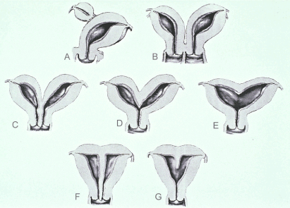
The uterine anomalies found were retrospectively grouped according to the classification of the American Fertility Society, AFS, now the American Society for Reproductive Medicine [6] (Table 1, Figure 1). The diagnostic methods used and the criteria for uterine malformations for the AFS system have been reported elsewhere [18].
Table 1. Renal tract findings in 376 women with uterine anomalies.
Class of uterine anomaly |
No. of patients |
No. of patients with renal tract status determined (%) |
Normal renal tract (%) |
Abnormal renal tract (%) |
I Agenesis-hypoplasia a. vaginal b. cervical c. tubal d. combined |
100 0 1 0 99 |
94 (94.0) 1 93 |
83 (88.3) 83 |
11 (11.7) 1 10 |
II Unicornuate a. communicating b. noncommunicating c. no cavity d. no horn |
56 0 21 27 8 |
44 (78.6) 17 19 8 |
31 (70.5) 15 12 4 |
13 (29.5) 2 7 4 |
III Didelphic |
61 |
55 (90.2) |
39 (70.9) |
16 (29.1) |
IV Bicornuate a. complete b. partial |
52 20 32 |
33 (63.5) 15 18 |
28 (84.8) 11 17 |
5 (15.2) 4 1 |
V Septate a. complete b. partial |
306 131 175 |
145 (47.4) 88 57 |
125 (86.2) 70 55 |
20 (13.8) 18 2 |
VI Arcuate |
17 |
5 (29.4) |
5 |
|
VII DES-related |
0 |
|
|
|
Total |
592 |
376 (63.5) |
311 (82.7) |
65 (17.3) |

Figure 1. Main groups of uterine anomalies. A) unicornuate uterus with rudimentary horn, B) didelphic uterus, C) complete bicornuate uterus, D) partial bicornuate uterus, E) arcuate uterus, F) septate uterus, G) subseptate uterus. Uterine agenesis and diethylstilbestrol (DES)-related T-shaped uterus are not presented.
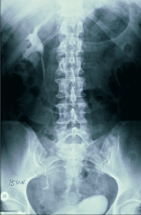
Retrospective analysis of hospital charts and imaging records revealed that in 376 (63.5 %) out of 592 patients with uterine anomalies renal tract status had been determined. Only subjects with reliable evaluation of the kidneys and lower urinary tract were accepted for the study. Renal tract status had been determined in 354 (94.1 %) cases using an intravenous pyelogram, 2D ultrasound scanning or both. Intravenous pyelography was the main method used to investigate urologic anomalies during the first phases of the present study (Figure 2). Ultrasonography has been the main means to study urological anomalies during the last three decades. Other diagnostic methods used were magnetic resonance imaging (MRI), computerized tomography imaging (CT), renography and angiography [19]. Imaging of the renal tract was not performed only during the diagnostic work-up for genital tract anomaly but also for evaluation of recurrent urinary tract infection, high blood pressure, ureterolithiasis or abdominal pain.

Figure 2. An intravenous pyelogram shows absence of the left kidney. The patient had a right-side unicornuate uterus without a rudimentary horn on the left side.
The main groups of renal tract malformations comprised unilateral renal agenesis (URA), horseshoe kidney, pelvic kidney, duplicated collecting system draining into a single ureter, bifid ureter: two ureters which unite before emptying into the bladder, or double ureters: two ureters draining separately into the bladder or genital tract [7, 10]. Findings pertaining to the renal tract were grouped according to the patient's uterine anomaly in the AFS classification [6]. The urologic findings in the survey have been partially reported in the European classification of female genital tract congenital anomalies covering malformations of the uterine cervix and vagina [18].
Qualitative data were assessed by Fisher’s exact test. The 95% confidence intervals (CI) were calculated for the estimated portions by confidence interval analysis. The institutional reviewing board in Tampere University Hospital approved the study protocols.
In all, 376 (63.5 %) out of 592 women with uterine anomalies had data on renal tract investigations (Table 1). When uterine anomalies were graded according to the AFS classification, patients with Müllerian agenesis (Class I) and patients with a didelphic uterus (Class III) had a high rate of urological investigation, 94 % and 90.2 %, respectively. Partial septate (Class Vb) and arcuate uterus (Class VI) involved the lowest rates of renal tract investigations, 32.6 % (57 of 175) and 29.4 %, respectively (Table 1).
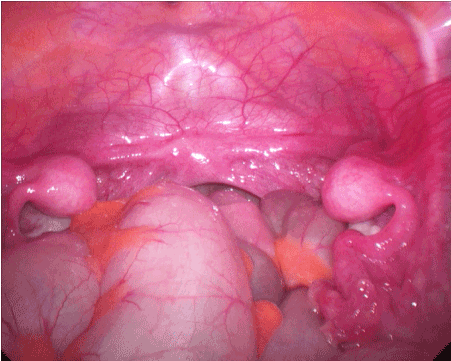
Anomalies of the kidneys and lower urinary tract were found in 65 (17.3 %, 95% CI 13.8 to 21.4)) out of the 376 women studied (Table 1). Urologic anomaly was encountered most frequently in patients with unicornuate (29.5 %) and didelphic uterus (29.1 %). Lower rates of abnormal renal tract (11.7-15.2 %) were noted in women with uterine agenesis, septate or bicornuate uterus (Figure 3). Common uterine anomalies as partial bicornuate (Class IVb), partial septate (Class Vb) and arcuate uterus (Class VI) were associated with a low rate (3.8 %, 3 of 80) of renal tract malformations (Table 1).

Figure 3. Laparoscopic view of the patient with congenital absence of the uterus and vagina shows bilateral uterine bulbs, Fallopian tubes and normal ovaries. Ultrasound revealed two normal kidneys.
Unilateral renal agenesis was the most common finding, 42 (64.6 %) out of all 65 renal tract malformations, being detected in 11.2 % (95 % CI 8.4 to 14.8) out of 376 women with uterine anomalies. Left-side renal defect occurred in 24 cases and right-side in 18 (P = 0.048). Unilateral renal agenesis was found in all main groups of uterine anomalies except for arcuate uterus (Table 2). This condition was associated with an ipsilateral obstructive vaginal septum in 22 (81.5 %) out of 27 cases with didelphic, complete bicornuate or septate uterus (Table 3). Two women with complete bicornuate uterus and 3 with complete septate uterus had no ipsilateral vaginal obstruction associated with unilateral renal agenesis. Compensatory enlargement of the solitary kidney was reported in 19 (45.2 %) cases.
Table 2. Renal tract malformations in 65 women with uterine anomalies.
Class of uterine anomaly |
No. of patients |
URAª |
Horseshoe kidney |
Pelvic kidney |
Duplex system |
Otherb |
I Agenesis b. cervical d. combined |
11 1 10 |
7 7 |
1 1 |
1 1 |
2 1 1 |
|
II Unicornuate b. noncommunicating c. no cavity d. no horn |
13 2 7 4 |
8 1 5 2 |
1 1 |
3 2 1 |
|
1 1 |
III Didelphic |
16 |
13 |
|
|
|
3 |
IV Bicornuate a. complete b. partial |
5 4 1 |
4 4 |
|
|
1 1 |
|
V Septate a. complete b. partial |
20 18 2 |
10 10 |
1 1 |
|
9 7 2 |
|
Total (%) |
65 |
42 (64.6) |
3 (4.6) |
4 (6.2) |
12 (18.5) |
4 (6.2) |
ªURA, unilateral renal agenesis.
bOther: malrotation (class II), fetal lobulation, malrotation, vascular anomaly (class III).
Table 3. Unilateral renal agenesis (URA) in women with uterine anomalies.
Uterine anomaly |
No. of patients |
URA (%) |
Left/Right |
Obstructed hemivagina |
Id Agenesis |
93 |
7 (7.5) |
6/1 |
- |
II Unicornuate |
44 |
8 (18.2) |
6/2 |
- |
III Didelphic |
55 |
13 (23.6) |
7/6 |
13 |
IVa Bicornuate |
15 |
4 (26.7) |
3/1 |
2 |
Va Septate |
88 |
10 (11.4) |
2/8 |
7 |
Total |
295 |
42 (14.2) |
24/18 |
22 |
|
|
|
|
|
The second largest group of the congenital renal tract abnormalities was duplication of the collective system. This was found in 12 (3.2 %, 95% CI 1.8 to 5.5) out of 376 women with uterine anomalies and in 18.5 % of the 65 cases with abnormal renal tract (Table 2). Nine out of 12 cases occurred in patients with a septate or subseptate uterus. Five out of 12 patients had a unilateral double pelvic system and seven also had a double ureter, one of them unilaterally two ureters emptying into the bladder. One patient with a subseptate uterus had an ectopic ureter opening into the vagina, demanding surgical treatment. Otherwise no surgical treatment for urinary tract was necessary and no severe renal diseases were found during the study period.
Eleven (16.9 %) out of 65 women with renal tract malformations had neither renal agenesis nor duplex collecting system. Sporadic urologic anomalies were horseshoe kidney (3 cases) and pelvic kidney (4 cases). Other renal tract abnormalities were renal rotation in two cases, fetal lobulation and vascular anomaly, both in one case (Table 2).
Renal tract anomalies were found in 17.3 % of 376 women with uterine anomalies. There are no previous studies comparing urologic anomalies in the main groups of the AFS classification. Oppelt and associates reported urologic anomalies in 42 (20.8 %) out of 202 patients with uterine anomalies when applying their own classification of the genital tract [20]. When vaginal abnormality alone was taken into consideration in their study, an associated developmental disturbance in the renal tract was detected in 30 % of 107 cases [20].
Unilateral renal agenesis was the most common (11.2 %) anomaly of the renal tract among all patients with a uterine anomaly as observed in previous studies [13,14,20]. The condition was mostly associated with concomitant vaginal and cervical anomalies, being found in patients with a didelphic uterus, complete bicornuate or septate uterus with obstructed hemivagina and with double or septate cervix and in those with Müllerian agenesis. Absence of one kidney was manifested in women with a unicornuate uterus without clinical signs of vaginal or cervical anomaly. Ipsilateral cervicovaginal atresia with renal agenesis in this group of the Müllerian duct anomalies has been suggested [21]. Its presence cannot be detected in clinical examination. Renal agenesis is found in women without uterovaginal anomalies [18], possibly indicating that there are different mechanisms to induce unilateral renal agenesis, although URA and concomitant vaginal and cervical anomalies are the most common manifestations.
The present study shows that common minor Müllerian duct anomalies such as partial bicornuate, subseptate and arcuate uterus are rarely associated renal tract anomalies. Concomitant cervical and vaginal anomalies are not involved in these uterine anomalies [18].
Malformations of the renal system has been the most frequent associated malformation with Müllerian duct agenesis [14,20]. German colleagues reported urologic malformations in 82 (29 %) out of 284 women with Müllerian duct agenesis [14]. Unilateral renal agenesis was the most common malformation (18.8 %) and a solitary kidney was associated with a duplex collecting system in 25.6 % of the patients [14]. The present study showed lower frequency (11.7 %) of renal tract malformations in this group (Class I). In one study 4 (10.8%) out of 37 patients with uterovaginal agenesis had absence of one kidney [22].
Urologic anomalies were found in 33 (36.3 %) out of 91 patients with a unicornuate uterus (Class II) when three studies were pooled [13,23,24]. Unilateral renal agenesis was the most frequent renal tract anomaly (17.6 %, 16/91) in all three. The corresponding figures in the present study were 29.5 % and 18.2 %, respectively. No differences were observed in the frequency of renal tract anomalies among the various subclasses of unicornuate uterus [13]. Absence of one kidney was always ipsilateral to the side of the uterine anlage.
Unilateral renal agenesis was commonly associated with a didelphic uterus (Class III) occurring in 23.6 % among 55 cases. Moutos and associates reported the presence of unilateral renal agenesis in 10 (47.6 %) out of 21 patients with a didelphic uterus [24]. The absent kidney was always ipsilateral to an obstructed hemivagina. This combination is known as the Herlyn-Werner-Wunderlich syndrome and is manifested in adolescence [17]. An obstructed hemivagina and ipsilateral renal agenesis are also found in patients with a complete bicornuate (Class IVa) and a septate uterus (Class Va) [12,15,25,26].
The left side is more commonly involved than the right in cases with renal agenesis [9]. In contrast, Vercellini and colleagues reported right-sided dominant (65 %) asymmetry in the lateral distribution of obstructed hemivagina and renal agenesis in a review of 179 women with a didelphic uterus [27]. Right- and left-side renal agenesis were equally involved in a study of 42 cases with didelphic or septate uterus and blind hemivagina [26]. The present cohort includes patients with Müllerian agenesis and unicornuate in addition to didelphic, bicornuate and septate uterus. This may be the explanation for the discrepancy in the lateral distribution of renal agenesis here compared to studies with didelphic uterus and obstructed hemivagina.
Renal agenesis commonly affects only one renal tract. A congenital solitary kidney opposite to an absent kidney is usually substantially enlarged, as in 45.2 % in the present material [7]. A congenital solitary kidney constitutes compensatory hypertrophy without renal dysfunction. Women with one kidney and a malformed uterus have more than twice the risk of the development of gestational hypertension and late-onset preeclampsia compared with those with a similar uterine anomaly but two normal kidneys [28].
Absence of one kidney and prenatal compensatory enlargement of a congenital solitary kidney are diagnosed with increasing frequency in utero with the advent of routine prenatal ultrasound screening [29,30]. The presence of a Müllerian duct anomaly should be borne in mind in these patients if they are symptomatic in adolescence, although unilateral renal agenesis is found in patients without genital tract anomalies [18].
Oppelt and associates found a double kidney or a double collecting system in 6 (3.0 %) out of 202 women with uterine malformations and in 12 (4.2 %) out of 284 patients with Müllerian agenesis [14,20]. Duplex collecting systems are seen in 0.7 % of the healthy adult population and 2-4 % of patients investigated for urinary tract symptoms [10]. In the present study duplication of the collecting system was found in 3.2 % of patients with uterine anomalies, and mostly among those with a complete or partial septate uterus. In one study, a double renal pelvis was found in 2 (5.4 %) out of 37 patients with a unicornuate uterus and none among 54 cases in two studies pooled [13, 23, 24]. Moutos and associates found a double collecting system in one (4.0 %) case among 25 patients with a didelphic uterus [24]. Two-dimensional ultrasonography may be suboptimal in the detection of trivial congenital renal tract malformations as a duplicated system and will be indefinite in distinguishing between partial and complete duplication.
The present study has some limitations. This is a retrospective study with a long enrolment period. The presence of renal tract malformations was not studied systematically and in some subgroups of uterine anomalies the number of patients undergoing urologic evaluation was low.
In conclusion, the absence of one kidney was the most frequent renal tract malformation in women with Müllerian duct anomalies. It occurred in the main groups of uterine anomaly and was manifested especially in patients with concomitant cervical and obstructive vaginal anomaly. Common minor uterine anomalies such as partial bicornuate, subseptate and arcuate uterus are rarely associated with a renal tract malformation.
The author reports no conflicts of interest. The author alone is responsible for the content and writing of the paper.
- Crouse GS (1986) Development of the female urogenital system. Semin Reprod Endocrinol 4: 1-11.
- Spencer TE, Dunlap KA, Filant J (2012) Comparative developmental biology of the uterus: insights into mechanisms and developmental disruption. Mol Cell Endocrinol 354: 34-53. [Crossref]
- Lawrence WD, Whitaker D, Sugimura H, Cunha G2021 Copyright OAT. All rights reservrastructural study of the developing urogenital tract in early human fetuses. Am J Obstet Gynecol 167: 185-193. [Crossref]
- Chan YY, Jayaprakasan K, Zamora J, Thornton JG, Raine-Fenning N, et al. (2011) The prevalence of congenital uterine anomalies in unselected and high-risk populations: a systematic review. Hum Reprod Update 17: 761-771. [Crossref]
- Dreisler E, Stampe Sørensen S (2014) Müllerian duct anomalies diagnosed by saline contrast sonohysterography: prevalence in a general population. Fertil Steril 102: 525-529. [Crossref]
- The American Fertility Society (1988) The American Fertility Society classifications of adnexal adhesions, distal tubal occlusion, tubal occlusion secondary to tubal ligation, tubal pregnancies, Müllerian anomalies and intrauterine adhesions. Fertil Steril 49: 944-955. [Crossref]
- Kerecuk L, Schreuder MF, Woolf AS (2008) Renal tract malformations: perspectives for nephrologists. Nat Clin Pract Nephrol 4: 312-325. [Crossref]
- Barakat AJ, Dougas JG (1991) Occurrence of congenital abnormalities of kidney and urinary tract in 13,775 autopsies. Urology 38: 347-350. [Crossref]
- Robson WLM, Leung AKC, Rogers RC (1995) Unilateral renal agenesis. Adv Pediatr 42: 575-592. [Crossref]
- Hartman GW, Hodson CJ (1969) The duplex kidney and related abnormalities. Clin Radiol 20: 387-400. [Crossref]
- Marshall FF, Beisel DS (1978) The association of the uterine and renal anomalies. Obstet Gynecol 51: 559-562. [Crossref]
- Li S, Qayyum L, Coakley FV, Hricak H (2000) Association of renal agenesis and Mullerian duct anomalies. J Comput Assist Tomogr 24: 829-834. [Crossref]
- Fedele L, Bianchi S, Agnoli B, Tozzi L, Vignali M (1996) Urinary tract anomalies associated with unicornuate uterus. Urology 155: 847-848. [Crossref]
- Oppelt PG, Lermann J, Strick R, Dittrich R, Strissel P, et al. (2012) Malformations in a cohort of 284 women with Mayer-Rokitansky-Küster-Hauser syndrome (MRKH). Reprod Biol Endocrinol 10: 57-63. [Crossref]
- Smith NA, Laufer MR (2007) Obstructed hemivagina and ipsilateral renal anomaly (OHVIRA) syndrome: management and follow-up. Fertil Steril 87: 918-922. [Crossref]
- Fedele L, Motta F, Frontino G, Restelli E, Bianchi S (2013) Double uterus with obstructed hemivagina and ipsilateral renal agenesis: pelvic anatomical variants in 87 cases. Hum Reprod 28: 1580-1583. [Crossref]
- Gholoum S, Puligandla PS, Hui T, Su W, Quiros E, et al. (2006) Management and outcome of patients with combined vaginal septum, bifid uterus and ipsilateral renal agenesis (Herlyn-Werner-Wunderlich syndrome). J Pediatr Surg 41: 987-992. [Crossref]
- Heinonen PK (2016) Distribution of female genital tract anomalies in two classifications. Eur J Obstet Gynecol Reprod Biol 206: 141-146. [Crossref]
- Heinonen PK (2013) Pregnancies in women with uterine malformation, treated obstruction of hemivagina and ipsilateral renal agenesis. Arch Gynecol Obstet 287: 975-978. [Crossref]
- Oppelt P, von Have M, Paulsen M, Strissel PL, Strick R, et al. (2007) Female genital malformations and their associated abnormalities. Fertil Steril 87: 335-342. [Crossref]
- Acién P, Acién M (2016) The presentation and management of complex female genital malformations. Hum Reprod Update 22: 48-69. [Crossref]
- Hall-Craggs MA, Kirkham A, Creighton SM (2013) Renal and urological abnormalities occurring with Mullerian anomalies. J Pediatr Urolog 9: 27-32. [Crossref]
- Donderwinkel PFJ, Dörr JPJ, Willemsen WNP (1992) The unicornuate uterus: clinical implications. Eur J Obstet Gynecol Reprod Biol 47: 135-139. [Crossref]
- Moutos DM, Damewood MD, Schlaff WD, Rock JA (1992) A comparison of the reproductive outcome between women with a unicornuate uterus and women with a didelphic uterus. Fertil Steril 58:88-93. [Crossref]
- Acién P, Acién M (2010) Unilateral renal agenesis and female genital tract pathologies. Acta Obstet Gynecol Scand 89: 1424-1431. [Crossref]
- Haddad B, Barranger E, Paniel BJ (1999) Blind hemivagina: long-term follow-up and reproductive performance in 42 cases. Hum Reprod 14: 1962-1964.
- Vercellini P, Daquati R, Somigliana E, Vigano P, Lanzani A, et al. (2007) Asymmetric lateral distribution of obstructed hemivagina and renal agenesis in women with uterus didelphys: institutional case series and a systematic literature review. Fertil Steril 87: 719-724. [Crossref]
- Heinonen PK (2004) Gestational hypertension and preeclampsia associated with unilateral renal agenesis in women with uterine malformations. Eur J Obstet Gynecol Reprod Biol 114: 39-43. [Crossref]
- Wiesel A, Queisser-Luft A, Clementi M, Bianca S, Stoll C, et al. (2005) Prenatal detection of congenital renal malformations by fetal ultrasonographic examination: an analysis of 709,030 births in 12 European countries. Eur J Med Genet 48: 131-144. [Crossref]
- van Vuuren SH, van der Doef R, Cohen-Overbeek TE, Goldschmeding R, Pistorius LR, et al. (2012) Compensatory enlargement of a solitary functioning kidney during fetal development. Ultrasound Obstet Gynecol 40: 665-668. [Crossref]


